




No CrossRef data available.
Article contents
Assessing the elusive cognitive deficits associated with ventromedial prefrontal damage: A case of a modern-day Phineas Gage
Published online by Cambridge University Press: 01 May 2004
Abstract
Cognitive deficits following ventromedial prefrontal damage (VM-PFD) have been elusive, with most studies reporting primarily emotional and behavioral changes. The present case illustrates the utility of a process approach to assessing cognitive deficits following VM-PFD. At age 26, C.D. acquired bilateral VM-PFD, more so in the left frontal region, following a penetrating head injury. Despite exemplary premorbid academic and military performances, his subsequent history suggests dramatic occupational and social changes, reminiscent of Phineas Gage. In fact, lesion analysis revealed similar structural damage to that estimated of Gage. C.D.'s scores on the vast majority of neuropsychological measures were average to superior (e.g., Verbal IQ = 119). However, on several new process measures, particularly those that quantify error rates on multilevel executive function and memory tasks, C.D. exhibited marked impairments. From his pattern of deficits, C.D. appeared to sacrifice accuracy for speed and to adopt liberal response strategies, implicating problems with cognitive inflexibility, impulsivity, and disinhibition. The current findings suggest that VM-PFD may be associated with a wider spectrum of cognitive deficits than previously characterized. (JINS, 2004, 10, 453–465.)
Information
- Type
- CASE STUDY
- Information
- Journal of the International Neuropsychological Society , Volume 10 , Issue 3 , May 2004 , pp. 453 - 465
- Copyright
- © 2004 The International Neuropsychological Society
INTRODUCTION
The cognitive architecture of the frontal lobes has remained one of the great enigmas of neuropsychology. Interest in the frontal lobes was sparked by the landmark case of Phineas Gage, who baffled scientists after a 13-pound tamping iron that measured approximately 3½ feet long and 2 inches in diameter shot straight through his left cheek bone, prefrontal cortex, and anterior dorsal skull (Harlow, 1848; see also Fleischman, 2002; MacMillan, 2000). Applying volumetric analysis using clues from the remains of Gage's skull, Damasio et al. (1994) reported that Gage's lesion likely involved bilateral anterior orbitofrontal cortex, polar and anterior mesial frontal cortices, and the rostral portion of the anterior cingulate gyrus, with underlying white matter involvement more extensive in the left hemisphere than the right. Gage not only survived this massive penetrating head injury, but he superficially appeared normal, particularly with respect to his intellect, language, memory, and motoric functions. However, upon returning to his preinjury work as the “most efficient and capable foreman” of the railroad track construction team, Gage displayed an array of emotional and behavioral problems that have come to be known as the archetypes of the frontal lobe syndrome. In describing these personality changes, Dr. John M. Harlow, Gage's physician, wrote in 1848, “He is fitful, irreverent, indulging at times in the grossest profanity (which was not previously his custom), manifesting but little deference for his fellows, impatient of restraint or advice when it conflicts with his desires” (Harlow, 1848). In other words, the most striking sequela of his prefrontal assault was that Gage “was no longer Gage.”
Since the famous case of Gage, theories of frontal-lobe functions have varied markedly in ascribing what role, if any, this vast cerebral region plays in mediating cognitive and behavioral functions. In the 1930s and 1940s, the predominant view was that the frontal lobes subserved the highest levels of mental functions, including the “abstract attitude,” “foresight,” and “intellectual synthesis” (Brickner, 1936; Goldstein, 1944; Goldstein & Scheerer, 1941). However, the pendulum of thought began to shift in the 1940s and 1950s, with claims of a lack of functional significance of the prefrontal cortex (Hebb, 1945; Hebb & Penfield, 1940; Teuber & Weinstein, 1954). At the forefront of this view was Hebb and Penfield's famous case study of “K.M.”, who underwent extensive, bilateral resection of the prefrontal cortex due to intractable seizures. Following the surgery, K.M. was thought to exhibit improvements in his cognitive and personality functioning. In addition, after failing to find consistent cognitive deficits in a large-scale study of over 90 World War II veterans who had suffered penetrating missile wounds to prefrontal cortex, Teuber (1968) proclaimed that the frontal lobes represented a “riddle” in terms of their role in subserving mental abilities.
Modern theories of frontal-lobe functions have attempted to resolve these early discrepancies by bifurcating the functions of prefrontal cortex into two major subregions. That is, higher-level cognitive skills, commonly referred to as executive functions, have been ascribed primarily to dorsolateral prefrontal regions, whereas emotional and behavioral regulation and control have been attributed primarily to ventromedial prefrontal cortex (Barch et al., 1997; Bechara et al., 1998; Chao & Knight, 1998; Damasio, 1995; Fuster, 2000; Goldman-Rakic & Leung, 2002; Tranel, 2002).
Support for the view that ventromedial prefrontal cortex (VM-PFC) may mediate primarily emotional rather than cognitive functions has come from both neuroanatomical and neuropsychological studies. For example, Nauta (1971) proposed that clues to the functions of VM-PFC in humans may be extracted from its vast interconnections with limbic structures, including the hypothalamus, dorsomedial thalamic nucleus, septal region, amygdala, and entorhinal and perirhinal cortices (see also Öngür & Price, 2000). Given this rich connectivity, Nauta (1971) postulated that VM-PFC serves as a link between the internal milieu via sensory and limbic inputs and behavioral responses via visceromotor effector systems. He speculated that damage to this region of cortex might alter affective and motivational associations with the environment, leading to maladaptive behavior.
Consistent with Nauta's (1971) predictions, neuropsychological case studies and experimental investigations with VM-PFD patients have also reported primarily emotional or behavioral changes but surprisingly few if any cognitive deficits. For example, several important case studies have highlighted discrepancies between the VM-PFD patient's relatively intact neuropsychological profile, including normal performances on tests of executive functions, in the face of marked emotional and behavioral changes (Angrilli et al., 1999; Dimitrov et al., 1999; Eslinger & Damasio, 1985). The case studies by Angrilli et al. (1999) and Dimitrov et al. (1999) also reported that, in spite of spared cognitive abilities, their patients were emotionally unresponsive to affectively laden stimuli as measured by electrodermal skin conductance. In addition, Rolls et al. (1994) reported an impairment in the emotional extinction of a previously rewarded stimulus in a group of VM-PFD patients who otherwise performed normally on a set of cognitive tests, including tests of verbal intelligence, paired-associate learning, and the Tower of London task. Bechara et al. (1994) designed a complex decision making task that simulates gambling within the affective context of making risk-taking judgments. Bechara et al. (1994) found that patients with VM-PFD tended to sustain heavy losses on this gambling task due to risky behavior (i.e., drawing cards from decks with an unfavorable profit-to-loss ratio due to the allure of cards in those decks with higher immediate gains). Based on these findings and the neuroanatomical connectivity of the VM-PFC, researchers have suggested that this region may play a vital role in the affective or emotional component of decision making and learning. According to the “somatic marker hypothesis” advanced by Damasio, Bechara, and others, patients with VM-PFD tend to lose the ability to guide their behavior with information from their internal milieu due to lost access to somatic feedback, which results in dysfunctional behavior (Bechara et al., 2000; Damasio, 1994).
The question remains, however, as to the role, if any, of the VM-PFC in the mediation of higher-level cognitive functions per se. On the one hand, patients with VM-PFD may truly be spared from cognitive deficits and suffer from isolated emotional and behavioral impairments. Another possibility is that VM-PFD does lead to cognitive deficits in addition to emotional/behavioral dysfunction, and traditional neuropsychological tests have generally failed to identify such deficits in these patients. In the present case study, we examined the utility of several new process-oriented neuropsychological measures designed to quantify subtle cognitive deficits that may exist over and above the emotional or behavioral changes typically found in VM-PFD patients. By process measures, we refer to the use of test conditions, scoring variables, and formulas that are designed to better elucidate the neurocognitive processes underlying aberrant test performance (Delis & Jacobson, 2000; Kaplan, 1988; Luria, 1969; Luria & Majovski, 1977; Milberg et al., 1996; White & Rose, 1997). For example, successful performance on most executive-function tasks depends upon a number of more fundamental cognitive skills; a process-oriented instrument is designed to provide separate measures reflecting the relative integrity of the component skills versus the higher-level cognitive functions. The new neuropsychological process measures discussed in this case were also designed to (1) increase the cognitive switching demands of traditional executive-function tasks; (2) quantify and provide normative data for error rates; and (3) measure the accuracy of memory recall in a novel way that factors in error (intrusion) rates (Delis et al., 2000; 2001). For example, a new cognitive-switching condition has been developed for the classic Stroop task in which the examinee must switch back and forth between naming the dissonant ink color and reading the printed word (Delis et al., 2001). This new switching condition not only taps the traditional executive function measured by this task (i.e., verbal inhibition), but it also simultaneously requires the examinee to engage in cognitive shifting while performing verbal inhibition. We hypothesized that these multilevel executive-function tasks, in conjunction with error analyses, may be more sensitive to the detection of cognitive impairment following VM-PFD than traditional executive-function tasks. We also examined whether a new memory measure called “recall discriminability,” which provides a single score of recall accuracy relative to intrusion rate (analogous to “recognition discriminability”), would be more sensitive to the detection of subtle memory impairment secondary to frontal damage than simply analyzing the level of recall accuracy alone.
The present case first caught our attention because the patient, C.D., bears striking similarities to behavioral and neuroanatomical descriptions of Phineas Gage. That is, C.D. was a relatively high-functioning man prior to acquiring VM-PFD (e.g., nearly a straight-A student in school; accelerated promotions in the military), and, in the present examination, he continued to exhibit high levels of intellectual and cognitive functioning on the vast majority of traditional neuropsychological tests administered to him (e.g., current Verbal IQ = 119). Following his brain injury while on active duty, however, C.D. displayed a precipitous decline in his social and behavioral functioning (e.g., discharge from military against his wishes; lifelong history of numerous low-level jobs such as newspaper delivery man; four marriages). Perhaps most intriguing was the fact that C.D.'s lesion site closely resembled that of Gage, including greater involvement in the left VM-PFC than the right. In this case study, we predicted that the new multilevel executive-function measures, combined with a quantification of error rates, would enhance the detection of cognitive deficits that, for decades, have been so elusive following VM-PFD.
CASE STUDY
C.D., a mixed-handed, Caucasian male, was 26 years old at the time of his accident, which occurred overseas during his military duty. This bright man (current Verbal IQ = 119), who reportedly received accelerated promotions during his military career, was involved in a motor vehicle accident in 1962. He was in the passenger-side front seat of a jeep that drove over a landmine that exploded. C.D. explained that, at the time of impact, his forehead was crushed by the metal upright on the rim of the jeep's windshield. By his account, he did not immediately lose consciousness from the blow to the head, as he recalled the explosion and someone questioning him immediately following the accident. He was reportedly placed in a drug-induced coma, and he believes he was in varying levels of a comatose state for 2–3 months following the accident. The driver of the jeep was killed in the accident.
When questioned about any physical symptoms caused by the accident, C.D. indicated that he had nerve damage in his left eye, a broken jaw, anosmia, aguesia, and diplopia. He recalled that, approximately 5 years following the accident, he underwent reconstructive surgery for placement of a metal plate in his forehead, and plastic surgery to repair facial damage from the impact of the blow. C.D. reported that he did not have any cognitive or emotional problems following the accident. In describing how his thinking skills were completely unaffected, C.D. stated that, “all the shattered bone was caught in the gray matter in front of the brain.” He added that his original doctors had explained to him that, since the skull fragments only penetrated the front part of his brain, his thinking skills were spared. During a routine physical examination at the San Diego Veterans Affairs Medical Center, C.D.'s primary care physician noted that the patient had a prior history of a head injury with placement of a metal plate in his forehead, and therefore ordered a computerized tomography (CT) head scan. When the CT scan results revealed that C.D. had bilateral prefrontal damage (see below), his physician referred the patient for neuropsychological testing.
CT Head Scan Analysis and Findings
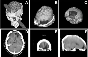
The patient could not undergo a magnetic resonance image (MRI) brain scan because of the prior surgical placement of a metal plate in his forehead. A CT head scan performed several months prior to our evaluation revealed encephalomalacia of the anterior inferior frontal region, with more involvement in the left frontal lobe than the right. This routine clinical axial head scan was acquired at a 90-deg angle to the orbitomeatal line, with various interslice distances of 5 mm to 10 mm. This clinical scan was then used to create a three-dimensional (3-D) representation of the structural brain (Figure 1). For purposes of visual comparison, panel A of Figure 1 depicts a 3-D reconstruction of the estimated damage sustained by Phineas Gage (reprinted with permission, Damasio et al., 1994). In Panels B–F, C.D.'s lesion is illustrated.

3-D depiction of likely damage in Phineas Gage (Panel A) [Reprinted with permission from H. Damasio, T. Grabowski, et al. “The return of Phineas Gage: Clues about the brain from the skull of a famous patient.” Science, 264, 1101–1105. Copyright (1994) American Association for the Advancement of Science.] and in C.D. (Panels B–F) using Analyze® (Robb, 2001) and Image (Rasband, 1996) software.
Using a combination of image analysis tools from Analyze® (Robb, 2001) and Image (Rasband, 1996) software, digital images captured from the clinical CT scan were sequentially stacked to create a 3-D reconstruction where transition points from one slice to the other were interpolated and surfaces smoothed. This permitted resectioning of the brain in any orientation, even though the original digital data were obtained only in the axial plane (as in Figure 1, panel D). As the clinical image acquisition was in the axial plane, panels E and F in Figure 1, which demonstrate the depth and extent of the left frontal lesion, were based on reslicing, and thus, these images appear somewhat fuzzy. Nonetheless, the 3-D representation of C.D.'s lesion clearly demonstrates a primarily deep, left frontal injury involving most of the inferior frontal region of the brain.
Panel B represents the 3-D model of C.D.'s brain, with skin, skull, and meninges stripped down to the frontal sinus area (same level as in panel D), with the orientation plane similar to that of Gage as depicted in panel A, where the presumed prominent area of damage was through the left frontal region. Panel C depicts C.D.'s brain, stripped of all bone from a left inferior frontal oblique view, highlighting the focal left frontal damage. Panel D represents the original clinical CT scan at the level of the frontal sinus and sphenoid depicting the extent of penetrating frontal lobe injury on the left. Of note, there is also some density change in the right frontal region, but with an absence of distinct margins needed for detailed outlining of the lesion. The patient was 65 years old at the time of the CT scan, and as expected with aging, but possibly associated with pathological changes from the injury, some generalized atrophy is evidenced by the presence of prominent temporal horns and subarachnoid cerebral spinal fluid. Panel E represents a resliced coronal view taken from the 3-D reconstruction based on axial imaging, and panel F represents a sagittal view through the mid-left frontal region. Due to the lack of distinct boundaries, the right frontal pathology is not illustrated in Figure 1, but is represented in Figure 2.

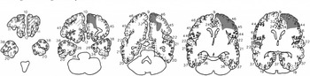
Standardized CT depiction of Brodmann's areas based on the schematic outlined by Damasio and Damasio (1989). Areas in solid red represent distinct density loss reflective of encephalomalacia. Red cross-hatched areas represent regions of likely abnormal density, but not as distinct because of artifact and resolution problems inherent to CT imaging at the level of the bone–brain interface.
Figure 2 plots the areas of distinct density change on a standardized CT schematic outlining Brodmann's areas (Damasio & Damasio, 1989). The right hemisphere is represented on the viewer's left, with all scans maintained in the standard radiographic orientation. Because of bone-hardening and volume-averaging artifact, as well as some distortion from prior facial surgeries, the most inferior sections were difficult to specifically interpret. Furthermore, we had no other imaging data other than from the clinical CT scan to assist in lesion identification. In light of these limitations, we used two methods to highlight abnormalities. Areas in solid red represent distinct density loss reflective of encephalomalacia. Red cross-hatched areas represent regions of likely abnormal density, but not as distinct because of artifact and resolution problems inherent to CT imaging at the level of the bone-brain interface. As can be seen in Figure 2, damage extended to Brodmann's areas 9 and 10 bilaterally and Brodmann's areas 44–46 in the left frontal region. As mentioned before, magnetic resonance imaging was not possible due to the metal plate, and it should be noted that the extent of the lesion may be underrepresented by the CT scan used in this analysis.
Patient Background Information
While C.D. did not acknowledge cognitive, emotional, or personality changes following his head injury, his description of his life from before and after this injury suggests a significant decline in functional status. C.D. stated that he was an excellent student who began school at age 4, skipped the 6th grade, and was “practically a straight-A student in high school.” His self-reported academic performance is consistent with his currently high Verbal IQ (119). At the age of 18, C.D. joined the military. He reportedly received accelerated promotion from the rank of Corporal to Sergeant, and he served most of his time in the Army as a boot-camp instructor for infantry training. Thus, his duties in the military called on leadership, firmness, teaching, and interpersonal skills in handling large groups of new recruits. In addition, while in the military, he stated that he completed the equivalent of 2 years of college. However, his military career, lasting 7 years, was curtailed prematurely by his head injury, and he received a medical discharge against his wishes from the Army in 1962.
After his military discharge, C.D. was never able to maintain the same job for longer than 2 or 3 years, with most jobs lasting 1–2 years. He worked in numerous, blue-collar vocations, including newspaper delivery, bartending, fiberglass boat repair, repair and sales of vacuum cleaners, and labor in a dye factory. He stated that the job he held most often was newspaper delivery. He often would return to newspaper delivery work after failing to maintain other jobs. Overall, his account of his occupational history suggested that he was a drifter, and that he was never able to achieve occupational stability or satisfaction, especially in light of his high level of intellectual functioning. Parallel to his vocational instability was his account of the breakup of three previous marriages, all occurring after his head injury, and estrangement from two of his three children.
C.D.'s medical history is otherwise notable for chronic obstructive pulmonary disease, peripheral vascular disease, hypertension, and high cholesterol. At the time of this evaluation, these medical conditions were successfully treated with medications, and he was receiving regular checkups from his primary physician. C.D. is a chronic cigarette smoker (up to 1½ packs per day over the past 45 years). He reported that he does not use illicit substances and has never been treated for or exhibited symptoms of alcohol dependence. He acknowledged moderate social alcohol consumption in the past but reported discontinuation of all alcohol use 7 years prior to this evaluation.
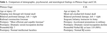
A comparison of the demographic, psychosocial, and neuroimaging findings in C.D. and Phineas Gage reveals a number of striking similarities between the two cases. These similarities are illustrated in Table 1.
Comparison of demographic, psychosocial, and neurological findings in Phineas Gage and C.D.

Neuropsychological Test Results
C.D.'s neuropsychological evaluation occurred over 40 years postinjury, which was likely due in part to (1) his having sustained his injury in the 1960s when neuropsychological services were less frequently provided in hospital settings; and (2) his being unaware of having cognitive or emotional problems. Testing was conducted on 2 separate days. On the first day, he was administered all of the neuropsychological tests, with the exception of the Delis-Kaplan Executive Function System (D-KEFS), by the first author (M.A.C.). Approximately 2 weeks later, he returned to complete the D-KEFS, which was administered by the second author (D.C.D). The second session was videotaped. Before the second session began, C.D. consented to participate in the present case study, to the release of his records, and to be videotaped.
Throughout testing, C.D. consistently put forth good effort. He was notably jocular and often would make light of tasks on which he believed he was performing poorly. He was adequately groomed and casually dressed, and he exhibited appropriate eye contact. Extensive interviewing and contact with this patient revealed the absence of overt signs of clinical depression or other major psychopathology. Facial scars were visible, especially around his forehead and eyes, as well as some asymmetry of the orbital bones, with his left eye appearing slightly lower than the right. During two clinical interviews and on a self-report measure (Beck Depression Inventory-Second Edition = 0) (BDI-2 = 0), C.D. did not acknowledge any psychiatric problems such as depressive or anxiety symptoms. Speech was normal for rate, rhythm, and articulation, with no evidence of word finding problems. His gait appeared unaffected.
Cognitive strengths
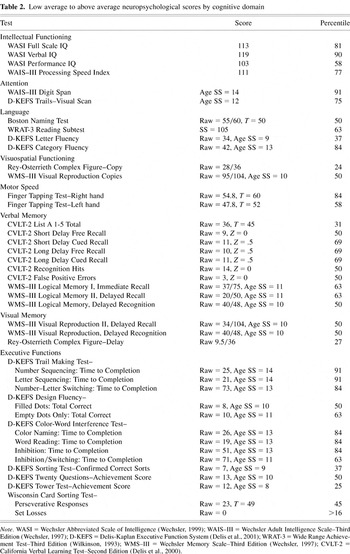
Table 2 lists those neuropsychological test scores obtained by C.D. that fell within the low average to superior ranges. As can be seen, he exhibited numerous areas of cognitive strength. Intellectual functioning as assessed by the Wechsler Abbreviated Scale of Intelligence (WASI; Wechsler, 1999) revealed overall intellectual functions in the high average range, with verbal abilities in the high average range and perceptual/organizational skills in the average range. Marked strengths included auditory attention span (Wechsler Adult Intelligence Scale–Third Edition; WAIS–III Digit Span = 91st percentile rank), knowledge of word definitions (WASI Vocabulary = 82nd percentile rank), verbal abstraction skills (WASI Similarities = 93rd percentile rank), nonverbal analysis (WASI Matrix Reasoning = 82nd percentile rank), and speed of information processing (WAIS–III Digit Symbol = 63rd percentile rank; WAIS–III Symbol Search = 84th percentile rank). In addition, C.D. performed in the average to superior ranges across a number of other cognitive domains, including word-finding skills, reading ability, drawing skills, block constructions, simple motor speed, and level of verbal and nonverbal memory.
Low average to above average neuropsychological scores by cognitive domain

C.D. also performed well on most traditional measures of executive functions. For example, his rate of perseverative responses and perseverative errors on the Wisconsin Card Sorting Test (Heaton et al., 1993) fell within the average range for his age, gender, and education. In addition, when analyzing only the traditional measure, time to completion, on the Switching Condition of the D-KEFS Trail Making Test (analogous to Trails B), his performance was above average (84th percentile rank). When examining only the traditional measure, time to completion, on the Inhibition Condition of the D-KEFS Color-Word Interference Test (analogous to the classic Stroop task), C.D.'s performance was also above average (84th percentile rank). His performances on the Verbal Fluency Test and on the two basic Design Fluency conditions of the D-KEFS were average to above average.
Cognitive deficits
On two executive-function tests—the D-KEFS Color-Word Interference Test and the D-KEFS Trail Making Test—C.D. exhibited a marked speed-accuracy tradeoff. That is, he was exceptionally fast in completing the tasks, but he exhibited a significantly elevated error rate. In addition, these errors occurred primarily on tests that required extensive cognitive switching.
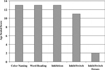
Specifically, Figure 3 shows C.D.'s results on the D-KEFS Color-Word Interference Test. This test is adapted from the classic Stroop procedure but consists of four conditions rather than three: Color Naming (a baseline condition of naming colored patches); Word Reading (a baseline condition of reading words denoting color names printed in black ink); Inhibition (the classic Stroop procedure of naming the dissonant ink color and not reading the word); and Inhibition/Switching (a new dual-level executive-function procedure that requires the examinee to switch between naming the dissonant ink color and reading the printed word). As can be seen in Figure 3, C.D.'s scaled scores on the traditional measure, time to completion, were high for all four conditions of this test, ranging from 11 (63rd percentile rank) to 13 (84th percentile rank). However, on the new Inhibition/Switching condition, C.D. made eight uncorrected errors (lower 8th cumulative percentile rank for his age group) and three corrected errors (lower 6th cumulative percentile rank for his age group). His total error score of 11 on the Inhibition/Switching condition was at the 1st percentile rank for his age (age scaled score = 2). Interestingly, his error rate was normal on the Inhibition condition, which is the classic Stroop procedure. Thus, C.D. displayed an impairment on this test, but only when (1) a switching procedure was added to the inhibition demands of the Stroop procedure; and (2) error data were quantified and normed in addition to the traditional measure of time-to-completion.

Performance on D-KEFS Color-Word Interference Test demonstrating intact speed of responding but with elevated error rates.
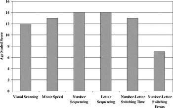
A similar finding emerged on the D-KEFS Trail Making Test. This test is comprised of five conditions: Visual Scanning (a baseline condition that involves a number cancellation task using visual stimuli the same size as those found on the other conditions); Number Sequencing (a baseline condition similar to the traditional Trails A task); Letter Sequencing (a baseline condition that evaluates alphabetical sequencing ability); Number–Letter Switching (analogous to the traditional Trails B task); and Motor Speed (a baseline condition assessing motor speed in tracing over a dotted line similar to the path found on the Number–Letter Switching condition). Figure 4 shows C.D.'s results on this test. As can be seen, C.D. achieved very high scores when his test performances were analyzed only in terms of the traditional measure, time to completion. That is, his scaled scores on the time-to-completion measures ranged from 12 (75th percentile rank) to 14 (91st percentile rank) across the five conditions. However, on the Number–Letter Switching condition, he again exhibited a significantly elevated error rate. On this condition, he made three set-loss errors (i.e., connecting two letters or two numbers; lower 5th cumulative percentile rank for his age group) and two sequencing errors (i.e., correctly switching symbol sets but connecting the wrong number or letter; lower 19th cumulative percentile rank). His total error rate was at the 16th percentile rank (age scaled score = 7). Thus, analogous to his performance on the D-KEFS Color-Word Interference Test, C.D. displayed a notable deficit on this test, but only when error data were analyzed on the multitask condition (sequencing plus switching). These findings again implicate a cognitive impairment in which accuracy is sacrificed for speed on a dual-level executive-function procedure.

Performance on D-KEFS Trail Making Test demonstrating intact speed of responding but with elevated error rates.
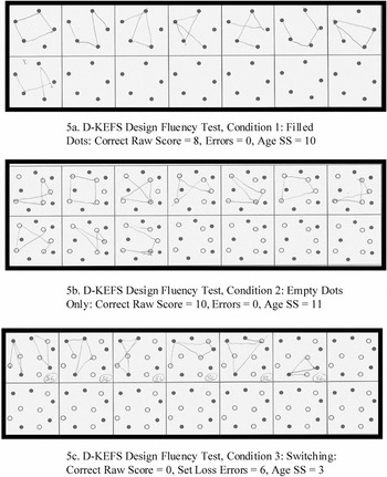
On the D-KEFS Design Fluency Test, C.D. exhibited impaired performance, but again only on a condition that required multiple executive-function skills, including cognitive switching. This test consists of three conditions: Filled Dots (which requires the examinee to draw in 60 s as many four-line designs as possible by connecting filled dots); Empty Dots Only (which requires the examinee to draw in 60 s as many four-line designs as possible by connecting only the empty dots and avoiding the filled dots); and Switching (which requires the examinee to draw in 60 s as many four-line designs as possible, only this time the examinee must switch each time by connecting a filled dot to an empty dot or vice versa). Figure 5 shows C.D.'s actual drawings on the test. As can be seen, C.D. performed well on the first two conditions of the test, which are similar to more traditional design-fluency tasks (Regard et al., 1982; Ruff et al., 1987) in that these conditions do not incorporate a switching procedure. However, on the new Switching condition, C.D. exhibited a moderate to severe impairment. Specifically, he failed to construct any designs correctly (age scaled score = 3), and he also exhibited an elevated set-loss error rate (age scaled score = 7). His set-loss errors consisted of either constructing five-line designs instead of four-line designs, or losing set by connecting two empty or two filled dots. Interestingly, his number of total attempted designs was average (age scaled score = 9). Thus, on this timed task, the accuracy of his responses again suffered at the expense of responding quickly, to the point where he failed to generate any correct designs. As with the other tests discussed above, it was the dual-task nature of Condition 3 of the D-KEFS Design Fluency test—requiring the examinee not only to generate designs but also to perform simultaneous switching operations—that proved necessary to elicit deficient performance in C.D.

Performance on Design Fluency Condition 1: Filled Dots (5a.), Condition 2: Empty Dots Only (5b.), and Condition 3: Switching (5c.).
On the D-KEFS Verbal Fluency Test, C.D. exhibited a pattern of scores often found in frontal patients in general. Specifically, his category fluency (e.g., generating names of animals) appeared intact (age scaled score = 13, a score consistent with his Verbal IQ). However, his performance in the letter condition (FAS), although average (age scaled score = 9), was significantly lower than his performance in the category condition. That is, his contrast score between letter fluency versus category fluency was at the 9th percentile rank (age scaled score = 6). On the new switching condition of the D-KEFS Verbal Fluency Test, which requires the examinee to switch between generating words from two semantic categories, C.D.'s performance was above average (age scaled score = 13). Thus, on this task, C.D. did not exhibit the switching deficit that he displayed on other switching tasks. It may be the case that C.D.'s high verbal intellectual skills assisted him in performing switching operations between two, over learned semantic categories.
The D-KEFS Sorting Test is a new problem-solving task that requires the examinee to sort six cards into two groups, three cards per group, according to eight possible sorting rules (three verbal and five nonverbal), and to describe the sorting rule after each sort made. After completing this Free Sort condition, the Sort Recognition condition is administered. In this condition, the examiner sorts the cards according to the eight target rules, and the examinee is asked to describe the sorting rule after each sort is generated by the examiner. C.D.'s ability to generate accurate sorts and descriptions of the sorting rules was average in the Free Sort condition, and his rule descriptions were average in the Sort Recognition condition. However, in the Sort Recognition condition, he also produced a significantly elevated number of incorrect descriptions of the sorting rules used (age scaled score = 6). Several of his incorrect rule descriptions reflected a loss of set in that he would describe how two rather than all three cards in a group were similar (e.g., “this group has two yellow cards and one blue card, and this group has two blue cards and one yellow card”). The Sort Recognition condition requires that the examinee have the cognitive flexibility to switch between eight possible sorting principles, which may have played a role in eliciting C.D.'s elevated error rate on this task.
On the Wisconsin Card Sorting Test (WCST), C.D. exhibited an unusual profile in that, at the start of the test, he spontaneously reported the three target rules (color, shape, and number), and his perseverative responses (23) fell within normal limits (T-score = 49; 45th percentile rank). However, he generated 30 “Other” responses (i.e., the sorted card did not match the key card on any of the three target rules). His Total Error rate was thus elevated (T-score = 30, 2nd percentile rank), and he successfully completed only one categorical sort (lower 11th to 16th cumulative percentile rank). His elevated “Other” responses likely reflected set-loss errors, similar to his errors on other executive-function tests. That the WCST places extensive demands on cognitive switching also likely played a role in eliciting these set-loss errors in C.D.
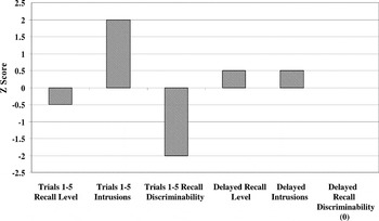
Finally, on the California Verbal Learning Test–Second Edition (CVLT–II), C.D.'s scores on the traditional measures of memory performance—level of correct responses—generally fell within the average range on all of the immediate and delayed recall and recognition trials (see Figure 6). Specifically, his immediate free-recall performance across the five learning trials (T-score = 45), his delayed free- and cued-recall scores (z-scores ranged from 0 to +0.5), and his delayed yes/no recognition discriminability (z-score = 0) all fell solidly within the average range. However, C.D. generated an elevated intrusion rate, but only on the immediate-recall trials (z-score = 2) and not on the delayed-recall trials (z-score = .5). In addition, on a new process measure called “Recall Discriminability” (Delis et al., 2000), which provides a single measure reflecting recall accuracy relative to intrusion rate (analogous to Recognition Discriminability, which reflects recognition hits relative to false positives), C.D.'s score was impaired (z-score = −2). Thus, analogous to his performance on several of the executive-function tests, C.D. had a tendency to spoil the accuracy of his recall with incorrect responses. As with many of the other tests discussed above, C.D.'s deficient performance on this memory test would have gone undetected if error measures had not been scored and normed.

Summary CVLT-II performances revealing impaired recall discriminability on immediate-recall trials.
DISCUSSION
Researchers have attempted to resolve long-standing enigmas surrounding the functions of the frontal lobes by demarcating two major subregions of the prefrontal cortex, with higher-level cognitive functions ascribed primarily to dorsolateral prefrontal regions, and emotional/behavioral regulation attributed primarily to ventromedial prefrontal cortex (VM-PFC; Barch et al., 1997; Bechara et al., 1998; Chao & Knight, 1998; Damasio, 1995; Fuster, 2000; Goldman-Rakic & Leung, 2002; Tranel, 2002). Consistent with this distinction, recent case studies and investigations of patients with VM-PFC damage have generally reported deficient emotional/behavioral functions in the face of intact cognitive skills, including normal performances on traditional tests of executive functions (e.g., Angrilli et al., 1999; Dimitrov et al., 1999; Eslinger & Damasio, 1985).
However, the present case study calls into question the view that VM-PFC fails to play a role in mediating cognitive functions. Our patient, C.D., whose VM-PFC damage was remarkably similar to that of Phineas Gage, exhibited the classic emotional and behavioral changes associated with damage to VM-PFC, including disinhibition, jocularity, and a decline in social and occupational functioning. In addition, on the vast majority of traditional neuropsychological measures administered to C.D., his scores generally fell within the average to above-average ranges (e.g., Verbal IQ = 119). In this sense, C.D.'s overall profile was consistent with other reported cases of VM-PFD, with the most salient problems appearing to be in the emotional/behavioral domain. However, on several new process-oriented neuropsychological tests, C.D. exhibited marked cognitive deficits. The tests that were most sensitive to the detection of cognitive deficits in C.D. appeared to share two common threads, including (1) task demands that tapped multiple executive functions simultaneously, including cognitive shifting; and (2) scoring systems that went beyond the traditional measures of time-to-completion or level of accuracy, and also quantified and normed error types.
For example, C.D. exhibited marked deficits on dual-level executive-function tasks that required both a traditional higher-level skill plus cognitive switching. One such task is the D-KEFS Color-Word Interference Test, which is a new Stroop procedure. This test contains four testing conditions instead of the traditional three conditions: (1) a baseline task of naming of color patches; (2) a baseline task of reading of words denoting colors but printed in black ink; (3) the classic Stroop inhibition condition of naming the dissonant ink color of color words; and (4) a new Inhibition/Switching condition that requires the examinee to switch between naming the dissonant ink color and reading the printed word. On this test, C.D.'s scores on the traditional “time-to-completion” measures were average to above average across all four conditions. However, he generated 11 errors on the new Inhibition/Switching condition, a performance that was at the 1st percentile rank for his age group. Thus, C.D. exhibited a significant impairment on this test, but only when (1) a switching procedure was added to the traditional inhibition task of the Stroop procedure; and (2) error data were scored and normed.
As another example, the D-KEFS Design Fluency Test contains two conditions that are similar to the more traditional procedures (i.e., drawing designs by connecting dots, with Condition 1 using filled dots and Condition 2 using empty dots). This test also contains a new third condition that simultaneously requires both design fluency and switching (i.e., drawing designs by always connecting a filled dot to an empty dot or vice versa). On the more traditional design-fluency tasks, C.D.'s performances were average, with normal error rates. However, on the new condition that taps both design fluency and shifting skills, C.D.'s performance was moderately to severely impaired. All of the designs he generated on this condition were inaccurate due to set-loss errors (e.g., failing to switch dots).
C.D. also exhibited elevated set-loss errors on other tasks assessing multiple executive-function and shifting skills, including the WCST, the D-KEFS Sorting Test, and the D-KEFS Trail Making Test. These findings suggest that, as task demands increasingly require higher-level processing plus cognitive flexibility, C.D. was prone to lose set and contaminate the accuracy of his responses with high error rates.
C.D.'s performances on memory tests were all in the average to above-average ranges in terms of the traditional measure of level of correct responses. On the CVLT–II, which is an unstructured memory task that taps both memory processes and higher-level organizational skills such as semantic clustering, his levels of correctly recalled responses across the various trials were also average. However, C.D. exhibited an elevated intrusion rate on the immediate-recall trials of the CVLT–II. His tendency to generate intrusion errors on the immediate-recall trials but not the delayed-recall trials may reflect problems with disinhibition, because the presented target words on immediate recall may pull for free-associative responses in such patients (see Delis et al., 2000). Analogous to his performance on the multistep executive-function tasks discussed above, C.D.'s memory deficiencies were revealed only on a memory task that (1) simultaneously taps multiple memory and organizational processes; and (2) provides scores and norms that factor in error types. Thus, on this multifactorial memory task, C.D.'s accuracy level was normal, but his correct responses were again contaminated by a high error (intrusion) rate.
The neurocognitive mechanisms that may underlie C.D.'s deficient performance on the multistep executive-function and memory tasks may be related to problems in speed/accuracy tradeoff and a liberal response style. C.D. performed well on several traditional executive-function tasks such as the classic Stroop procedure and basic design-fluency tasks; however, as task complexity was increased with the addition of switching procedures to these tests, C.D. continued to respond quickly but at the expense of generating elevated error rates. His continued rapid response style, even when presented with increasingly complex tasks, suggests an inflexibility in behavior. That is, C.D. did not appear to be able to modify his problem-solving strategy by slowing his response rate on the more complex tasks in order to provide more accurate responses. In addition, C.D. may have exhibited a subtle form of disinhibition that surfaced only on the most complex executive-function and memory tasks and resulted in a liberal response style. Such a style may explain why C.D.'s accurate responses were often contaminated by set-loss errors on executive-function tasks and by intrusion errors on memory testing.
The profile of cognitive strengths and weaknesses exhibited by C.D. may explain in part the nature of his occupational difficulties following his brain injury. The fact that he performed so well on so many cognitive tests suggests that, on first impression, C.D. may appear to others to be highly competent and capable of handling many tasks and situations. However, with time, the accuracy of his work performance may be vulnerable to erroneous behaviors, particularly when assigned a larger number or more complex tasks. This may be one reason why C.D. could typically obtain a job but had difficulty maintaining it for longer than 1 or 2 years. In addition, his behavioral features of jocularity, impulsivity, and disinhibition, accompanied with a lack of awareness of these deficits, also likely contributed to his problems in maintaining consistent occupational and psychosocial functioning (see also Crosson et al., 1989; Toglia & Kirk, 2000).
In light of the current findings, further exploration of the nature of cognitive deficits associated with VM-PFD seems warranted. For example, poor performance on Bechara's gambling task by patients with VM-PFD has been attributed to a deficit in the affective component of decision making due to a loss of somatic feedback during rewards and punishments. However, cognitive disinhibition might also explain the poor performance of VM-PFD patients on this task and would be consistent with observations of C.D., who generated more errors on increasingly complex tasks. The present case study also illustrates the utility of going beyond traditional scoring methods often used in executive-function tasks and incorporating additional process measures. For example, traditional executive-function tasks often use only time-to-completion scores (e.g., as found on the traditional Trail Making and Stroop instruments). This case highlights the enhanced sensitivity afforded when error measures and norms are incorporated into executive-function testing procedures. In addition, this case illustrates how the use of multilevel executive-function tasks (e.g., inhibition plus switching) may capture higher-level deficits that may otherwise be missed by traditional procedures. Additional cases with VM-PFD should be examined with the same types of process measures that captured C.D.'s cognitive deficits to determine if VM-PFD leads to a consistent pattern of deficits, including difficulties with complex switching tasks and disinhibition.
CONCLUSION
The case of C.D. invites the hypothesis that the ventromedial prefrontal cortex may not be as “silent” with respect to mediating cognitive functions as previously thought. That is, the bifurcation of prefrontal cortex into two functional subregions, with higher-level cognitive functions mediated primarily by dorsolateral prefrontal regions and emotional/behavioral regulation subserved primarily by ventromedial prefrontal cortex, may be inaccurate. Rather, it may be the case that both regions mediate cognitive functions, but that the nature of the processes subserved by these regions may differ. One possibility is that cognitive functions are organized in the frontal lobes along a continuum, with increasingly higher-order processes represented in the most anterior regions of the frontal lobes (i.e., areas within VM-PFC). Whereas single-level or less complex executive-function tasks (e.g., the classic Stroop procedure) may be sensitive to damage in dorsolateral prefrontal regions, multilevel executive-function tasks (e.g., adding a switching procedure to the classic Stroop task) may be needed to detect cognitive deficits following VM-PFC lesions. That is, the highest levels of cognitive and emotional integration may occur in ventromedial prefrontal cortex, which, when compromised, may affect an individual's ability to perform multilevel executive-function and memory tasks.
ACKNOWLEDGMENTS
We thank Wes Houston, Karyn Moriel, and Mindy Kane for their assistance with this case.
References
REFERENCES
Angrilli, A.,
Palomba, D.,
Cantagallo, A.,
Maietti, A., &
Stegagno, L.
(1999).
Emotional impairment after right orbitofrontal lesion in a patient
without cognitive deficits.
NeuroReport,
10,
1741–1746.CrossRefGoogle Scholar
Barch, D.M.,
Braver, T.S.,
Nystrom, L.E.,
Forman, S.D.,
Noll, D.C., &
Cohen, J.D.
(1997).
Dissociating working memory from task difficulty in human
prefrontal cortex.
Neuropsychologia,
35,
1373–1380.CrossRefGoogle Scholar
Bechara, A.,
Damasio, A.R.,
Damasio, H., &
Anderson, S.W.
(1994).
Insensitivity to future consequences following damage to human
prefrontal cortex.
Cognition,
50,
7–15.CrossRefGoogle Scholar
Bechara, A.,
Damasio, H.,
Tranel, D., &
Anderson S.W.
(1998).
Dissociation of working memory from decision making within the
human prefrontal cortex.
Journal of Neuroscience,
18,
428–437.CrossRefGoogle Scholar
Bechara, A.,
Damasio, H., &
Damasio, A.R.
(2000).
Emotion, decision making and the orbitofrontal cortex.
Cerebral Cortex,
10,
295–307.CrossRefGoogle Scholar
Brickner, R.M.
(1936).
The intellectual functions of the frontal lobes: A study based upon
observation of a man after partial bilateral frontal lobectomy.
New York:
The Macmillan Company.
Chao, L. &
Knight, R.T.
(1998).
Contribution of human prefrontal cortex to delay performance.
Journal of Cognitive Neuroscience,
10,
167–177.CrossRefGoogle Scholar
Crosson, B.,
Barco, P.P.,
Velozo, C.A., &
Bolesta, M.M.
(1989).
Awareness and compensation in postacute head injury rehabilitation.
Journal of Head Trauma Rehabilitation,
4,
46–54.CrossRefGoogle Scholar
Damasio, A.R.
(1994).
Descartes' error: Emotion, reason, and the human brain.
New York:
Grosset/Putnam.
Damasio, A.R.
(1995).
On some functions of the human prefrontal cortex.
New York:
New York Academy of Sciences.
Damasio, H. &
Damasio, A.R.
(1989).
Lesion analysis in neuropsychology.
New York:
Oxford University Press.
Damasio, H.,
Grabowski, T.,
Frank, R.,
Galaburda, A.M., &
Damasio, A.R.
(1994).
The return of Phineas Gage: Clues about the brain from the skull of
a famous patient.
Science,
264,
1102–1105.CrossRefGoogle Scholar
Delis, D.,
Kaplan, E., &
Kramer, J.
(2001)
Delis-Kaplan Executive Function System manual.
San Antonio, Texas:
The Psychological Corporation.
Delis, D.C. &
Jacobson, M.
(2000).
Neuropsychological testing.
Encyclopedia of Psychology.
New York:
Oxford University Press and the American Psychological Association.
Delis, D.C.,
Kramer, J.H.,
Kaplan, E., &
Ober, B.A.
(2000).
California Verbal Learning Test–Second Edition, Adult Version
manual.
San Antonio, Texas:
The Psychological Corporation.
Dimitrov, M.,
Phipps, M.,
Zahn, T.P., &
Grafman, J.
(1999).
A thoroughly modern Gage.
Neurocase,
5,
345–354.CrossRefGoogle Scholar
Eslinger, P.J. &
Damasio, A.R.
(1985).
Severe disturbance of higher cognition after bilateral frontal lobe
ablation: Patient EVR.
Neurology,
35,
1731–1741.CrossRefGoogle Scholar
Fleischman, J.
(2002).
Phineas Gage: A gruesome but true story about brain science.
New York:
Houghton Mifflin Co.
Fuster, J.M.
(2000).
Prefrontal neurons in networks of executive memory.
Brain Research Bulletin,
52,
331–336.CrossRefGoogle Scholar
Goldman-Rakic, P.S. &
Leung, H.
(2002).
Functional architecture of the dorsolateral prefrontal cortex in
monkeys and humans. In
D.T. Stuss &
R.T. Knight (Eds.),
Principles of frontal lobe function (pp.
85–95).
London:
Oxford University Press.
Goldstein, K.
(1944).
The mental changes due to frontal lobe damage.
Journal of Psychology,
17,
187–208.CrossRefGoogle Scholar
Goldstein, K. &
Scheerer, M.
(1941).
Abstract and concrete behavior: An experimental study with special
tests.
Psychological Monographs,
53,
151.CrossRefGoogle Scholar
Harlow, J.M.
(1848).
Passage of an iron rod through the head.
Boston Medical Surgery Journal,
39,
389–393.Google Scholar
Heaton, R.K.,
Chelune, G.J.,
Talley, J.L.,
Kay, G.G., &
Curtiss, G.
(1993).
Wisconsin Card Sorting Test manual.
Odessa, Florida:
Psychological Assessment Resources, Inc.
Hebb, D.O.
(1945).
Man's frontal lobes.
Archives of Neurology and Psychiatry,
54,
10–24.CrossRefGoogle Scholar
Hebb, D.O. &
Penfield, W.
(1940).
Human behavior after extensive bilateral removal from the frontal
lobes.
Archives of Neurology and Psychiatry,
43,
421–438.CrossRefGoogle Scholar
Kaplan, E.
(1988).
A process approach to neuropsychological assessment. In
T. Boll &
B.K. Bryant (Eds.),
The Master lecture series: Vol. 7. Clinical neuropsychology and
brain function: Research, measurement, and practice (pp.
127–167).
Washington, DC:
American Psychological Association.
Luria, A.R. &
Majovski, L.V.
(1977).
Basic approaches used in American and Soviet clinical
neuropsychology.
American Psychologist,
32,
959–968.CrossRefGoogle Scholar
MacMillan, M.
(2000).
An odd kind of fame: Stories of Phineas Gage.
Cambridge:
Cambridge University Press.
Milberg, W.P.,
Hebben, N., &
Kaplan, E.
(1996).
The Boston process approach to neuropsychological assessment. In
I. Grant &
K. M. Adams (Eds.),
Neuropsychological assessment of neuropsychiatric disorders
(2nd ed., pp.
58–80).
New York:
Oxford University Press.
Nauta, W.J.H.
(1971).
The problem of the frontal lobe: A reinterpretation.
Journal of Psychiatric Research,
8,
167–187.CrossRefGoogle Scholar
Öngür, D. &
Price, J.L.
(2000).
The organization of networks within the orbital and medial
prefrontal cortex of rats, monkeys and humans.
Cerebral Cortex,
10,
206–219.CrossRefGoogle Scholar
Rasband, W.
(1996).
IMAGE (Version 1.60).
Washington D.C.:
National Institute of Health.
Regard, M.,
Strauss, E., &
Knapp, P.
(1982).
Children's production on verbal and non-verbal fluency tasks.
Perceptual and Motor Skills,
55,
839–844.CrossRefGoogle Scholar
Robb, R.A.
(2001).
ANALYZE: The biomedical imaging resource at Mayo Clinic.
IEEE Transactions on Medical Imaging,
20,
854–867.CrossRefGoogle Scholar
Rolls, E.T.,
Hornak, J.,
Wade, D., &
McGrath, J.
(1994).
Emotion-related learning in patients with social and emotional
changes associated with frontal lobe damage.
Journal of Neurology, Neurosurgery, and Psychiatry,
57,
1518–1524.CrossRefGoogle Scholar
Ruff, R.M.,
Light, R.H., &
Evans, R.W.
(1987).
The Ruff Figural Fluency Test: A normative study with adults.
Developmental Neuropsychology,
3,
37–51.CrossRefGoogle Scholar
Teuber, H.L.
(1968).
Disorders of memory following penetrating missile wounds of the
brain.
Neurology,
18,
287–288.Google Scholar
Teuber, H.L. &
Weinstein, S.
(1954).
Performance on a formboard task after penetrating brain injury.
Journal of Psychology,
38,
177–190.CrossRefGoogle Scholar
Toglia, J. &
Kirk, U.
(2000).
Understanding awareness deficits following brain injury.
NeuroRehabilitation,
15,
57–70.Google Scholar
Tranel, D.
(2002).
Emotion, decision making, and the ventromedial prefrontal cortex.
In
D.T. Stuss &
R.T. Knight (Eds.),
Principles of frontal lobe function (pp.
338–352).
London:
Oxford University Press.
Wechsler, D.
(1997).
Wechsler Adult Intelligence Scale–Third Edition and Wechsler
Memory Scale–Third Edition technical manual.
San Antonio, Texas:
The Psychological Corporation.
Wechsler, D.
(1999).
Wechsler Abbreviated Scale of Intelligence manual.
San Antonio, TX:
The Psychological Corporation.
White, R.F. &
Rose, F.E.
(1997).
The Boston process approach: A brief history and current
practice. In
G. Goldstein &
T.M. Incagnoli (Eds.),
Contemporary approaches to neuropsychological assessment. Critical
issues in neuropsychology (pp.
171–211).
New York:
Plenum Press.
Wilkinson, G.S.
(1993).
The Wide Range Achievement Test–Third Edition administration
manual.
Wilmington, Delaware:
Wide Range, Inc.
3-D depiction of likely damage in Phineas Gage (Panel A) [Reprinted with permission from H. Damasio, T. Grabowski, et al. “The return of Phineas Gage: Clues about the brain from the skull of a famous patient.” Science, 264, 1101–1105. Copyright (1994) American Association for the Advancement of Science.] and in C.D. (Panels B–F) using Analyze® (Robb, 2001) and Image (Rasband, 1996) software.
Standardized CT depiction of Brodmann's areas based on the schematic outlined by Damasio and Damasio (1989). Areas in solid red represent distinct density loss reflective of encephalomalacia. Red cross-hatched areas represent regions of likely abnormal density, but not as distinct because of artifact and resolution problems inherent to CT imaging at the level of the bone–brain interface.
Comparison of demographic, psychosocial, and neurological findings in Phineas Gage and C.D.
Low average to above average neuropsychological scores by cognitive domain
Performance on D-KEFS Color-Word Interference Test demonstrating intact speed of responding but with elevated error rates.
Performance on D-KEFS Trail Making Test demonstrating intact speed of responding but with elevated error rates.
Performance on Design Fluency Condition 1: Filled Dots (5a.), Condition 2: Empty Dots Only (5b.), and Condition 3: Switching (5c.).
Summary CVLT-II performances revealing impaired recall discriminability on immediate-recall trials.
 You have
Access
- 75
- Cited by