Introduction
Chronic rhinosinusitis with nasal polyps is a common chronic rhinological disease that causes nasal obstruction, a runny nose, headache, chronic sinus infection and diminishment of the sense of smell. It can significantly affect the quality of life of patients and contributes considerably to medical costs.Reference Fokkens, Lund, Mullol, Bachert, Alobid and Baroody 1 In the general population, the prevalence of nasal polyps is reported as ranging from 2 to 4 per cent.Reference Fokkens, Lund, Mullol, Bachert, Alobid and Baroody 1
Nasal polyps are characterised by inflammatory cell accumulation and marked tissue remodelling.Reference Fokkens, Lund, Mullol, Bachert, Alobid and Baroody 1 – Reference Li, Zhang, Li, Lin, Li and Curotto de Lafaille 3 However, nasal polyps in patients from Western countries predominantly show T helper type 2 (Th2) cytokine skewing and abundant eosinophilic infiltration,Reference Fokkens, Lund, Mullol, Bachert, Alobid and Baroody 1 whereas growing evidence suggests that nasal polyps in patients from Asian countries exhibit a mixed T cell immune response and more neutrophilic infiltration.Reference Baba, Kagoya, Kondo, Suzukawa, Ohta and Yamasoba 4 , Reference Gao, Ng, Li, Li, Duan and Shen 5 Even so, nasal polyps from both Western and Asian countries show similar epithelial remodelling, including epithelial hyperplasia, goblet cells hyperplasia and squamous metaplasia.
It is well known that a close relationship exists between upper and lower respiratory tract diseases: the presence of allergic rhinitis has been confirmed as one of the risk factors for the development of asthma, and the link between allergic rhinitis and asthma is explained by the ‘united airway disease’ hypothesis.Reference Fokkens, Lund, Mullol, Bachert, Alobid and Baroody 1 , Reference Brozek, Bousquet, Baena-Cagnani, Bonini, Canonica and Casale 6 Recently, nasal polyps patients without clinically diagnosed lung disease were found to have latent lung obstruction;Reference Kariya, Okano, Oto, Higaki, Makihara and Haruna 7 chronic rhinosinusitis patients were reported to have significant obstructive lung function changes, regardless of the presence of asthma;Reference Kariya, Okano, Higaki, Noyama, Haruna and Ishihara 8 and decreased lung functions were found in rhinitis patients with sputum eosinophilia, in the absence of airway hyperresponsiveness.Reference Yang, Lee, Kim, Song, Kim and Kwon 9 However, the pulmonary function of patients with nasal polyps is not yet fully understood, and the relationship between histopathological features in tissue and lower airway disease manifestations remains largely unclear.
Against this background, we conducted a comprehensive histological study of a large sample size of Chinese nasal polyps patients, to determine: their demographic and clinical factors, the main features of inflammatory cellular infiltration, epithelial remodelling patterns, and their association with lung function parameters.
Materials and methods
Patients and samples
A total of 99 patients with chronic rhinosinusitis with nasal polyps, who underwent endoscopic sinus surgery at The Second Hospital of Shandong University and the Jinan Central Hospital Affiliated to Shandong University, China, were recruited between 2014 and 2016, after giving informed consent. The diagnosis of chronic rhinosinusitis with nasal polyps was made based on the definition provided in the European Position Paper on Rhinosinusitis and Nasal Polyps 2012.Reference Fokkens, Lund, Mullol, Bachert, Alobid and Baroody 1 Patients with nasal polyps associated with chronic obstructive pulmonary disease, diffuse panbronchiolitis or cystic fibrosis were excluded from this study. Smokers were defined as current cigarette smokers who consume one or more packs of cigarettes a day, averaged over one year. Atopy was confirmed by either a serum-specific immunoglobulin E test or a skin prick test for common allergens. Asthma was defined as self-reported, physician-diagnosed asthma. The study protocol was approved by the Institutional Review Board of the Second Affiliated Hospital, Shandong University, China.
Blood samples were collected prior to all endoscopic sinus surgical procedures and the eosinophil count in peripheral blood was determined. An eosinophil percentage in the total white cell count in peripheral blood that exceeded 5.0 per cent was defined as peripheral blood eosinophilia. A neutrophil percentage in the total white cell count in peripheral blood that exceeded 70.0 per cent was defined as peripheral blood neutrophilia.
The radiographic severity of the nasal polyps was assessed prior to endoscopic sinus surgery according to the Lund–Mackay computed tomography (CT) staging system.
Pulmonary function was tested with the MasterScreen pulmonary function testing system (Jaeger, Würzburg, Germany). The following parameters were measured or calculated: per cent predicted forced vital capacity; per cent predicted forced expiratory volume in 1 second; forced expiratory volume in 1 second/forced vital capacity ratio; per cent predicted maximal expiratory flow rate at 25, 50 and 75 per cent of vital capacity; and per cent predicted maximal mid-expiratory flow.
Histological staining
The intra-operative biopsy specimens of the nasal polyps were embedded in paraffin, and sectioned at 4 µm with a microtome (Leica, Wetzlar, Germany). Haematoxylin and eosin staining was used to highlight the eosinophils. The neutrophils were stained with mouse anti-human neutrophil elastase monoclonal antibody (clone NP57; Dako, Glostrup, Denmark). Immunohistochemical staining was performed with a modified horseradish peroxidase technique using the Dako Cytomation EnVision+ System, HRP. A 0.3 per cent hydrogen peroxide solution was used to block endogenous peroxidase activity for 5 minutes. Sections were then stained with mouse primary antibody and incubated at 4 °C overnight. Slides were subsequently incubated using the Dako system at room temperature for 30 minutes; this was followed by an application of horseradish peroxidase substrate (diaminobenzidine) for colour development. After this, all slides were counterstained with haematoxylin.
Immunofluorescence staining was also performed to assess the epithelium of the nasal polyps. The antibodies of goblet cell marker (mucin 5AC (MUC5AC), an oligomeric mucus/gel-forming protein coding gene), ciliated cell marker (α-tubulin and βIV-tubulin) and squamous cell marker (involucrin) used were: rabbit anti-human MUC5AC polyclonal antibody (Santa Cruz Biotechnology, Dallas, Texas, USA); mouse anti-human βIV-tubulin monoclonal antibody (clone ONS.1A6; Abcam, Cambridge, Massachusetts, USA); rabbit anti-human acetyl-α-tubulin monoclonal antibody (Cell Signaling Technology, Boston, Massachusetts, USA); and mouse anti-human involucrin monoclonal antibody (clone SY5; Abcam). The deparaffinised sections were processed with target retrieval buffer (Dako). The slides were subsequently incubated with primary antibodies at 4 °C overnight. They were then incubated with Alexa Fluor 488 or 594 conjugated secondary antibodies (goat anti-mouse or anti-rabbit immunoglobulin G, heavy and light chains; Molecular Probes, Carlsbad, California, USA) at 1:400 in the dark at room temperature for 1 hour. This was followed by mounting with anti-fade reagent using 4′, 6-diamidino-2-phenylindole (DAPI) (Molecular Probes).
Histological evaluation
All samples were coded confidentially and assessed by two independent examiners who followed the same protocol. Three individual fields with high-density inflammatory cell infiltration were randomly selected. Eosinophils and neutrophils were counted at ×400 magnification (high-power field). The proportion of either type of cells in the three high-power fields with respect to the total number of inflammatory cells in each high-power field was calculated using the following formula: proportion of cell type = (n1 + n2 + n3) / (m1 + m2 + m3) × 100 per cent, where n1, n2 and n3 are the numbers of the inflammatory cell type in question in three high-power fields, and m1, m2 and m3 are the total number of inflammatory cells in these three high-power fields. The specimens were categorised as eosinophilia or neutrophilia when the proportion of eosinophils or neutrophils, respectively, exceeded 10 per cent of the total number of inflammatory cells.
In the evaluation of the epithelium, epithelial hyperplasia was defined as epithelium with more than four layers of cells. Goblet cell hyperplasia was defined as two or more layers of goblet cells in the epithelium. Squamous metaplasia was identified in specimens where the epithelium had lost its pseudostratified columnar epithelial structure, and showed an absence of goblet cell and cilia, and this was replaced by squamous epithelium. Where the presence of squamous metaplasia was uncertain, staining for involucrin, a marker of squamous metaplasia, was performed.
Statistical analysis
Statistical analyses were conducted using SPSS statistical software (IBM, Armonk, New York, USA). The Mann–Whitney U test was employed to assess for differences between groups. Correlations were analysed using the Pearson correlation test. Values of p < 0.05 were considered statistically significant.
Results
Clinical characteristics
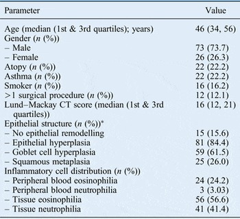
The demographic data and clinical characteristics of the study subjects are presented in Table I. There was a male predominance of 73.7 per cent. Of the 99 nasal polyps available for analysis, 22 (22.2 per cent) were from patients suffering from allergic rhinitis at the same time, and 22 (22.2 per cent) had a history of physician-diagnosed asthma. Sixteen per cent of the patients were smokers. Of the patients, 87.9 per cent were diagnosed with nasal polyps for the first time, whereas 12.1 per cent had previous nasal polyps surgical history. The median Lund–Mackay CT score was 16.
Clinical characteristics and histopathological findings

Total n = 99. *Three tissue samples with poor epithelial structure were not evaluated. CT = computed tomography
Histopathological features
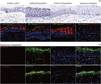
A total of 96 nasal polyps specimens were analysed for epithelial structure. Three specimens were not analysed because of the poor quality of the epithelial cells, caused by biopsy or processing techniques. Three patterns of epithelial remodelling were identified: epithelial hyperplasia, goblet cell hyperplasia and squamous metaplasia. Figure 1 shows the typical histological appearance of epithelial remodelling. The most common finding was epithelial hyperplasia (84.4 per cent), followed by goblet cell hyperplasia (61.5 per cent) and squamous metaplasia (26.0 per cent). All three types of epithelial remodelling were present in 18.8 per cent of the specimens. In 15.6 per cent of the samples, epithelial remodelling was not found (Table I).
(a) Haematoxylin and eosin and immunofluorescence staining (βIV-tubulin, MUC5AC and 4′,6-diamidino-2-phenylindole (DAPI)) from the healthy control and three types of epithelial remodelling in nasal polyps; and (b) immunofluorescence staining of specimens with squamous metaplasia. Note the complete loss of goblet cells (stained by MUC5AC) and cilia (stained by α-tubulin). All photographs were taken with a light microscope at ×400 magnification (scale bar = 20 µm).

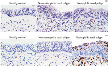
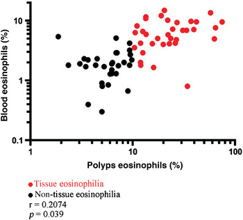
Eosinophilic infiltration in the tissue was the most common finding (n = 56, 56.6 per cent). Up to 41.4 per cent of nasal polyps showed neutrophilic infiltration (Table I). Moreover, 26.3 per cent of nasal polyps showed both eosinophilic infiltration and neutrophilic infiltration. Figure 2 shows the typical staining of eosinophils and neutrophils in inferior turbinates from healthy controls, and patients with non-eosinophilic nasal polyps, eosinophilic nasal polyps, non-neutrophilic nasal polyps and neutrophilic nasal polyps. Of the study subjects, 24.2 per cent showed peripheral blood eosinophilia, while only 3.03 per cent showed peripheral blood neutrophilia. As shown in Figure 3, eosinophils in nasal polyps tissue revealed a significantly positive correlation with eosinophils in peripheral blood in both non-eosinophilic and eosinophilic nasal polyps patients (r = 0.2074, p = 0.039).
(a) Haematoxylin and eosin staining for eosinophils and (b) immunohistochemistry staining for neutrophils in the healthy control, and in eosinophilic and non-eosinophilic nasal polyps cases. All photographs were taken with a light microscope at ×400 magnification (scale bar = 20 µm).

Graph showing a significantly positive correlation between the infiltration of eosinophils in nasal polyps tissue and the percentage of eosinophils in peripheral blood.

Clinical characteristics and pulmonary function
In regard to all the pulmonary function parameters investigated (predicted forced vital capacity, predicted forced expiratory volume in 1 second, forced expiratory volume in 1 second/forced vital capacity ratio, predicted maximal expiratory flow rate at 25, 50 and 75 per cent of vital capacity, and predicted maximal mid-expiratory flow), no significant differences were found between: atopy and non-atopy, first surgical procedure and recurrence, or non-smokers and smokers. However, when compared to the non-asthma group, the asthma group had significantly lower: predicted forced expiratory volume in 1 second (p < 0.01), forced expiratory volume in 1 second/forced vital capacity ratio (p < 0.001), predicted maximal expiratory flow rate at 25, 50 and 75 per cent of vital capacity (p < 0.001, p < 0.001 and p < 0.01, respectively), and predicted maximal mid-expiratory flow (p < 0.001) (Table II). Moreover, no significant correlation was found between the Lund–Mackay CT scores and pulmonary function parameters.
Comparison of lung function parameters between clinical characteristics subgroups

Data are presented as medians (1st and 3rd quartiles). *p < 0.01; † p < 0.001. FVC pred% = per cent predicted forced vital capacity; FEV1 pred% = per cent predicted forced expiratory volume in 1 second; FEV1/FVC = forced expiratory volume in 1 second/forced vital capacity ratio; FEF25% pred% = per cent predicted maximal expiratory flow rate at 25 per cent of vital capacity; FEF50% pred% = per cent predicted maximal expiratory flow rate at 50 per cent of vital capacity; FEF75% pred% = per cent predicted maximal expiratory flow rate at 75 per cent of vital capacity; MMEF pred% = per cent predicted maximal mid-expiratory flow
Epithelial remodelling and pulmonary function
In regard to epithelial hyperplasia and goblet cell hyperplasia, no significant changes were found in the lung function parameters, except for reduced predicted maximal expiratory flow rate at 25 per cent of vital capacity (p < 0.05) in nasal polyps patients with epithelial hyperplasia, and reduced predicted maximal expiratory flow rate at 50 per cent of vital capacity (p < 0.05) in nasal polyps patients with goblet cell hyperplasia. No significant differences were found between the squamous and non-squamous metaplasia groups in all the pulmonary function parameters assessed in our study (Table III).
Comparison of lung function parameters between epithelial structures subgroups*

Data are presented as medians (1st and 3rd quartiles). *Three tissue samples with poor epithelial structure were not evaluated. † p < 0.05. FVC pred% = per cent predicted forced vital capacity; FEV1 pred% = per cent predicted forced expiratory volume in 1 second; FEV1/FVC = forced expiratory volume in 1 second/forced vital capacity ratio; FEF25% pred% = per cent predicted maximal expiratory flow rate at 25 per cent of vital capacity; FEF50% pred% = per cent predicted maximal expiratory flow rate at 50 per cent of vital capacity; FEF75% pred% = per cent predicted maximal expiratory flow rate at 75 per cent of vital capacity; MMEF pred% = per cent predicted maximal mid-expiratory flow
Inflammatory cells and pulmonary function
When compared to the non-eosinophilic nasal polyps patients, the eosinophilic nasal polyps patients demonstrated significant reductions in: predicted forced expiratory volume in 1 second (p < 0.05), forced expiratory volume in 1 second/forced vital capacity ratio (p < 0.001), predicted maximal expiratory flow rate at 25, 50 and 75 per cent of vital capacity (p < 0.05, p < 0.05 and p < 0.01, respectively), and predicted maximal mid-expiratory flow (p < 0.01). Compared to nasal polyps patients with a peripheral blood eosinophil percentage of less than 5 per cent, nasal polyps patients with peripheral blood eosinophilia showed significant reductions in: predicted forced expiratory volume in 1 second (p < 0.05), forced expiratory volume in 1 second/forced vital capacity ratio (p < 0.01), predicted maximal expiratory flow rate at 25, 50 and 75 per cent of vital capacity (p < 0.01, p < 0.01 and p < 0.05, respectively), and predicted maximal mid-expiratory flow (p < 0.01). However, no significant differences were found between non-neutrophilic and neutrophilic nasal polyps patients (Table IV).
Comparison of lung function parameters between inflammatory cell infiltration subgroups

Data are presented as medians (1st and 3rd quartiles). *p < 0.05; † p < 0.01; ‡ p < 0.001. FVC pred% = per cent predicted forced vital capacity; FEV1 pred% = per cent predicted forced expiratory volume in 1 second; FEV1/FVC = forced expiratory volume in 1 second/forced vital capacity ratio; FEF25% pred% = per cent predicted maximal expiratory flow rate at 25 per cent of vital capacity; FEF50% pred% = per cent predicted maximal expiratory flow rate at 50 per cent of vital capacity; FEF75% pred% = per cent predicted maximal expiratory flow rate at 75 per cent of vital capacity; MMEF pred% = per cent predicted maximal mid-expiratory flow
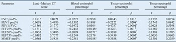
The eosinophils in the peripheral blood showed significantly negative correlations with: predicted maximal expiratory flow rate at 25 per cent (r = −0.2443, p < 0.05) and 50 per cent (r = −0.2099, p < 0.05) of vital capacity, and predicted maximal mid-expiratory flow (r = −0.2381, p < 0.05). The eosinophils in tissue demonstrated significantly negative correlations with: predicted forced expiratory volume in 1 second (r = −0.2322, p < 0.05), forced expiratory volume in 1 second/forced vital capacity ratio (r = −0.4787, p < 0.001), predicted maximal expiratory flow rate at 25 per cent (r = −0.3263, p < 0.01), 50 per cent (r = −0.3288, p < 0.001), and 75 per cent (r = −0.3439, p < 0.001) of vital capacity, and predicted maximal mid-expiratory flow (r = −0.3588, p < 0.001). No significant correlation was found between neutrophils in tissue and the pulmonary function parameters (Table V).
Association of lung function parameters with CT scores, peripheral blood eosinophils, tissue eosinophils and tissue neutrophils

*p < 0.05; † p < 0.001; ‡ p < 0.01. CT = computed tomography; FVC pred% = per cent predicted forced vital capacity; FEV1 pred% = per cent predicted forced expiratory volume in 1 second; FEV1/FVC = forced expiratory volume in 1 second/forced vital capacity ratio; FEF25% pred% = per cent predicted maximal expiratory flow rate at 25 per cent of vital capacity; FEF50% pred% = per cent predicted maximal expiratory flow rate at 50 per cent of vital capacity; FEF75% pred% = per cent predicted maximal expiratory flow rate at 75 per cent of vital capacity; MMEF pred% = per cent predicted maximal mid-expiratory flow
Discussion
Chronic rhinosinusitis with nasal polyps is a high prevalence disease among all races.Reference Fokkens, Lund, Mullol, Bachert, Alobid and Baroody 1 However, the predominant inflammatory cell type varies considerably between different ethnicities. It has been reported that the percentage of tissue eosinophilia in Asian patients with polyps is much lower than that in Caucasian patients with polyps. In contrast, neutrophil infiltration is apparently increased when compared to that in Caucasian patients with polyps.Reference Fokkens, Lund, Mullol, Bachert, Alobid and Baroody 1 , Reference Li, Zhang, Li, Lin, Li and Curotto de Lafaille 3 – Reference Gao, Ng, Li, Li, Duan and Shen 5 While approximately 80 per cent of Caucasian patients with polyps are eosinophilic,Reference Fokkens, Lund, Mullol, Bachert, Alobid and Baroody 1 about half of Asian patients with polyps demonstrate tissue eosinophilia,Reference Li, Zhang, Li, Lin, Li and Curotto de Lafaille 3 – Reference Gao, Ng, Li, Li, Duan and Shen 5 and the proportion of tissues with neutrophilia ranges from 36 to 57 per cent.Reference Li, Zhang, Li, Lin, Li and Curotto de Lafaille 3 , Reference Gao, Ng, Li, Li, Duan and Shen 5 In our study, 56.6 per cent of our samples showed tissue eosinophilia and up to 41.4 per cent of all the tissues were neutrophilic, which is consistent with outcomes from most Asian studies. The amount of eosinophils in nasal polyps tissues in our study was significantly correlated with eosinophils in the peripheral blood, which is also consistent with previous studies.Reference Kim, Hong, Kim, Lee, Min and Rhee 2 , Reference Baba, Kagoya, Kondo, Suzukawa, Ohta and Yamasoba 4
The sub-classification of nasal polyps into eosinophilic nasal polyps and non-eosinophilic nasal polyps was thought to have clinical importance in predicting the prognosis and selecting an effective therapeutic strategy for patients.Reference Alatas, Baba, San and Kurcer 10 , Reference Soler, Sauer, Mace and Smith 11 Soler et al. reported that mucosal eosinophilia correlated with objective disease severity, as defined by CT, endoscopy and smell identification test scores.Reference Soler, Sauer, Mace and Smith 11 In addition, eosinophilic nasal polyps patients are more likely to respond to steroid therapy, whereas non-eosinophilic nasal polyps patients usually respond to macrolide treatment.Reference Alatas, Baba, San and Kurcer 10 However, the relationship between inflammatory cell infiltration and pulmonary function in nasal polyps patients is not yet fully understood.
Kariya et al. reported that chronic rhinosinusitis patients have latent obstruction of the small airway.Reference Kariya, Okano, Oto, Higaki, Makihara and Haruna 7 Our research took some steps forward, and hypothesised that eosinophils in peripheral blood and tissue, but not neutrophil infiltration, play an important role in nasal polyps patients’ pulmonary function, as nasal polyps patients with peripheral blood eosinophilia or tissue eosinophilia showed reduced lung function. From an immunological perspective, eosinophilic nasal polyps is associated with enhanced Th2 function and a higher interleukin (IL)-5 level, whereas non-eosinophilic nasal polyps is believed to be related to the combination of T helper types 1, 2 and 17 cell polarisation.Reference Fokkens, Lund, Mullol, Bachert, Alobid and Baroody 1 , Reference Li, Zhang, Li, Lin, Li and Curotto de Lafaille 3 , Reference Gao, Ng, Li, Li, Duan and Shen 5 In eosinophil biology, IL-5 plays a central role in the production, activation, mobilisation, recruitment, survival, proliferation and suppression of apoptosis in eosinophils at the site of inflammation. Thus, IL-5 in nasal discharge may be related to damaged lung function in nasal polyps patients.Reference Kariya, Okano, Oto, Higaki, Makihara and Haruna 7 It has been suggested that eosinophils have a significant role in tissue remodelling.Reference Lee, Jacobsen, McGarry, Schleimer and Lee 12 Hence, the pulmonary impairment in patients with epithelial hyperplasia and goblet cell hyperplasia observed in our study might be explained by the connection between eosinophilia and goblet cell hyperplasia.
Although the link between allergic rhinitis and asthma has been well established, and the presence of allergic rhinitis has been confirmed as one of the risk factors for the development of asthma,Reference Fokkens, Lund, Mullol, Bachert, Alobid and Baroody 1 , Reference Brozek, Bousquet, Baena-Cagnani, Bonini, Canonica and Casale 6 the effect of allergic rhinitis on lung function remains controversial. No significant differences between allergic rhinitis patients and control subjects were found in spirometric parameters in Kariya and colleagues’ study.Reference Kariya, Okano, Oto, Higaki, Makihara and Haruna 7 In our study, no significant differences between the non-atopic group and the atopic group were found for any of the pulmonary function parameters investigated, which is consistent with Kariya and colleagues’ findings. In a recent study, asymptomatic obstructive lung function changes were observed in patients who had never smoked and had chronic rhinosinusitis.Reference Kariya, Okano, Higaki, Noyama, Haruna and Nishizaki 13 Gao et al. found that smoking is an independent predictor of squamous metaplasia in Chinese nasal polyposis.Reference Gao, Ng, Li, Li, Duan and Shen 5 Smoking has been suggested as being related to goblet cell hyperplasia in both nasal and bronchial mucosa.Reference Lapperre, Sont, van Schadewijk, Gosman, Postma and Bajema 14 – Reference Elwany, Ibrahim, Mandour and Talaat 16 We did not, however, find that smoking had any statistically significant effects on the representative pulmonary function parameters in our study. Although the eosinophilic nasal polyps phenotype was believed to have a higher recurrence,Reference Ikeda, Shiozawa, Ono, Kusunoki, Hirotsu and Homma 17 – Reference Nakayama, Yoshikawa, Asaka, Okushi, Matsuwaki and Otori 19 and the eosinophilic nasal polyps patients showed worse pulmonary function, no statistically significant changes were found in lung function, regardless of recurrence status.
-
• Upper and lower respiratory diseases are closely related
-
• Nasal polyps patients with decreased pulmonary function may be at risk of developing lower respiratory disease
-
• Nasal polyps patients with peripheral blood eosinophilia and tissue eosinophilia showed poorer pulmonary functions in our study
-
• Clinicians should be aware of lung function decline in nasal polyps patients, especially those with tissue eosinophilia
The effect of nasal polyps on lower respiratory function is currently under debate. Several research papers report that nasal polyps patients’ lung function is affected.Reference Kariya, Okano, Oto, Higaki, Makihara and Haruna 7 , Reference Kariya, Okano, Higaki, Noyama, Haruna and Ishihara 8 , Reference Kariya, Okano, Higaki, Noyama, Haruna and Nishizaki 13 , Reference Promsopa, Kansara, Citardi, Fakhri, Porter and Luong 20 Our study clearly shows that nasal polyps patients with peripheral blood eosinophilia and tissue eosinophilia have poorer pulmonary functions, and it considers the effects of nasal polyps on the pathogenesis of pulmonary dysfunction. Further investigation on the causes of lung function deterioration in patients with nasal polyps is required to clarify the relationship between upper and lower respiratory tract disorders.
Conclusion
A close relationship between upper and lower respiratory diseases has been reported. The nasal polyps patients with decreased pulmonary function may be in danger of developing lower respiratory diseases. Nasal polyps patients with peripheral blood eosinophilia and tissue eosinophilia showed poorer pulmonary functions in our study. Our findings suggest that patients with nasal polyposis should be followed carefully to quickly detect any lung diseases that might develop. Clinicians should be aware of the decline of lung function and lung condition in patients with nasal polyps, especially those with tissue eosinophilia.
Acknowledgements
This study was supported by grants from the National Nature Science Foundation of China (grant number 81670909) and the Department of Science and Technology of Shandong Province (grant number 2016GSF201113).