Introduction
Radiotherapy (RT), especially in combination with chemotherapy, is becoming increasingly important as definitive treatment in patients with locally advanced intrathoracic tumours, for example, lung cancer. As techniques evolve, and patients can expect longer progression-free survival, the issue of serious long-term side effects, for example, cardiac complications, becomes more important. Reference Beukema, van Luijk, Widder, Langendijk and Muijs1,Reference Hardy, Liu, Cormier, Xia and Du2 A meta-analysis of ten randomised studies on post-operative RT in lung cancer showed shorter survival for patients undergoing such adjuvant treatment, even though the incidence of local tumour recurrence was significantly lower in the RT treatment arms. 3 The reason for these contradictory findings is assumed to be due to RT-induced side effects, for example, cardiotoxicity.
Radiation-induced heart disease (RIHD) can be acute or develop years or even decades after treatment. Radiation may affect all anatomical and functional components of the heart, that is, the pericardium, myocardium, coronary arteries and valves. Accordingly, RIHD includes a wide variety of pathological conditions, for example, pericarditis, ischemic disease, rhythm abnormalities, valve defects and myocardial fibrosis. Common risk factors for heart disease such as diabetes, obesity and smoking increase the risk of RIHD. Reference Heidenreich and Kapoor4,Reference Lancellotti, Nkomo and Badano5
The nuclear imaging method myocardial perfusion scintigraphy (MPS) assesses myocardial perfusion and function. Reference Patel and Beller6 Results of several studies in breast cancer show a decrease in myocardial perfusion corresponding to the irradiated volume of the left ventricle (LV) as measured by MPS after post-operative RT. Reference Gyenes, Fornander, Carlens, Glas and Rutqvist7–Reference Seddon, Cook and Gothard9 The irradiated heart volume is normally much smaller in adjuvant RT to the breast compared to the majority of cases of definitive RT in lung cancer. Presently, there is limited data on assessing RT-induced myocardial perfusion defects by MPS for the latter diagnosis.
Cardiac magnetic resonance imaging (CMR) can produce detailed information regarding heart anatomy, morphology, ventricular, valvular function and myocardial perfusion. Reference Finn, Nael, Deshpande, Ratib and Laub10,Reference Kolentinis, Le, Nagel and Puntmann11 Late gadolinium enhancement cardiac magnetic resonance imaging (LGE CMR) can be used to detect increased extracellular space in the myocardium such as oedema and fibrosis/scar tissue following infarction. Although the method has a wide range of clinical applications, it has yet to find its role in assessing cardiac changes in patients undergoing RT. Reference Lancellotti, Nkomo and Badano5
A prospective study comparing MPS with stress perfusion CMR in detecting coronary heart disease in non-irradiated patients showed a significant advantage in favour of CMR, which is now considered gold standard in evaluating acute and chronic myocardial infarction. Reference Greenwood, Maredia and Younger12
Although there is no consensus on how to screen patients for RIHD, both CMR and MPS are considered possible alternatives. Reference Lancellotti, Nkomo and Badano5 CMR offers two advantages compared to MPS; first, patients are not exposed to radiation, and second, CMR is not sensitive to attenuation effects. To date, no study comparing these modalities for detection of signs of radiation-induced myocardial damage in patients treated with RT for lung cancer has been published.
The aim of the present study was to determine whether RIHD changes can be detected and measured with MPS and LGE CMR in patients treated with RT for inoperable lung cancer. As a secondary outcome, the CMRs were also analysed for changes in cardiac function.
Patients and Methods
Set-up and patient characteristics
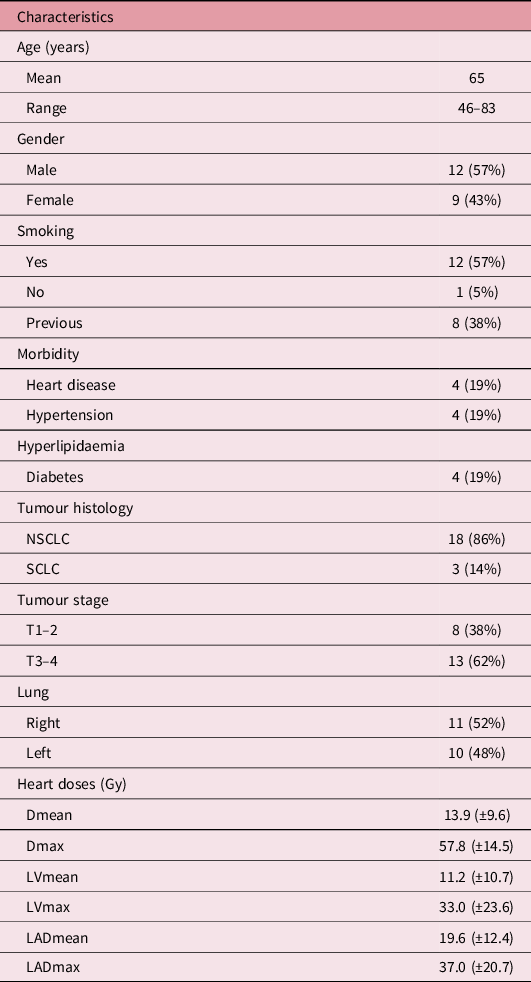
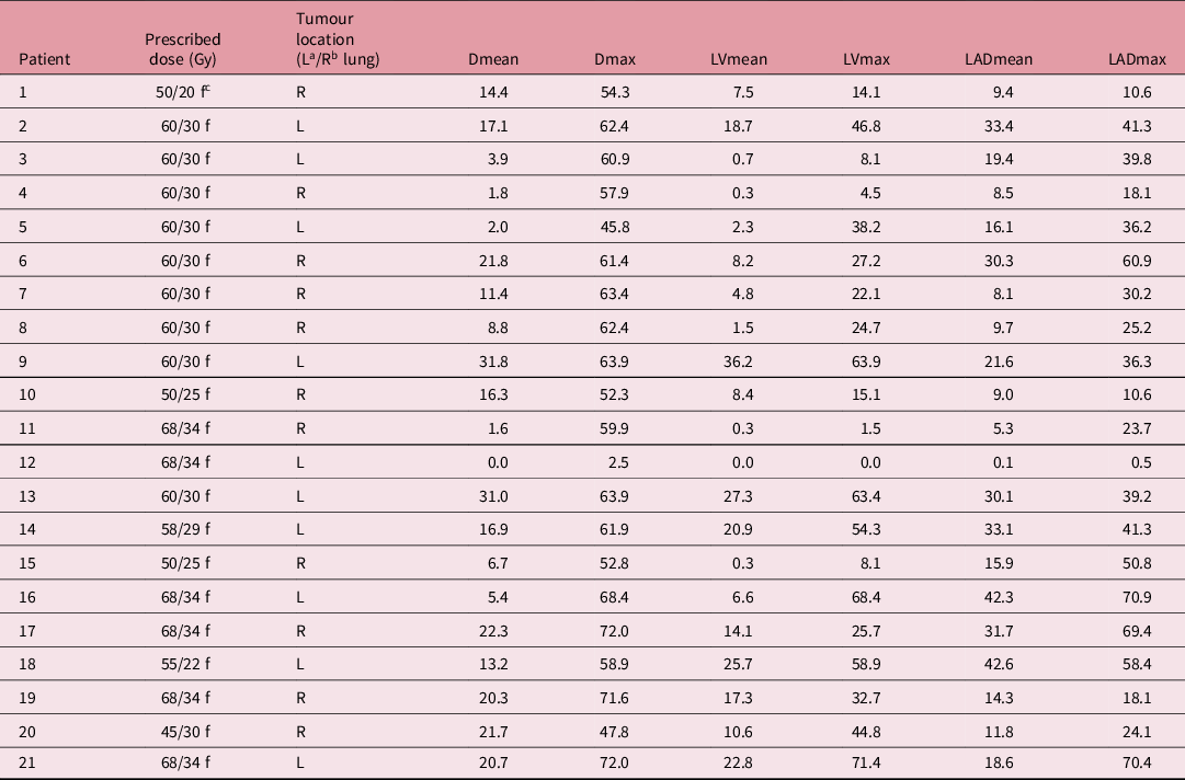
Twenty-one patients with biopsy-proven inoperable lung cancer undergoing definitive three-dimensional conformal radiotherapy (3D-CRT) treatment were evaluated with MPS and LGE CMR pre-RT and at 2 and 6 months post-RT follow-ups (FUs). Patient and tumour characteristics and heart RT doses are presented in Table 1. All patients received induction and/or concomitant chemotherapy (i.e., gemcitabine in combination with carboplatin as induction chemotherapy plus docetaxel concomitantly with RT in non-small-cell lung cancer (NSCLC), and etoposide in combination with carboplatin concomitantly with RT in small-cell lung cancer (SCLC). RT target doses varied depending on the specific diagnosis, that is, 50 Gy/20 fractions (f), 50 Gy/25 f, 55 Gy/22 f, 58 Gy/29 f, 60 Gy/30 f, or 68 Gy/39 f in NSCLC and 45 Gy/30 f or 50 Gy/25 f in SCLC (Table 2).
Patient and tumour characteristics and mean RT doses to heart

Prescribed RT doses, tumour location and doses to the heart in all patients

Note: aLeft, bright, cfractions.
3D-CRT plans
For each patient, a pre-treatment planning CT scan was performed in a standard supine position. A radiologist outlined the heart, LV and the left anterior descending coronary artery (LAD). Heart: The right ventricle and the right atrium defined the upper border of the heart. The caudal myocardial border determined the lower border of the heart. The great vessels were excluded. LV: In all patients, a diagnostic contrast-enhanced pre-treatment CT scan was used to discriminate the myocardial wall from the blood-filled ventricular cavity. LAD: The LAD was followed from its aortic origin on its route along the anterior left ventricular wall. The Pinnacle® planning system version 2.2 (ADAC Laboratories, Milpitas, CA, United States) was used for RT planning. All treatments were delivered with 6 and/or 18 MV photons on Varian linear accelerators (Varian Medical Systems, Palo Alto, CA, United States) with three horizontal beams in a 3D-conformal fashion. Heart doses were calculated using dose–volume histograms. Prescribed doses and doses to heart, LV and LAD for each patient are presented in Table 2.
Myocardial perfusion scintigraphy (MPS)
All patients were scheduled to undergo MPS before and at 2 and 6 months post-RT. Before RT and at 6 months FU, both stress and rest tests were performed. At 2 months FU, only a rest test was performed. All patients underwent myocardial perfusion-gated MPS using technetium-99 m as a tracer as described in detail by Zuber et al. Reference Zuber and Rosfors13 The studies were performed after approximately 12 hours of fasting. The stress study was performed on day 1 and the rest study on day 2. On day 1, adenosine was infused for 5 minutes and 260 to 500 MBq of Tc-99 m was injected intravenously, and all patients performed bicycle exercise during the infusion. On day 2, 360 to 700 MBq of Tc-99 m was injected after 15 minutes of rest. The severity of MPS changes in the LV was analysed using the semi-quantitative summed rest score (SRS) method (20 segments). The severity of the perfusion defects within each segment was separately scored through consensus by two experienced nuclear medicine specialists (T.L and A.T.N) as normal (0 points), minimal (1 point), moderate (2 points), significant (3 points) or no perfusion (4 points). The score for each segment was added to form a summed stress score (SSS) and a SRS. As a measure of reversibility, the difference between the two was calculated as a summed difference score (SDS). An SSS or SRS ≥ 4 or an SDS ≥ 2 was considered abnormal. Reference Czaja, Wygoda and Duszanska14,Reference Knollmann, Knebel and Koch15 The combination of an SSS ≥ 4 and an SRS ≥ 4 was considered as evidence of an irreversible defect, that is , scar tissue/fibrosis. The extent of perfusion defects was further quantified with a quantitative computer-assisted polar map reconstruction technique, that is, the bull’s eye view method as volume % (cut-off level for 1.5 SD below the mean). Reference Lind, Pagnanelli and Marks8
Late gadolinium enhancement cardiac magnetic resonance imaging (LGE CMR)
All patients were scheduled to undergo LGE CMR before and at 2 and 6 months post-RT. The LGE CMR protocol used was previously described by Engblom et al. Reference Engblom, Wagner and Setser16 Patients were studied in a 1.5-T clinical MR scanner (Siemens Symphony, Siemens, Munich, Germany) using an intravenously administrated gadolinium-based (gadoteric acid) contrast agent (Dotarem, Guerbet, Paris, France) as enhancer.
Two experienced readers (T.L and A.T.N) analysed the LGE CMRs. A qualitative examination of visual signs of myocardial damage in the LV was assessed after administration of contrast agent. Stroke volume (SV), ejection fraction (EF), end-diastolic volume (EDV), end-systolic volume (ESV), cardiac output (CO) and left ventricular mass (LVM) were all calculated by outlining the epi -and endocardial borders in systole and diastole by use of ARGUS (Siemens version 2002B, Siemens, Munich, Germany).
Statistics
All data were tested for normal distribution and were found to be non-normally distributed. McNemar test (without Yate’s correction) was used for repeated comparison for nominal data. A p < 0.05 was considered statistically significant. A Wilcoxon signed-rank test or a Friedman test was used to compare repeated measurements for continuous data. A p < 0.05 was considered statistically significant. For the Friedman test, a post hoc analysis using Wilcoxon signed-rank test was performed with a Bonferroni correction applied, resulting in a significance level set at p < 0.017.
Results
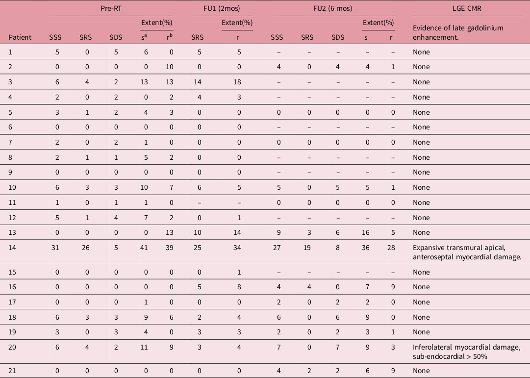
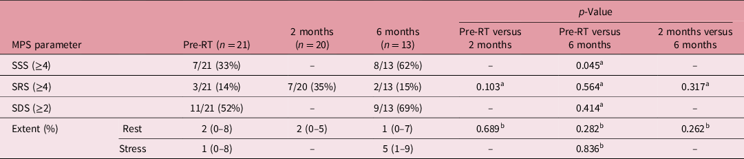
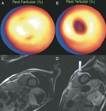
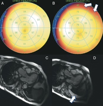
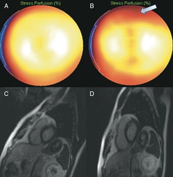
The results are presented in Tables 3–5. Twenty and 13 patients were assessable at 2 and 6 months FU, respectively. Significant semi-quantitative MPS changes (SSS ≥ 4 and/or SRS ≥ 4) were detectable in 7 out of 20 patients at 2 months FU and in 8 out of 13 patients at 6 months FU (Table 3). Three out of seven MPS changes at 2 months FU and four out of eight MPS changes at 6 months FU were determined to be new. The only significantly changed MPS parameter at a group level was the presence of SSS ≥ 4, which increased from 33% at baseline to 62% at 6 months FU (p = 0.045) (Table 4). At the 2 months FU, one patient (patient no 3) had developed a new deep apical perfusion MPS defect at rest (Figure 1). There was, however, no corresponding defect on LGE CMR. Furthermore, a new mass in the thoracic wall was visible on LGE CMR in this patient (Figure 1). This mass may have caused a false-positive MPS defect due to attenuation. The patient died of causes unrelated to the diagnosis and treatment before the 6 months FU, and no postmortem examination was performed. Out of the eight patients with perfusion defects at the 6 months FU, only two patients (patients no 16 and 21) were presumed to have new irreversible defects. Patient no 21 had pleural effusions on LGE CMR, which probably caused attenuation on MPS (Figure 2), leaving only patient no 16, in whom the new perfusion defect had no other explanation than having been caused by the radiation treatment (Figure 3). The newly developed perfusion defect on MPS in this patient was located basal anterior, that is, within the LAD distribution. This patient had the highest LADmax of all patients in the study (Table 2). All new MPS defects were minor, and no corresponding signs of myocardial damage/fibrosis were evident on LGE CMR. According to CMR measurements, there were no statistically significant changes in left ventricular size or function as assessed by CO, EF, EDV, ESV, LVM or SV at 2 or 6 months FU as compared to baseline (Table 5).
Changes in MPS parameters in all patients at pre-RT, 2 and 6 months FU with corresponding LGE CMR findings

Note: aStress, brest.
Change in proportions of significant MPS defects, and of extent of perfusion defects at pre-RT, 2 and 6 months FU. Extent is presented as median and interquartile range

Note: aMcNemar test, bWilcoxon signed-rank test.
Changes in cardiac function and morphology assessed with MRI. All values are presented as median values and interquartile range (IQR)

Note: aFriedman test.
A–D. Patient 3. A. Rest MPS at baseline showing no signs of apical perfusion defects. B. Rest MPS at 2 months FU showing a perfusion defect corresponding to the apical part of the anterior left ventricular (LV) wall. C. LGE CMR at baseline (short-axis plane) showing no signs of LGE/myocardial fibrosis. D. LGE CMR at 2 months FU (short-axis plane) showing no sign of fibrosis that would correspond to the perfusion defect detected on MPS. A new extracardial mass is evident (arrow).

A–D. Patient 21. A. Stress MPS at baseline showing no signs of perfusion defects. B. Stress MPS at 6 months FU showing newly developed basal anterior minor perfusion defects (arrows). No corresponding LGEs are evident on CMR (C and D). Newly developed pleural effusion (arrow) (D) may have caused attenuation on MPS in B.

A–D. Patient 16. A: Stress MPS at baseline showing no signs of perfusion defects. B. Stress MPS at 6 months FU showing newly developed basal anterior minor perfusion defects. No corresponding LGEs are evident on LGE CMR at baseline (C) or at 6 months FU (D).

Discussion
In the present study, radiation doses to the heart were clinically significant (Tables 1 and 2). Despite this, the incidence of newly developed irreversible defect on MPS at FU was lower than expected, compared to previous reports. Reference Lind, Pagnanelli and Marks8,Reference Marks, Yu and Prosnitz17 Also surprisingly was the fact that, in two of the three patients that developed new irreversible perfusion defects on MPS, attenuation effects caused by a new thoracic mass or pleural effusion were the most probable causes for the MPS defects. No patient in the study had any evidence of post-irradiatory LGE on MRI.
For many years, MPS has been regarded as the gold standard in detecting RT-induced myocardial perfusion defects, especially in breast cancer. Reference Kaidar-Person, Zagar and Oldan18 Whether LGE CMR can be used to detect RT-induced heart damage has been the subject of only a few studies. In a study by Umezawa et al, 24 patients with oesophageal cancer were analysed with contrast-enhanced LGE CMR at a median FU of 23.5 months after definitive RT. Reference Umezawa, Ota and Takanami19 LGE was detected in 12 patients and the incidence of LGE increased with increasing RT doses within the specific segments that were analysed. The LGEs were likely related to the RT, but a limitation of the study was the lack of pre-treatment baseline exams. In a small pilot study, Huang et al. Reference Huang, Harrison and Sarkar20 investigated the feasibility of using LGE CMR in detecting radiation damage to the left atrium in seven patients treated with external RT for different thoracic tumours. A linear relationship between mean dose to the left atrium and the LGE CMR detected scar volume was found. Neither of these two studies compared LGE CMR to MPS.
Why did not the present study reproduce these findings? Several explanations should be considered. First, there could be a time factor in favour of MPS. Gadolinium is an extracellular agent and late enhancement is a sign of increased extracellular space. Reference Doltra, Amundsen, Gebker, Fleck and Kelle21 In the situation of cardiac infarction, the increased extracellular space is caused by inflammation and oedema in the acute phase and by scar tissue in the late phase. The situation could be similar in the context of RT-induced cardiac damage, that is, inflammation and oedema in the acute phase and scar tissue as a late radiation effect. The time frame from the beginning to the end of the inflammation/oedema phase and the formation of definitive scar tissue in RT is, to our knowledge, essentially unknown. Theoretically, the LGE CMRs in our study could have been performed too late (viz. 2 months) to detect the acute oedema phase and too early (viz. 6 months) to detect the late scar formation. In our study, the time from completion of RT to the final LGE CMR FU was only 6 months. In the study by Umezawa et al, the median time from completion of RT to the LGE CMR FU in the 12 patients with LGEs was 19 months and in the study by Huang et al, the time from completion of RT to LGE CMR FU was 3.1 ± 1.9 years.
Second, some diffusion defects detected by MPS could be false and caused by enhanced density in the thoracic wall or tissues surrounding the heart, as was evident in two patients in our study. A study by Lawrence and colleagues may contradict this theory. Reference Lawrence, Saynak and Fried22 In 23 patients treated with RT for breast or lung cancer, pre -and post-treatment CT scans were analysed for enhanced density in the thoracic wall or tissues surrounding the heart. Modest enhancements in soft tissue density were detected but they were, according to the authors, not likely to cause false perfusion defects. False MPS defects caused by pleural effusion have, however, previously been described. Reference Shih and Justice23
Third, the sample size of our study was quite small and the dropout at 6 months FU, mostly due to death or progressive disease impeding further participation, was large. This may also have diminished our chances of detecting RT-induced cardiac pathology.
The frequency of MPS changes in our study was low; however, since RIHD may be a cause of morbidity and mortality even in patients treated with RT for locally advanced lung cancer, it is still vital to try to minimise the incidental radiation to the heart. Reference Atkins, Rawal and Chaunzwa24,Reference Wang, Eblan and Deal25 In our study, all patients were treated with 3D-CRT. The evaluation of alternative techniques, for example, intensity-modulated RT (IMRT) or volumetric-modulated arc therapy (VMAT), with the potential of reducing doses to the heart, should be studied in future trials. The only randomised prospective study to date, comparing 3D-CRT with IMRT in the treatment of locally advanced NSCLC, reports significantly lower heart doses in favour of IMRT. Reference Chun, Hu and Choy26 Furthermore, small number of dosimetric studies comparing VMAT with IMRT in lung cancer RT have shown mixed results with regard to doses to the heart. Reference Della Gala, Dirkx and Hoekstra27,Reference Jiang, Li and Liu28
All patients in our study were treated with chemotherapy in addition to RT. None of the chemotherapeutic agents used were typically cardiotoxic. It is worth noticing, however, that some chemotherapeutic agents, for example, anthracyclines, may in themselves cause cardiac changes detectable with both MPS and CMR. Reference Drafts, Twomley and D’Agostino29,Reference Wakabayashi, Taki, Inaki, Sumiya, Tsuchiya and Kinuya30 LGE is, however, uncommon as a result of chemotherapy-induced cardiotoxicity, Reference Neilan, Coelho-Filho and Shah31 which may indicate that LGE in patients treated with chemoradiation is caused by the radiation.
Further studies aiming to investigate the optimal timing for CMR FU to detect early signs of RIHD should be considered. Studies to correlate early CMR-detected findings of RIHD with future cardiac events are also warranted.
Conclusion
In the present study, in contrast to two previous reports, Reference Umezawa, Ota and Takanami19,Reference Huang, Harrison and Sarkar20 we could not detect signs of RT-induced myocardial damage/fibrosis using LGE CMR. A significant minority of patients treated with definitive RT in locally advanced intrathoracic tumours will be cured or benefit by a long-term stable disease. Further studies on the concept of RIHD in this patient group are therefore warranted. Despite the low frequency of MPS changes observed in our trial, incidental irradiation to the heart should still be minimised. Reference Atkins, Rawal and Chaunzwa24,Reference Wang, Eblan and Deal25 Attenuation caused by pleural effusion is an important factor to account for in the FU of RT for lung cancer with MPS.
Acknowledgements
The authors wish to thank Dr A.T.Nygren for help with analysing MPS and LGE CMR data. The authors would also like to thank the staff of the Department of Clinical Physiology and Nuclear Medicine at Södersjukhuset, Stockholm, Sweden.
Financial Support
This work was supported by grants from the Swedish Medical Society and from the Swedish Cancer Society (Cancerfonden).
Conflicts of Interest
None.
Ethical Standards
The authors assert that all procedures contributing to this work comply with the ethical standards of the “Good Research Practice” guidelines of the Swedish Research Council, 2017, and with the Helsinki Declaration of 1975, as revised in 2008, and have been approved by the Regional Medical Ethics Committee in Stockholm (now the Swedish Ethical Review Authority).