Cardiovascular magnetic resonance imaging provided definitive diagnosis of anomalous origin of the left coronary artery from the pulmonary artery in this 17-year-old basketball player with 6 years of intermittent chest pain.
Repeat echocardiography demonstrated multiple collateral vessels (cine loop 1), with fistulous communications between the left and right coronary artery circulations, normal right coronary artery origin, and apparently normal left main coronary artery origin (cine loop 2). An exercise stress test was inconclusive. A Thallium stress test revealed a moderately reversible myocardial perfusion defect in the left ventricular anterior and lateral wall, with a corresponding left main coronary artery territory S-T depression on the electrocardiogram.
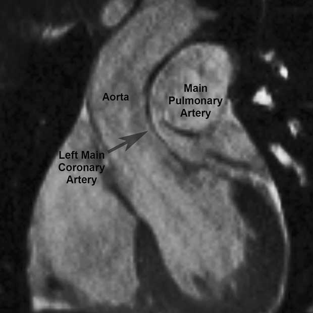
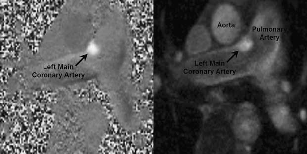
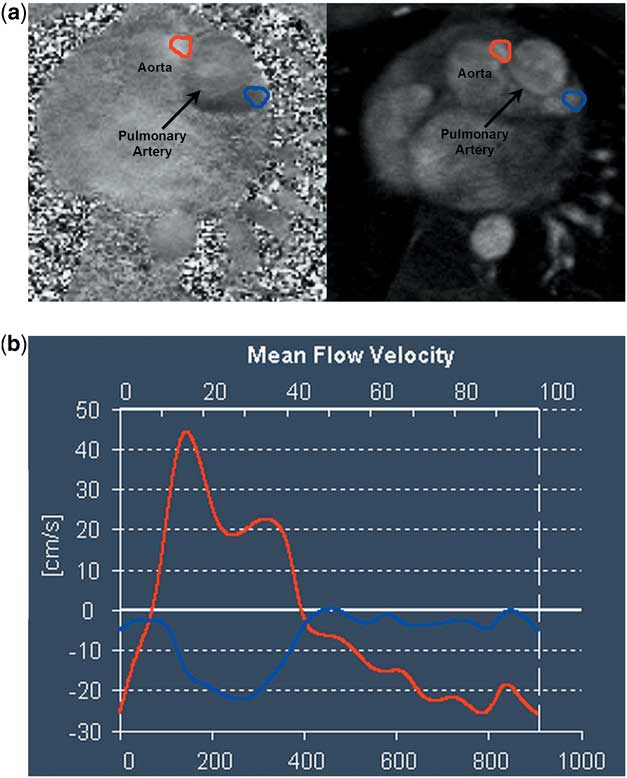
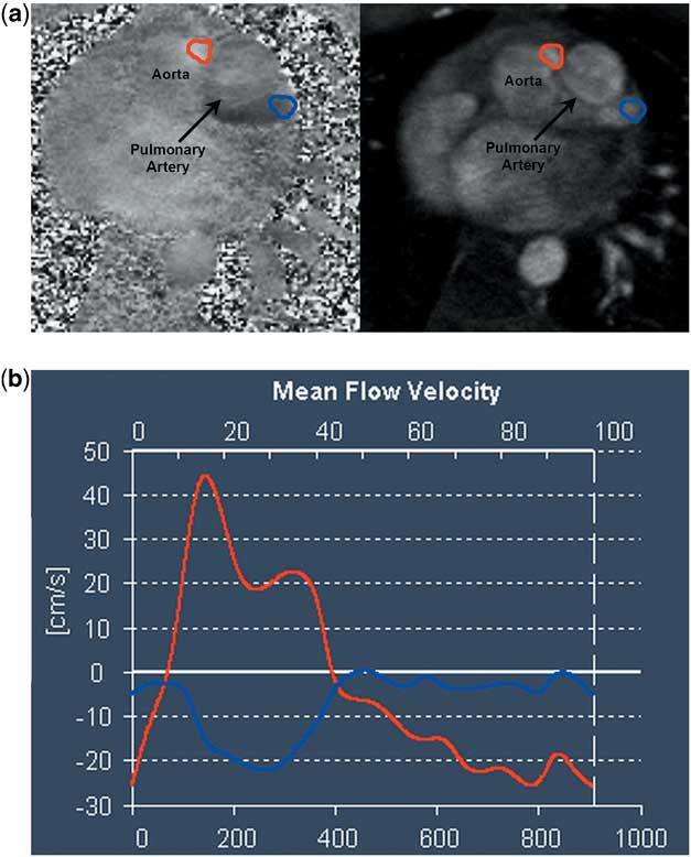
We found that two-dimensional cine steady-state free-precession imaging of the aorta delineated anomalous origin of the left main coronary artery from the distal main pulmonary artery (Fig 1), whereas three-dimensional steady-state free-precession respiratory navigator-gated and electrocardiogram-triggered imaging better depicted right coronary artery origin. Velocity-encoded phase contrast imaging showed retrograde flow from the left main coronary artery into the pulmonary artery (Fig 2), with opposite directions of flow in the right coronary artery (red) and left main coronary artery (blue) in
Figure 3. There was mild left ventricular dilatation, no regional wall motion or late gadolinium enhancement abnormality. Stress magnetic resonance imaging was not performed. Surgery confirmed anomalous origin of the left coronary artery from the pulmonary artery, with direct re-implantation of the coronary artery into the aorta. Potential low flow in the left coronary circulation necessitated long-term anticoagulant therapy.
Figure 1
Figure 2
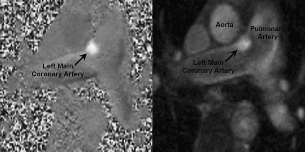
Figure 3
Cardiovascular magnetic resonance imaging provides non-invasive anatomic and functional information, without the echocardiography limitation of poor windows or ionising radiation of nuclear medicine and digital subtraction angiography. Phase contrast and steady-state free-precession sequences delineate aberrant vessel origin and flow direction, and late gadolinium enhancement assessment of myocardial viability, which is of prognostic importance in guiding surgery.Reference Peña, Nguyen, Merchant and Dennie1