Introduction
Alzheimer's disease (AD) is the most common age-related neurodegenerative disorder. Symptoms result in progressive deterioration of cognitive function, particularly those related to memory. Both genetic and environmental factors appear to play a role in the onset of AD. One pathological hallmark of the disease is the formation and extracellular aggregation of amyloid β in senile plaques and cerebral blood vessels. This is likely a key aspect in the pathogenesis of AD, given that all mutations that underlie familial cases of AD are implicated in the processing and accumulation of amyloid β. The mechanism of amyloid β generation is fairly well known, whereas its catabolism and degradation are not well understood.
Matrix metalloproteinases can degrade components of the extracellular matrix in a variety of physiological and pathophysiological conditions such as stroke, intracerebral hemorrhage (Montaner et al., Reference Montaner2001; Horstmann et al., Reference Horstmann, Kalb, Koziol, Gardner and Wagner2003; Alvarez-Sabin et al., Reference Alvarez-Sabin2004) amyotrophic lateral sclerosis, and multiple sclerosis (Campbell and Pagenstecher, Reference Campbell and Pagenstecher1999). There is growing evidence that matrix metalloproteinases play an important role in the pathogenesis of AD, and, in particular, may be involved in the processing pathway of amyloid β. In this regard, it is not known whether their functions in the pathogenesis of AD are protective or destructive.
In this study, we investigated MMP-2, MMP-3, MMP-9 and MMP-10 in plasma and cerebrospinal fluid (CSF) of AD patients compared to gender- and age-matched healthy controls. Given the prior evidence of genetic alterations in MMP encoding genes in AD, we hypothesized that there would be significant differences in levels of circulating MMPs between these two groups.
Methods
Patients
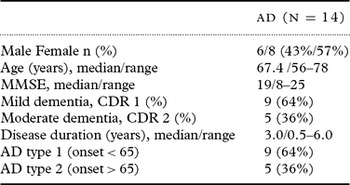
Subjects with AD enrolled in this study were recruited from the population seen in the memory clinic at the Central Institute of Mental Health, Mannheim, Germany. Diagnosis of AD was made according to medical history, clinical examination, and the CERAD test battery (Consortium to Establish a Registry for Alzheimer's Disease). Classification of disease was made according to the Clinical Dementia Rating (CDR) scale. Cranial magnetic resonance imaging (MRI) was performed in all patients to rule out other causes of dementia, e.g. vascular disorders. Patients who were included in our study had (1) no previous history of transient ischemic attack or stroke; (2) no history of recent head trauma; and (3) no major cardiac, renal, hepatic or malignant disease. Fourteen patients (8 female and 6 male, median age 67.4 years) fulfilled these strict criteria. Cognitive function of all participants was studied by using neuropsychological testing – Mini-mental State Examination (MMSE), CDR, CERAD. Disease duration in these patients varied from 0.5 to 6 years (mean 3.0 years). Patient characteristics are shown in Table 1. The study was approved by the ethics committee of the Medical School, University of Heidelberg, and all patients or their designated caregivers gave informed consent.
Characteristics of AD patients

Healthy controls
Fourteen gender- and age-matched healthy individuals without any history of ischemic stroke, recent head trauma or major cardiac, renal, hepatic or malignant disease, were used as controls.
Samples
Venous blood samples and cerebrospinal fluid samples were drawn from each patient at time of admission into the study. Samples were centrifuged (3000g/10 minutes) and stored at minus 80°C until analysis.
MMP-3 and MMP-10 were analyzed using casein zymography; MMP-2 and MMP-9 were analyzed by gelatin zymography. Plasma samples were prepared by dilution (plasma 1:5) into PBS, and protein content of each sample was measured by Bradford Reagent (Bio-Rad). Sample volume was corrected for protein concentration. A volume corresponding to 55μg (plasma) and 12μg (CSF) protein concentration was subjected to electrophoresis; the electrophoresis gels were then scanned (UMAX UC 1200 S Ultra Vision) into a Macintosh Power PC computer. Lysis zones of enzymatic activity were quantified with NIH Image 1.6.
Zymography
Casein and gelatin zymograms were performed as previously described (Pozzi et al., Reference Pozzi, Moberg, Miles, Wagner, Soloway and Gardner2000, Horstmann et al., Reference Horstmann, Kalb, Koziol, Gardner and Wagner2003). In brief: Gel electrophoresis was performed with the use of 7.5% polyacrylamid gels containing 2 mg of casein/gelatin, Sigma. 2μl of a loading buffer consisting of 1.25 ml 0.5 M Tris/HCl ph 6.8, 2 ml 10% sodium dodecyl sulfate, 1 ml Glycerol and 0.001% bromphenol blue was added to the samples and gels were loaded. After electrophoresis gels were incubated in renaturating solution (2.5% Triton X 100, 0.1 M NaCl, 50 mM, Tris ph 7.5) for 2 hours and then for 17 hours at 37°C in a developing buffer containing 10 mM CaCl2, 50 mM Tris ph 7.5 and 0.02% sodium azide. Gels were then stained with Coomassie blue (Sigma), and destained in a destaining solution. Gels were scanned under standardized conditions (UMAX UC 1200 S ultra vision) and image analysis software (NIH image 1.62) was used to analyze gels on a Macintosh Power PC. Standardization was done with an optical step tablet. To ensure inter-gel reproducibility, electrophoresis running times and staining and destaining procedures were standardized. Gels displayed clear bands of MMP-2, MMP-3, MMP-9 and MMP-10 (Figure 1).
Casein Zymogram showing MMP-3 and MMP-10 in plasma of AD patients compared to controls.

Statistical analyses
Summary statistics relating to levels of MMP-2, MMP-3, MMP-9, and MMP-10 are reported with means and standard deviations (SD), as well as quartiles and ranges. Comparisons between AD patients and controls relative to levels of MMP-2, MMP-3, MMP-9, and MMP-10 were made with Wilcoxon rank statistics. Pearson correlations were computed as measures of association. Wilcoxon statistics were also used to compare MMP levels between individuals with mild versus moderate dementia.
Results
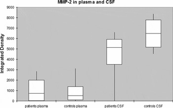
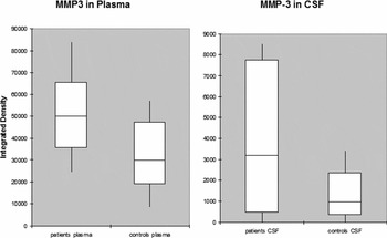
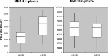
Levels of MMP-2, MMP-3, MMP-9, and MMP-10 in AD patients and controls are summarized in Figures 2, 3, and 4, reported with medians, quartiles and ranges. Salient findings are:
Box-and-whisker-graph showing MMP-2 values indicated in integrated density. No difference in plasma levels between the two groups; significantly decreased CSF levels in AD patients compared to controls.

Box-and-whisker-graphs showing MMP-3 values indicated in integrated density. Significantly increased plasma levels in AD patients compared to controls; elevated but not significantly increased CSF levels in AD patients compared to controls.

Box-and-whisker-graphs showing MMP-9 and MMP-10 values indicated in integrated density. Significantly decreased plasma levels of MMP-9 in AD patients relative to controls; no detection of MMP-9 in CSF. No difference in plasma levels between the two groups; no detection of MMP-10 in CSF.

MMP-2: no difference in plasma levels between the two groups (AD patients: mean = 976.3, SD = 1039.0; controls: mean = 910.8, SD = 997.5; p = 0.95); significantly decreased CSF levels in AD patients (mean = 4336.6, SD 2071.0) compared to controls (mean = 6383.8, SD 1341.2; p = 0.021).
MMP-3: significantly increased plasma levels in AD patients (mean = 51799, SD = 18342) compared to controls (mean = 31489, SD = 15753; p = 0.0063); elevated CSF levels not reaching statistical significance in AD patients (mean = 3789.6, SD = 3277.6) compared to controls (mean = 1233.6, SD = 1155.5; p = 0.053).
MMP-9: significantly decreased plasma levels of MMP-9 in AD patients (mean = 4948.3, SD = 3264.9) relative to controls (mean = 8274.3, SD 3262.2; p = 0.02); no detection of MMP-9 in CSF.
MMP-10: no difference in plasma levels in plasma between the two groups (AD patients: mean = 51094, SD = 23434; controls: mean = 49449, SD = 20162; p = 0.85); no detection of MMP-10 in CSF.
We examined pairwise correlations among MMP-2, -3, -9, -10 levels, in both AD patients and controls, and in both plasma and CSF. Among the AD patients, only two pairwise correlations were of absolute magnitude exceeding 0.50: plasma MMP-2 vs. plasma MMP-10, r = −0.58; and, plasma MMP-2 vs. CSF MMP-2, r = 0.69. There were no pairwise correlations among the controls of absolute magnitude 0.50 or greater. Only MMP-3 levels were weakly related to duration of disease in AD patients, r = 0.33. Nor did we find any significant differences in MMP levels between patients with mild vs. moderate dementia. MMSE was not strongly associated with levels of MMPs.
Discussion
In this study, we investigated levels of MMP-2, -3, -9 and -10 in plasma and CSF of patients with AD and matched healthy controls. MMP-3 levels in plasma of AD patients were significantly increased compared to controls, and showed a trend towards an elevation in CSF of AD patients. We observed significantly decreased levels of MMP-2 in CSF of AD patients while MMP-2 in plasma of AD patients did not show any difference from controls. MMP-9 in patient plasma was significantly decreased and MMP-10 was unchanged compared to controls. We could not detect MMP-9 and MMP-10 in CSF.
We focused our studies on MMPs because they have been shown to play an important role in the pathophysiology of AD (Yoshiyama et al., Reference Yoshiyama, Asahina and Hattori2000; Saarela et al., Reference Saarela2004; Lorenzl et al., Reference Lorenzl2003b; Reference Lorenzl, Buerger, Hampel and Beal2008). MMP-3 might play an interesting role in the pathogenesis of AD. Known polymorphisms in the MMP-3 encoding gene are implicated in the development of AD (Flex et al., Reference Flex2006). MMP-3 is an important activator of several metalloproteinases. To our knowledge, MMP-3 and MMP-10 have not been examined in AD.
MMP-3 levels were correlated to the duration of disease. Previous studies have shown that MMP-3 is selectively detected in brain white matter and on senile plaques in the cortex of AD patients. It plays an important role in activating MMP-9, which is involved in amyloid β degradation (Yoshiyama et al., Reference Yoshiyama, Asahina and Hattori2000) as well as being directly involved in amyloid β degradation itself (White et al., Reference White2006). These findings suggest a protective role for MMP-3 in the pathogenesis of AD. On the other hand, genetic findings indicate a potential role of MMP-3 in the promotion of AD. In previous studies, the 5A allele of a functional polymorphism (5A/6A) of MMP-3 showed an increased risk of AD in association with the apolipoprotein E ɛ4 allele, which is one gene shown to increase the risk of developing AD (Flex et al., Reference Flex2006). It also has an impact on the risk of dementia in apolipoprotein E ɛ4 non carriers (Helbecque et al., Reference Helbecque, Cottel, Hermant and Amouyel2007). Other studies reported no impact of this polymorphism on the risk of AD (Reitz et al., Reference Reitz2007), and a modulation of the risk by the apolipoprotein E genotype in a Finnish population (Saarela et al., Reference Saarela2004).
MMP levels and activity are tightly regulated. Growth factors and inflammatory cytokines regulate MMP expression at the level of transcription (Beuche et al., Reference Beuche, Yushchenko, Mader, Maliszewska, Felgenhauer and Weber2000). Another level of regulation is the activation of the latent enzyme by proteolytic cleavage and the inhibition of the active enzyme by their natural inhibitors, tissue inhibitors of metalloproteinase (TIMP). Previous studies showed elevated expression of inflammatory cytokines in brains of AD patients (Mrak and Griffin, Reference Mrak and Griffin2007), which could be responsible for release of MMP-3. Amyloid β is also capable of increasing the production of MMP-3 in cultured astrocytes (Deb et al., Reference Deb, Wenjun Zhang and Gottschall2003). TIMP-1 is a direct inhibitor of MMP-3 (Woessner, Reference Woessner1991); and, TIMP-1 has been shown to be elevated in CSF of AD patients, possibly as a reaction to the elevated MMP-3 levels (Lorenzl et al., Reference Lorenzl2003a).
These might be possible explanations for the increased MMP-3 levels we found in CSF and plasma. Furthermore, MMP-3 is associated with the risk of atherosclerosis and cardiovascular disease, which in turn have been associated with the risk of AD (Hofman et al., Reference Hofman1997; Pollanen et al., Reference Pollanen2002; Reference Pollanen2005; Beyzade et al., Reference Beyzade, Zhang, Wong, Day, Eriksson and Ye2003). Although there are certainly functions of MMP-3 in the pathogenesis of AD which are not yet known, our finding of elevated levels in plasma and CSF suggests a rather devastating role of MMP-3. This view is supported by the positive correlation of MMP-3 to the duration of disease.
In the central nervous system, MMP-10 is implicated in the pathogenesis of multiple sclerosis (Bar-Or et al., Reference Bar-Or2003); and, it contributes to a worse prognosis in tumors of the central nervous system (Thorns et al., Reference Thorns, Walter and Thorns2003). We found no significant difference in the plasma levels of MMP-10 between AD patients and controls, and could not detect MMP-10 in CSF.
One of the main pathological findings in AD patients is the aggregation and deposition of amyloid β in senile plaques in the brain. Amyloid β is generated by proteolytic processing of the amyloid precursor protein (APP). MMP-2 plays a role in amyloid β degradation (Roher et al., Reference Roher, Kasunic, Woods, Cotter, Ball and Fridman1994). In our study, we observed significantly decreased levels of MMP-2 in CSF of AD patients while there were no differences in plasma compared to controls. These findings of unchanged levels in plasma of AD patients are consistent with the observation of a previous study (Lorenzl et al., Reference Lorenzl2003b; Martin-Aragon et al., Reference Martin-Aragon2009). Decreased levels of MMP-2 in AD CSF might permit amyloid β formation by the β-secretase pathway, and could be one reason for increased amyloid β generation in AD patients. In the same way, the failure of amyloid β degradation can be a reason of the augmented aggregation of amyloid β. Thus, MMP-2 might be assumed to have a protective role in AD. It is also possible that the decreased levels of MMP-2 in CSF of AD patients might be explained pathophysiologically, as a failure of detection of circulating MMP-2 because of an accelerated MMP turnover, or binding of MMP-2 to brain tissue, or increased utilization of MMP-2 for local matrix remodeling. This hypothesis is supported by Yoshiyama et al. (Reference Yoshiyama, Asahina and Hattori2000), who found elevated levels of MMP-2 in brain tissues of AD patients compared to controls.
MMP-9 is another metalloproteinase implicated in the pathophysiology of AD. Our analyses showed significantly decreased MMP-9 values in plasma of AD patients, while we could not detect MMP-9 in CSF. MMP-9 is detected in regions near senile plaques in AD brains and is capable of degrading the amyloid β peptide in vitro. This finding suggests a protective role also for MMP-9 in AD (Backstrom et al., Reference Backstrom, Lim, Cullen and Tokes1996). The inability of MMP-9 to cleave the amyloid β peptide could be one reason for amyloid aggregation in AD brain. A protective role of MMP-9 is also assumed by Helbecque et al., who analyzed a promoter polymorphism of the MMP-9 encoding gene that affects transcription. They found a small protective effect on the risk of dementia by subjects carrying one high-activity allele (Helbecque et al., Reference Helbecque, Hermant, Cottel and Amouyel2003). In contrast to our findings, Lorenzl et al. (Reference Lorenzl2003b) observed elevated levels of circulating latent MMP-9 in AD patients, and Martín-Aragón and co-workers did not find any change in MMP-9 activity in plasma of AD patients (Martin-Aragon et al., Reference Martin-Aragon2009). A variety of mechanisms may activate MMP-9 such as circulating amyloid β, proinflammatory cytokines and homocysteine which is elevated in plasma of AD patients (McGeer and McGeer, Reference McGeer and McGeer1998). Backstrom et al. argued that the inflammatory protein α1-antichymotrypsin, which accumulates in amyloid plaques and which is a known inhibitor of serine proteinases, may prevent activation of MMP-9 and thereby contribute to reduced cleavage of amyloid β (Backstrom et al., Reference Backstrom, Lim, Cullen and Tokes1996). Both are possible explanations of the contradictory findings of MMP-9 levels in plasma of AD patients. We could not detect MMP-9 in the CSF of either AD patients or controls via zymography. This parallels findings of another group (Lorenzl et al., Reference Lorenzl2003b). In ELISA assays MMP-9 could be detected by Lorenzl et al. in complex with tissue inhibitor of metalloproteinase 1 (TIMP-1). Immediately after secretion MMP-9 may be complexed to TIMP-1 and thus fails to be detected in the zymographically expected 92 kDa lysis zone. In another study of Adair and co-workers MMP-9 could be detected in CSF but there was no difference compared to controls (Adair et al., Reference Adair2004).
Although the study is limited by a small sample size due to very strict inclusion criteria, the finding of an increase of MMP-3 is novel and of potential therapeutic relevance.
In summary, we found significant changes in levels of metalloproteinases in plasma and CSF of AD patients. We could show correlations of MMP abundance to clinical findings, which underlines a potential role of MMPs in the pathogenesis of AD.
Conflict of interest
None.
Description of authors’ roles
Solveig Horstmann and Leila Budig were responsible for the analysis of MMP-levels and assisted with writing the paper. Michael Deuschle and Claudia Schilling recruited patients and controls and collected the samples. Jim Koziol carried out the statistical analysis of the study. Humphrey Gardner served as a scientific expert and advisor and helped with editing the paper. Simone Wagner designed the study and supervised the data collection.