Introduction
Odontogenic sinusitis is a recognised but frequently overlooked and undiagnosed cause of rhinosinusitis, both acute and chronic. The reported estimated incidence of odontogenic sinusitis varies between 10 and 40 per cent, but data are old and limited.
Sinonasal symptoms dominate over dental symptoms, compounding differentiation from other causes of sinus disease. Furthermore, there is a lack of association between dental and sinus disease in the radiology literature. This has resulted in poor recognition, and thus under-reporting of associated dental disease, on computed tomography (CT) imaging by radiologists.Reference Whyte and Boeddinghaus1 Specific clinical indicators, including unilateral foul discharge and facial pain, are associated but not specific to sinusitis of dental origin. Furthermore, radiological imaging, if dependent on plain dental radiography, can fail to identify periapical disease.
We present four cases of chronic sinusitis that had an odontogenic origin; all four patients had a delayed diagnosis. We review the literature and highlight the clinical features that should alert the physician to investigate and identify odontogenic sinusitis. We also discuss the specific radiological and management features of this subgroup of patients. Informed consent was obtained from the patients for publication of the case details and accompanying images.
Case series
Case one
A 48-year-old man was referred for a second opinion following an acute episode of rhinosinusitis complicated by left pre-septal cellulitis. He had a background of left chronic frontal headaches and marked associated epiphora, including ptosis and altered left V1 sensory disturbance which had been diagnosed as cluster-type headaches. This, however, had not responded to standard therapies. His headaches had increased in both frequency and severity in the lead up to this event, which he described as being the worst of his life.
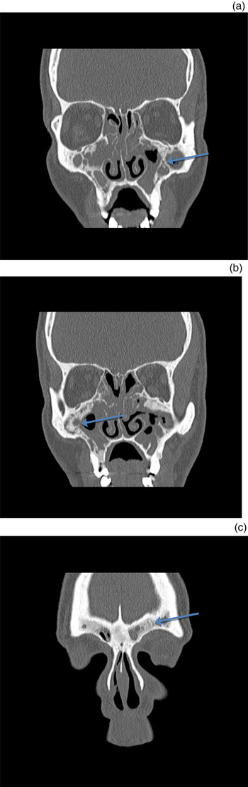
On examination, mucopurulent secretions were visible within the nose, with significant congestion and evidence of previous sinus surgery. The CT scans conducted at this time showed sinusitis, with a Lund–McKay score of 13 out of 24. Significantly, the patient was noted to have extensive neo-osteogenesis of both maxillary sinuses and frontal sinuses. There was also an area of dehiscence laterally, on the floor of the left frontal sinus (Figure 1a, b). Specific questioning about his dental background indicated a correlation with dental work performed 15 years earlier. A loculated fistula was noted on review of his left maxillary sinus (Figure 1c).
Case one. Coronal computed tomography scans of the sinuses, showing: (a) extensive neo-osteogenesis of both maxillary sinuses with significant opacity of the remaining paranasal sinuses (arrow); (b) further neo-osteogenesis of the maxillary sinuses (arrow); and (c) neo-osteogenesis of the frontal sinuses bilaterally (arrow).

The patient was admitted for exploration and sinus revision surgery. Swabs for microbiology were taken. The patient required two dental extractions. This was followed by a three-month course of antibiotics, which was guided by the microbiology team. The swab result showed a polymicrobial picture. He was originally started on clindamycin but was switched to doxycycline because of poor tolerance.
As a result of these interventions, the patient has had a significant response to this treatment. His headaches have now almost completely resolved, with only mild intermittent headaches every three to four months. These last a few hours, and have been graded 1–2 out of 10. Flexible nasendoscopy has not shown any active inflammatory process or mucopurulent secretions.
Whilst the aetiology for his sinus disease was likely multifactorial in nature, the temporal relationship with his dental treatment and the onset of his symptoms cannot be ignored in conjunction with the significant neo-osteogenesis. This highlights the fact that these cases are frequently unclear, and in many instances the diagnosis may be multifactorial and often subtle.
Case two
The second case is that of a 55-year-old woman, referred for a second opinion, with marked neo-osteogenesis of her left frontal sinus and chronic recurrent headaches, who had experienced two recurrent episodes of left pre-septal cellulitis. Clinical history revealed chronic sinonasal symptoms of nasal obstruction and discharge, refractory to maximum medical and surgical management.
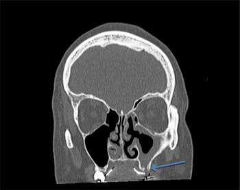
Review of the patient's CT scans identified unilateral maxillary sinus disease and extensive neo-osteogenesis of her frontal sinus on the ipsilateral side. Close review also revealed a dehiscence in the floor of the maxillary sinus in keeping with an oroantral fistula. This had not been identified by radiology in the preliminary report (Figure 2).
Case two. Coronal computed tomography scan of the sinus showing an oroantral fistula and resulting left maxillary sinus opacification (arrow).

She underwent revised left maxillary antrostomy and repair of her oroantral fistula, and has remained symptom-free since. There was significant anaerobic growth on the microscopic analysis of samples taken intra-operatively. This is to be expected given the open communication with the oral cavity.
Case three
An 82-year-old man presented acutely unwell to the emergency department with confusion and agitation 1 week following sinus surgery. A magnetic resonance imaging scan showed focal cerebritis and revealed significant sinus disease. Maximum medical treatment was instigated; however, his condition deteriorated, with the development of seizures and a significant progressive deterioration. He had undergone multiple sinus surgical procedures in the preceding months for chronic nasal obstruction and foul-smelling nasal discharge which failed to respond to treatment. Significantly, he had previously undergone bisphosphonate treatment for multiple myeloma. In addition, he was a type II diabetic.
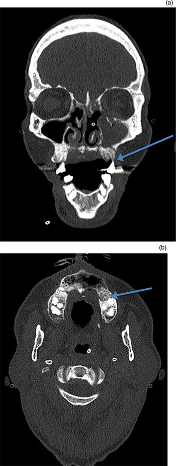
An ENT opinion was sought, and the patient subsequently underwent emergency exploration and drainage of all sinuses. Negative biopsy findings ruled out invasive fungal sinus disease. Cultures for microbiological guidance were also taken. The next day, the patient had a foul smell emanating from his mouth, whereupon his premaxilla was noted to be necrotic on imaging (Figure 3a, b). He underwent exploration and removal of a significant amount of necrotic infected bone. This was felt to be a consequence of his previous bisphosphonate therapy for myeloma and the presumed source of his chronic sinusitis.
Case three. (a) Coronal computed tomography (CT) scan of the sinuses showing a necrotic premaxilla and resulting sinus opacification (arrow). (b) Axial CT scan of the sinuses showing a necrotic premaxilla (arrow). A = anterior

The patient's recovery was slow; it was complicated by the development of an intracranial abscess, which required drainage. Two years later, after a prolonged hospital course and rehabilitation, he is living independently and remains free of any disease.
Case four
A 53-year-old woman was referred to our service by our respiratory physician colleagues. She suffered from severe unstable asthma, requiring multiple courses of steroids, triggered by recurrent sinus infections. These infections were characterised by recurrent unilateral foul taste, cheek swelling and mucopurulent discharge, all of which were refractory to maximum medical therapy.
Flexible nasendoscopy revealed mucopurulent secretions pouring from the left maxillary sinus. On examination of her oral cavity, a decayed upper left molar was identified, thought to be the trigger of the sinus infections.
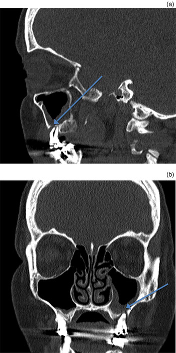
A review of the patient's CT scan showed a thickening of the left maxillary sinus, with a breach of the Schneiderian membrane serving as a tract for infection; this had not previously been commented on by radiology (Figure 4a, b). This breach appeared related to the aforementioned infected tooth.
Case four. (a) Sagittal computed tomography (CT) scan of the sinus showing left maxillary sinus mucosal thickening as a result of a breach in the Schneiderian membrane due to dental pathology (arrow). (b) Coronal CT scan of the sinus showing a breach in the Schneiderian membrane with associated mucosal thickening (arrow). L = left; A = anterior

The patient was referred to the maxillofacial surgery department for further assessment and management of her dentition. The decayed upper left molar was extracted and a short course of antibiotics was prescribed. She was reviewed in the out-patient clinic three months post extraction, and has had no further episodes of sinusitis, with much better control of her asthma.
Discussion
Odontogenic sinusitis can be responsible for both acute and chronic sinusitis, and needs to be considered when evaluating patients with sinusitis. The incidence is thought to be increasing; this may be attributable to rising costs for dental care in some countries as well as an increase in complex dentistry.Reference Little, Long, Loehrl and Poetker2 Odontogenic sinusitis more often presents with chronic symptoms.Reference Troeltzsch, Lohse, Moser, Kauffmann, Cordesmeyer and Aung3–Reference Melén, Lindahl, Andréasson and Rundcrantz5 However, in a recent large study, acute sinusitis, with or without intracranial complications, was attributable to an odontogenic cause in 15 per cent of cases.Reference Wuokko-Landén, Blomgren and Välimaa6 Another recent study suggested an odontogenic cause of sinusitis in 72.6 per cent of patients with unilateral paranasal sinusitis.Reference Matsumoto, Ikeda, Yokoi and Kohno7 We reviewed the literature to identify the specific features of odontogenic sinusitis that need to be recognised in the clinical setting.
Clinical history
To date, no one symptom has been shown as specific for odontogenic sinusitis; nevertheless, sinonasal symptoms predominate.Reference Costa, Emanuelli, Franz, Tel and Robiony8–Reference Rosenfeld, Piccirillo, Chandrasekhar, Brook, Ashok Kumar and Kramper10 Dental symptoms are often minimal or absent, although Pokorny and Tataryn reported facial or maxillary pain in 40 per cent of cases.Reference Pokorny and Tataryn11 The extent of maxillary sinus involvement seen radiologically correlated with sinonasal symptoms.Reference Costa, Emanuelli, Franz, Tel and Robiony8 Nasal obstruction, post-nasal drip and foul smell were the most common symptoms,Reference Longhini and Ferguson4,Reference Pokorny and Tataryn11,Reference Lee and Lee12 and were more commonly unilateral. A review and comparison of 22-item Sino-Nasal Outcome Test (SNOT-22) scores between non-odontogenic and odontogenic sinusitis identified malodour as a distinguishing feature of odontogenic sinusitis.Reference Simuntis, Vaitkus, Kubilius, Padervinskis, Tušas and Leketas13
Facial pain and headache also appear to be among the more prominent symptoms that patients can present with.Reference Pokorny and Tataryn11 This was seen in two of our four cases. Three of our cases had a history of complicated acute chronic sinusitis; two of the patients presented with peri-orbital cellulitis and one unfortunately had an intracranial abscess.
Iatrogenic causes account for 65.7 per cent of odontogenic sinusitis cases; hence, a dental history should be an important inclusion in the history-taking process, but is often neglected.Reference Costa, Emanuelli, Franz, Tel and Robiony8,Reference Lechien, Filleul, Costa de Araujo, Hsieh, Chantrain and Saussez14 A wide range of odontogenic pathologies can be responsible,Reference Mehra and Murad15 including periodontitis, peri-implantitis and medication complications, as with one of our cases. It is also prudent to take a targeted history regarding prior sinus symptoms or treatments.
Examination
Nasendoscopy identified mucopurulent secretions in all four of our cases. Mucopus in the middle meatus without oedema of the uncinate process was the most common finding, reported in 66.3 per cent of cases, and correlated with nasal symptoms in one retrospective study.Reference Costa, Emanuelli, Franz, Tel and Robiony8
Examination should include an inspection of dentition and consideration of a specialist opinion, to rule out a dental cause in suspected cases. This would be helpful given the range of pathologies, which more frequently relate to maxillary molars and premolars rather than wisdom teeth and other teeth.Reference Costa, Emanuelli, Franz, Tel and Robiony8 A specialist opinion should be considered where there is a significant relevant dental background, and in cases where history or radiological findings support an odontogenic cause. In the absence of this, otolaryngologists should be adept, and examination should incorporate, at the very least, a basic inspection of the teeth and palate, especially in cases of recalcitrant disease.
Radiology
Radiological imaging is crucial to the evaluation of this patient group. There has been scant acknowledgement of the relationship between dental and sinus disease in imaging literature, even in the last few years. Thus, this relationship has received very little attention, and consequently radiologists frequently do not address it when reporting on sinus imaging. A failure by clinicians to suspect a dental aetiology, and thus guide the radiological study, can therefore be contributory.Reference Whyte and Boeddinghaus1,Reference Craig, McHugh, Griggs and Peterson16 In our series, none of the underlying dental issues had been identified on radiological reports for these patients. As stated, this can be attributed to a lack of acknowledgement or awareness for a dental aetiology in presentations such as this.
Computed tomography and cone-beam CT are preferred over plain X-ray in the evaluation of cases of this nature.Reference Shahbazian and Jacobs17 Unilateral radiological findings are more common. Such findings suggested a dental cause in 72.6 per cent of cases in a recent retrospective study.Reference Costa, Emanuelli, Franz, Tel and Robiony8 However, bilateral sinus opacification does not exclude a dental cause and needs to be considered in relation to clinical history.Reference Caruso, Watkins, Suwansaard, Yamamoto, Durand and Romo18 Maxillary molars are the most common teeth involved;Reference Matsumoto, Ikeda, Yokoi and Kohno7,Reference Andric, Saranovic, Drazic, Brkovic and Todorovic9,Reference Lechien, Filleul, Costa de Araujo, Hsieh, Chantrain and Saussez14 thus, maxillary disease is the most frequent finding on CT. However, we frequently see mild mucosal maxillary disease on CT imaging, reported in 29–53 per cent of cases,Reference Vidal, Coutinho, Carvalho Ferreira, Souza and Gonçalves19 and determining its significance can be difficult. Maxillary mucosal thickening is more common with increasing age, and in overlying edentulous regions, but it is also associated with dental disease. The presence of maxillary teeth restorations correlates strongly with mucosal thickening on routine CT imaging.Reference Connor, Chavda and Pahor20
After iatrogenic causes (67 per cent), the most common dental pathologies are periapical inflammation, reported in 29 per cent, and periodontitis, in 8 per cent.Reference Lechien, Filleul, Costa de Araujo, Hsieh, Chantrain and Saussez14 In the literature, a consensus acknowledges mucosal thickening of more than 10 mm as severe.
Where the history or findings are supportive, or when conventional management fails, a dental opinion and/or cone-beam CT should be included in the investigation, as cone-beam CT is more sensitive than plain X-ray imaging.Reference Shahbazian and Jacobs17 Cone-beam CT, with a spatial resolution of less than 0.2 mm, can identify early signs of periapical inflammation. This develops at the tooth apex, causing subtle widening of the periodontal ligament space. Expansion of this space can be the result of an abscess, cyst or granuloma, but these are not indistinguishable, and are observed as periapical inflammatory lucencies on imaging. Whilst an abscess is often symptomatic, cysts or granulomas are often not.Reference Boeddinghaus and Whyte21
• Odontogenic sinusitis is a recognised but frequently overlooked and undiagnosed cause of rhinosinusitis, both acute and chronic
• No single sign or symptom is specific to odontogenic sinusitis, and failure to focus on specific radiological features can delay diagnosis
• It is important to recognise the association between dental pathology and both acute and chronic sinusitis
• Collaboration with a maxillofacial specialist is important for diagnosis and management of odontogenic sinusitis
• Unilateral radiological findings and/or the presence of neo-osteogenesis should increase the suspicion for an odontogenic cause
• Basic dental history and examination, and awareness of specific imaging features are crucial in cases of recalcitrant disease
Several studies note a correlation between mucosal thickening and the presence of periapical inflammatory lesions, with reports of evidence in 80 per cent of cases.Reference Vidal, Coutinho, Carvalho Ferreira, Souza and Gonçalves19,Reference Nascimento, Pontual, Pontual, Freitas, Perez and Ramos-Perez22–Reference Eggmann, Connert, Bühler, Dagassan-Berndt, Weiger and Walter24 Periodontitis represents loss of the osseous and ligamentous dental root structure, which results from the progression of gingivitis. It is graded radiologically by the depth of the periodontal pocket relative to the length of the involved tooth. A correlation between severity of periodontitis and mucosal thickening has been reported in two large studies as 42 per cent and 49 per cent respectively.Reference Phothikhun, Suphanantachat, Chuenchompoonut and Nisapakultorn25,Reference Ren, Zhao, Liu, Wang and Pan26 Eighty-eight per cent of patients with severe periodontitis have mucosal thickening, however, in contrast to periapical pathology; this is associated with mucosal thickening of less than 10 mm, and is frequently bilateral.Reference Whyte and Boeddinghaus1
Overall, periodontal disease is more commonly the cause of mucosal thickening.Reference Vidal, Coutinho, Carvalho Ferreira, Souza and Gonçalves19,Reference Nascimento, Pontual, Pontual, Freitas, Perez and Ramos-Perez22–Reference Eggmann, Connert, Bühler, Dagassan-Berndt, Weiger and Walter24,Reference Hoskison, Daniel, Rowson and Jones27 Mucosal disease of dental origin can be defined as a spectrum, ranging from mild mucosal thickening to extensive anterior sinus disease involving the maxillary, anterior ethmoid and frontal sinuses.Reference Boeddinghaus and Whyte21 The presence of unilateral disease with such involvement should alert the otolaryngologist, and, indeed, the radiologist, to review the floor of the maxillary sinus and associated alveolar process, and posterior maxillary teeth.
In two of our four cases, radiologically, neo-osteogenesis was a significant feature. This has been reported in the literature previously; however, it is not commonly referred to.Reference Huang, Xu, Xiao, Li, Dong and Li28 Of note, both cases were also associated with facial pain and headaches.
Microbiology
Bacteriology is usually polymicrobial, although anaerobes feature significantly in dental sources of infection, because of maxillary sinus contamination with oral cavity pathogens.Reference Saibene, Pipolo, Lozza, Maccari, Portaleone and Scotti29 These reports are consistent with the findings from our case series. Samples from all of the cases grew multiple pathogens, with anaerobes featuring frequently. Cases with oroantral fistulae showed heavy anaerobic growth on microscopy.
Recurrent acute infective sinusitis, a subgroup of chronic rhinosinusitis, is more commonly associated with Streptococcus pneumoniae, Moraxella catarrhalis and Haemophilus influenzae, whereas cultures from multiple studies of odontogenic sinusitis have shown an increased incidence of polymicrobial infections with oral anaerobes such as Peptostreptococcus and Prevotella spp., as well as aerobic bacteria such as Staphylococcus aureus featuring. For this, respiratory quinolones may be favoured as antibiotic therapy, and the length of therapy based on symptom duration. In their study comparing the microbiology of rhinogenic and odontogenic sinus disease, Saibene et al. found that only 60 per cent of chronic sinusitis samples grew microbes, whereas all of the odontogenic sinusitis samples had a large microbial burden and bacterial growth.Reference Saibene, Pipolo, Lozza, Maccari, Portaleone and Scotti29
Conclusion
It is important to recognise the association between dental pathology and both acute and chronic sinusitis. Collaboration with a maxillofacial specialist is important for diagnosis and management of odontogenic sinusitis, and should be considered where standard management fails, or where certain clinical features or radiological signs are present. Often dental symptoms are not present and there can be a wide range of dental causes, including periodontitis which would require dental diagnosis and management.
Nasal obstruction and significant mucopurulent discharge, especially if foul-smelling and accompanied by facial pain or headaches, are common nasal symptoms that can be highly suggestive of a dental cause, as is the presence of mucopurulent secretions in the middle meatus, especially if unilateral. Patients with odontogenic sinusitis often first present to the ENT clinic; therefore, otolaryngologists should incorporate an examination of the teeth and palate as part of their assessment, especially in cases with the previously mentioned features. Bilateral disease does not exclude an odontogenic cause, but is less frequently due to odontogenic sinusitis.Reference Saibene, Pipolo, Lozza, Maccari, Portaleone and Scotti29 Recalcitrant, non-eosinophilic sinus disease should also prompt consideration of a dental pathology as the underlying cause.
Radiological investigation is best conducted via CT imaging.Reference Shahbazian and Jacobs17 Unilateral radiological findings and/or the presence of neo-osteogenesis should increase the suspicion for an odontogenic cause.Reference Huang, Xu, Xiao, Li, Dong and Li28 Radiological under-reporting is common and thus CT imaging should be reviewed specifically for features suggestive of dental pathology. Finally, a high index of suspicion should be present in those cases of sinusitis that do not respond to standard management, such as with our four cases.
Competing interests
None declared