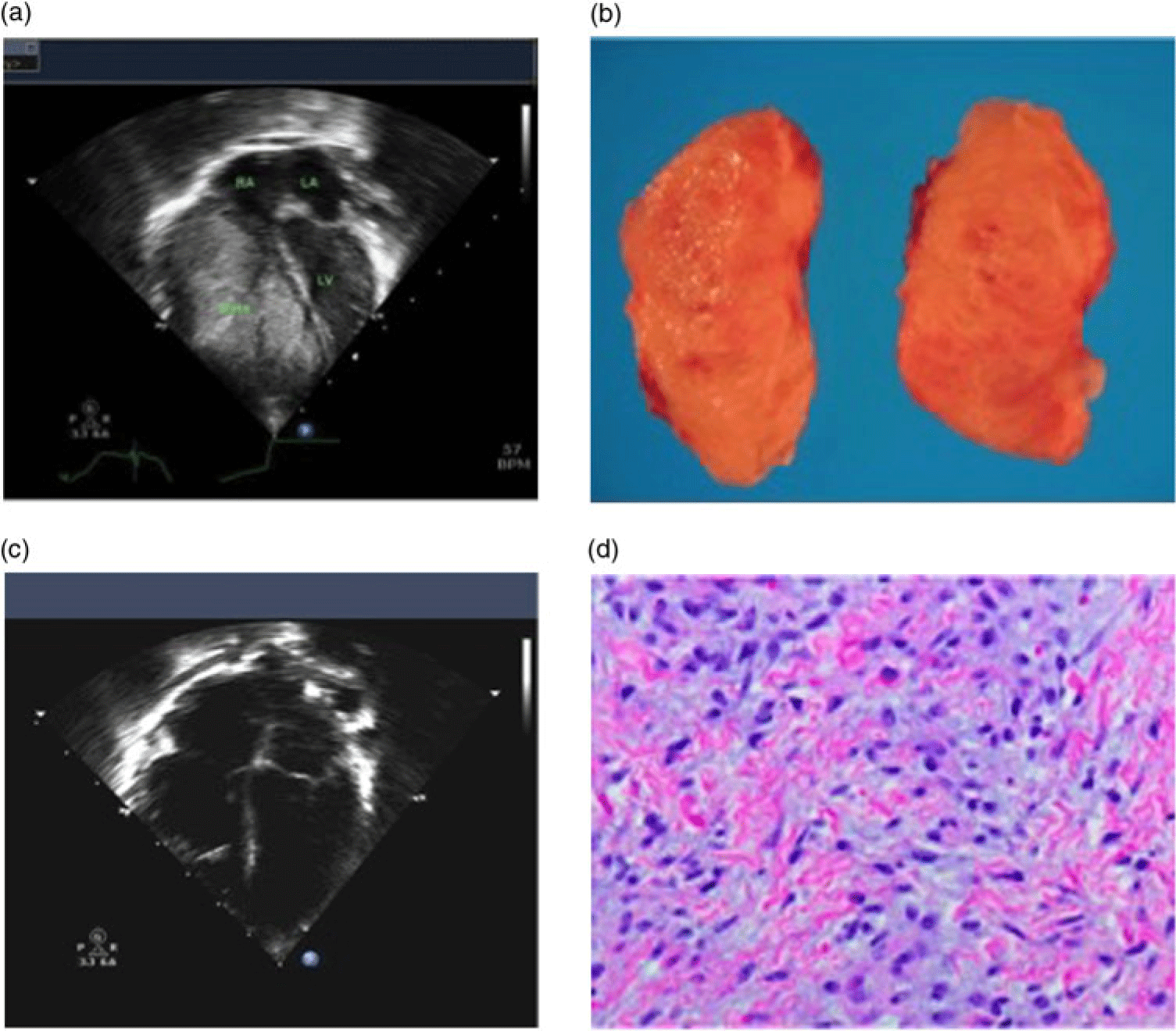
A 3-month-old infant was transferred for escalation in care and surgical management of an intra-cardiac tumour. Prenatal ultrasound was concerning for a right ventricular intra-cardiac mass. This was confirmed on postnatal ultrasound following caesarean section at 34 weeks gestational age, revealing a 45 mm × 29 mm mass originating from the anterior wall, completely obliterating the right ventricular cavity (Fig 1a). A diagnosis of cardiac fibroma was made on biopsy during pericardial window placement for a pericardial effusion at 1 month of life. Electrocardiogram and telemetry monitoring revealed no arrhythmias; however, symptoms of low cardiac output with failure to gain weight and feeding intolerance prompted pursuit of surgical intervention.
(a) pre-operative transthoracic echocardiogram showing a fibroma obscuring the right ventricle (LV- left ventricle, LA- left atrium, RA- right atrium). (b) Gross images of the resected fibroma, measuring 66 × 46 × 33 mm and weighing 46.4 g. (c) Post-operative thransthoracic echocardiogram following fibroma resection. (d) Pathology of the resected mass showing fibroblasts interspersed amongst a collagen matrix, consistent with a fibroma.

Given the child’s progressive symptomatology and propensity for ongoing fibroma enlargement, multi-disciplinary discussion considered total resection, single ventricle palliation with aortapulmonary shunt or bidirectional Glenn, or cardiac transplantation as therapeutic options. The decision was made for total surgical resection via right ventriculotomy of the massive 66 mm × 46 mm × 33 mm fibroma (Fig 1b). The patient received a standard sternotomy and was cannulated via her right atria after cooling prior to bypass. Piecemeal dissection via a right ventricle incision resulted in total mass resection after 93 minutes bypass time. She was extubated on post-operative day 3. After an uneventful convalescence, post-operative echocardiogram revealed qualitatively normal biventricular function (Fig 1c). She was discharged on post-operative day 11 on full oral feeds.
Discussion
Management of massive cardiac fibroma obliterating the ventricular cavity can be difficult. Various strategies include the use of single ventricle palliation, cardiac transplantation, and total/subtotal resection.Reference Lee, Kim and Kim1 There are limited reports of undertaking complete resection of a fibroma of this size at this young age. This case highlights the rare presentation of a massive cardiac fibroma as well as the ability to completely resect the mass, restoring normal right ventricular cavity size and function.
Limited data quantifying size of resected cardiac masses in children and adult patients. One review of 94 patients reveals an average resected fibroma size of 20–30 mm, about half that of our patient’s resected specimen.Reference Patel and Sheppard2 Very few reports exist of fibroma resection of this size in neonates and infants.Reference Jha, Kiraly and Tamas3 Key limitation in massive tumour resection and surgical planning is the presence or absence of sufficient myocardial tissue and lack of tumour involvement of the endocardial surface allowing for total or subtotal tumour resection and closure.Reference Lee, Kim and Kim1
Primary cardiac tumours are rare in children, making up 3% of all tumours in patients younger than 18.Reference Yadava4 The type of tumour, aside from identifying benign versus malignant, is important because each is associated with their own set of operative and non-operative complications. In the case of fibromas, mortality is more frequent compared to that of rhabdomyomas or myxomas that required surgical interventions. A case series of 120 children operated on for their cardiac tumours found that patient’s with fibromas accounted for nearly half of total deaths before discharge, with a much higher surgical mortality.Reference Bielefeld and Moller5 The aetiology of this mortality difference is thought to be their larger size and unencapsulated nature resulting in invasion of the adjacent myocardium making excision difficult.
One-third of patients with cardiac fibromas are asymptomatic upon discovery. Fibromas are usually isolated single tumours, most commonly located in the free ventricular wall or the inter-ventricular septum.Reference Shi, Wu and Fang6 Architecturally, the fibroblasts and collagen matrices that make up fibromas as seen in our patient’s specimen are similar to that of purkinje cellsReference Uzun, Wilson, Vujanic, Parsons and De Giovanni7 (Fig 1d). This is most evident in that patients with cardiac fibromas are pre-disposed to arrhythmias at a higher rate compared to other cardiac tumours.
Miyake et al. report an incidence of ventricular arrhythmias as high as 64% in patients with high-risk cardiac tumours such as fibromas.Reference Miyake, Del Nido and Alexander8 While evidence regarding management of intra-cardiac tumours in children is sparse, some data point towards resection significantly reducing arrhythmia burden. A 19 patient multi-centre study in British hospitals followed patients with pathology confirmed ventricular fibromas. The most common indication for surgical intervention in this population was out-of-hospital arrest presumably due to ventricular arrhythmia. The majority of patients were weaned off of their anti-arrhythmic therapies following resection.Reference Jones, Ramcharan and Chaudhari9
In summary, symptomatic cardiac fibromas should be considered for complete resection due to arrhythmia burden and impedance of blood flow due to tumour mass effect. This allows for resolution of symptomatology without the need for other surgical palliative strategies or cardiac transplantation. The patient was doing well at follow-up 2 years after her operation, with no clinical signs of heart failure and effectively normal cardiac function on interval echocardiogram.
Acknowledgements
Special thanks to Dr Robert Romp and Dr Luz A. Padilla for assistance in editing and image acquisition.
Financial Support
This research received no specific grant from any funding agency, commercial or not-for-profit sectors.
Conflicts of Interest
None.