INTRODUCTION
The parasites of phylum Haplosporidia Caullery and Mesnil, 1899 can be amongst the most pathogenic of all bivalve parasites (Burreson et al. Reference Burreson, Stokes and Friedman2000). Several species of Haplosporidia have been associated with significant epizootic mortalities and economic losses in commercially important shellfish aquaculture around the world (Burreson and Ford, Reference Burreson and Ford2004). Haplosporidium nelsoni (MSX) has impacted the production of the Eastern Oyster (Crassostrea virginica) on the eastern seaboard of the United States and Bonamia ostrea has severely affected the culture of the flat oyster (Ostrea edulis) in Western Europe (Carnegie et al. Reference Carnegie, Barber and Distel2003).
The Haplosporidia are characterized morphologically as protists possessing ovoid, walled spores lacking polar filaments or polar tubes, and with an orifice at one pole (Cavalier-Smith and Chao, Reference Cavalier-Smith and Chao2003). The orifice is covered either externally by a hinged lid or internally by a flap of wall material. Currently, there are four genera allocated to the phylum; Urosporidium Caullery and Mesnil, 1905; Minchinia Labbe, 1895; Haplosporidium Caullery and Mesnil, 1899 and Bonamia Pichot, Comps, Tige, Grizel and Rabouin, 1980 (Reece et al. Reference Reece, Siddall, Stokes and Burreson2004). However, species identification and genus assignment has been confounded since spore ornamentation is not known for many haplosporidian species (Burreson and Reece, Reference Burreson and Reece2006). The taxonomy of Haplosporidia may remain confused until spore ornamentation has been characterized for a large number of species. DNA sequence analysis of a number of these species will also be required to determine the relationships among the different morphological types.
The primary taxonomic feature used to differentiate species and genera within Haplosporidia is the morphology and origin of spore ornamentation, usually described as filaments, tails, wrappings or extensions. Currently, the species within the genera Haplosporidium and Bonamia are separated from those of Minchinia on the basis of the origin of the spore ornamentation. For the members of Haplosporidium and Bonamia in which spores have been detected (i.e Bonamia perspora), ornamentation originates from the spore wall while in species of Minchinia it originates from the epispore cytoplasm (Ormieres, Reference Ormieres1980; Hine and Thorne, Reference Hine and Thorne1998; Azevedo et al. Reference Azevedo, Montes and Corral1999; Burreson, Reference Burreson2001; Carnegie et al. Reference Carnegie, Burreson, Hine, Stokes, Audemard, Bishop and Peterson2006). Thus, for descriptions of new species it is important to describe the spore ornamentation.
Mortalities (up to 80%) among rock oysters Saccostrea cuccullata were first recognized by energy companies operating on the North West Gas Shelf of Western Australia in the early 1990s (Hine and Thorne, Reference Hine and Thorne2002). The companies submitted samples for diagnosis, and a haplosporidian species parasitizing rock oysters was subsequently morphologically described by Hine and Thorne (Reference Hine and Thorne2002) using light and transmission electron microscopy. Samples obtained by Hine and Thorne (Reference Hine and Thorne2002) contained single, bi-nucleate and multinucleate plasmodia as well as spore stages. At least four attempts to sample more oysters from within the parasite's range were made but infected oysters could not be detected (Hine and Thorne, Reference Hine and Thorne2002). A Minchinia species, based on DNA sequence analysis, was described by Bearham et al. (Reference Bearham, Spiers, Raidal, Jones and Nicholls2007) from within the range and from the same host species as described by Hine and Thorne (Reference Hine and Thorne2002). Morphologically, the parasite described by Bearham et al. (Reference Bearham, Spiers, Raidal, Jones and Nicholls2007) consisted of uni-nucleated naked cells that resembled a Bonamia species. No spores were observed so the parasite is not able to be described following the established morphological criteria. It was possible that the parasite was not detected in previous sampling expeditions because of the cryptic nature of the uni-nucleate stages. It is therefore possible that this stage may be the typical state of the parasite and those stages described by Hine and Thorne (Reference Hine and Thorne2002) are unusual. A section of the organism's rRNA gene was sequenced and an in situ hybridization assay was developed in our laboratory (Bearham et al. Reference Bearham, Spiers, Raidal, Jones and Nicholls2007, Reference Bearham, Spiers, Raidal, Jones and Nicholls2008b). The parasite fell within the Minchinia clade in a phylogenetic analysis of the parasite's SSU rRNA gene and was a sister taxon to a clade composed of M. chitonis, M. teredinis and Minchinia sp. of Cyrenoida floridana (Bearham et al. Reference Bearham, Spiers, Raidal, Jones and Nicholls2007).
Rock oysters are currently commercially harvested only in the Australian state of Queensland (Anon, 2002). Minchinia sp. could be a major impediment to the development of tropical rock oyster aquaculture in northern Western Australia (Anon, 2004) given that it is associated with significant mortality. A Minchinia sp. along with Haplosporidium hinei (Bearham et al. Reference Bearham, Spiers, Raidal, Burreson, Jones and Nicholls2008a) has also been detected in pearl oyster spat in Australia's South Sea pearl industry although it has not been associated with any mortality. Sporadic reports of a Bonamia-like infection have also been reported for pearl oysters (SCFH, 1993; Norris, Reference Norris1996; Humphrey et al. Reference Humphrey, Norton, Jones, Barton, Connell, Shelley and Creeper1998). It's possible that these parasites were actually Minchinia occulta n.sp. since the uni-nucleate naked cells described by Bearham et al. (Reference Bearham, Spiers, Raidal, Jones and Nicholls2007) resemble a Bonamia sp.
This study describes the spore ornamentation of a Minchinia species parasitizing rock oysters (Saccostrea cuccullata) using both scanning and transmission electron microscopy. The parasite is named in this paper as Minchinia occulta n. sp.
MATERIALS AND METHODS
Archived formalin-fixed, paraffin-embedded rock oyster tissues infected with Minchinia occulta n. sp. were obtained from the Western Australian Department of Fisheries. All of the material was collected from the same sample of infected oysters from Varanus Island on the north coast of Western Australia (co-ordinates 20°39′3″ S 115°34′27″ E) and was fixed whole in 10% formalin made up with seawater. Material specifically fixed for electron microscopy was not available. Tissue sections were processed for histological examination and stained with haematoxylin and eosin using conventional techniques. The unstained sections were cut at a thickness of 4 μm and placed on slides. The sections were de-waxed in xylene and rehydrated in an ethanol series (2 changes for 22 min each). The slides were then placed in haematoxylin for 5 min, rinsed in water and then placed in Scott's tap water substitute until blue. Each section was again rinsed in water followed by 95% ethanol (1 change for 30 sec). The sections were stained with 1% eosin for 30 sec and finally dehydrated in 95% ethanol (2 changes 30 sec each), absolute ethanol (2 changes 30 sec each) and xylene (3 changes 30 sec each). Ornamentation on the spores of M. occulta n. sp. was assessed through light microscopy, transmission electron microscopy (TEM) and scanning electron microscopy (SEM).
In situ hybridization
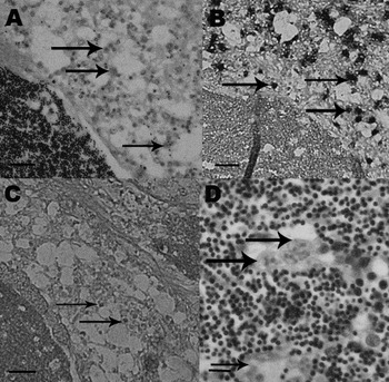
In situ hybridization (ISH) was performed to confirm the identity of the parasite as the same species sequenced by Bearham et al. (Reference Bearham, Spiers, Raidal, Jones and Nicholls2007). An oligonucleotide probe was used for this purpose. The SSRDb probe used in the study (5′ GTTAGCCTTGCGCGCAGCCGATACG 3′) was obtained pre-labelled from Operon Biotechnologies GmbH (Kohl, Germany). The presence and location of M. occulta n. sp. was confirmed using haematoxylin – eosin (H&E) stained serial sections (Fig. 1). The in situ hybridization procedure was performed as described by Bearham et al. (Reference Bearham, Spiers, Raidal, Jones and Nicholls2007).
Rock oyster connective tissue and reproductive tissue containing single, bi- and tri-nucleate stages of Minchinia occulta n. sp. (A) Haematoxylin-eosin-stained section containing the parasites (arrows). (B) Serial section containing the parasites in an in situ hybridization. Parasites are identified by a darker colouration. Examples are indicated with an arrow. (C) Negative control serial section from the same hybridization. Example parasites are indicated with an arrow. (D) Haematoxylin-eosin-stained section containing the parasites in reproductive tissue of the host. The small arrow indicates a uni-nucleate stage similar to that observed in Bearham et al. (Reference Bearham, Spiers, Raidal, Jones and Nicholls2007) while the larger arrows indicate multi-nucleate stages. Scale bars=15 μm. All samples were taken from the same sample of infected oysters.

Electron microscopy
Formalin-fixed, paraffin-embedded samples observed as possessing spores in histological sections were processed for electron microscopy. These oysters were obtained from the same sample of oysters that underwent in situ hybridization. Material specifically fixed for electron microscopy was not available. Rock oyster tissue samples containing M. occulta spores were removed from the paraffin block and prepared for TEM by de-waxing overnight in xylene. The tissue samples were then hydrated through 3 changes of 100, 90 and 70% ethanol. A 1 mm3 cube of digestive gland tissue was dissected in a puddle of 5% Sorensen's phosphate-buffered glutaraldehyde and stored in 5% glutaraldehyde overnight. The samples were then washed in 0·07 m Sorensen's phosphate buffer and immersed in Dalton's Chrome Osmic Acid for 60–90 min at 4°C. Following this, the samples were then dehydrated through an ethanol series and immersed in propylene oxide, a 60:40 propylene oxide/Epon mixture. The samples were finally embedded in pure ‘Epon’ overnight. The tissues were sectioned with an mU3/C (Reichert, Austria) ultramicrotome and stained with uranyl acetate. Analysis was performed with a Philips CM100 Bio TEM.
For SEM, infected oyster tissue was removed from the paraffin block and deparaffinized in xylene for 3 days. The samples were rehydrated through a graded series of ethanol (100%, 90%, 70%, 50% and 30%) and then sonicated in pure water. A puddle of the resulting suspension was placed onto 6×12 mm diameter cover-slips coated with poly- L-lysine. M. occulta spores in the suspension were allowed to settle for 1 h in a moist chamber. Following this, the cover-slips were dipped in water to remove the excess material and then dehydrated through an ethanol series. The affixed spores were subjected to critical-point drying in liquid CO2 and coating with gold: palladium. Observations of the spore ornamentation were made on a Zeiss Leo 435vp SEM. The measurements of 16 parasites are given in micrometres with the range in parentheses.
RESULTS
Light microscopy
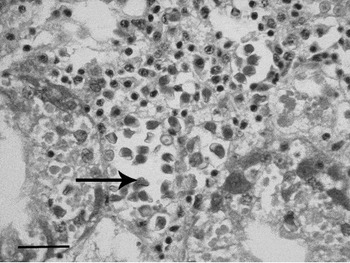
Histopathological examination of 16 positive oysters from Varanus Island, Western Australia revealed large numbers of parasites present usually as focal lesions in the connective tissue of the gills or disseminated around the connective tissue between the digestive diverticulae in the digestive gland, and in the mantle (Figs 1 and 2). Parasites were also observed between the epithelial cells of the gut but not in the epithelium of the digestive diverticulae. Large numbers of parasites were also present in the reproductive follicles of the host of some samples (Fig. 1). All parasite stages including uni-nucleate and bi-nucleate naked cells were extracellular. Uni-nucleate cells were 4·5 to 5·8 μm (mean=5 μm) in diameter with a generally central nucleus. Multi-nucleate plasmodia with between 2 and 24 nuclei were also present in infected oysters. Sporulation was confined to the connective tissue between the digestive diverticulae in the digestive gland. Mature spores with a yellow refractile wall enclosing an eosinophilic sporoplasm were also observed (Fig. 2). Spore ornamentation was not apparent with light microscopy and parasites were not observed in the epithelium of the digestive diverticulae. Phagocytosis of parasites was not observed.
Rock oyster digestive gland containing large numbers of the pre-sporulation and sporulation stages of Minchinia occulta n. sp. in a haematoxylin-eosin-stained section. Arrow indicates an example of the refractile mature spores. Scale bar=20 μm.

In situ hybridization
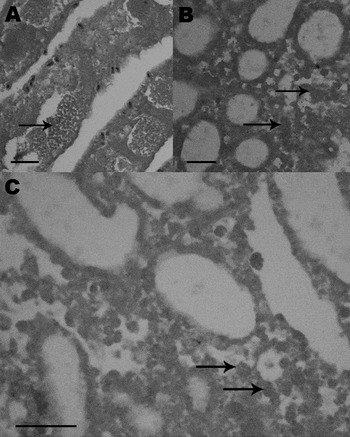
Parasite identity was confirmed using in situ hybridization. The SSRDb probe produced strong hybridization signals with little background staining (Fig. 1). The signal was not reproduced in tissues produced from uninfected oysters or in negative control material (Fig. 1). Sections of Minchinia teredinis, Haplosporidium nelsoni and H. costale included in the assay did not produce a positive signal (Fig. 3).
Haplosporidian parasites used to assess the specificity of the ISH assay. Example parasites are indicated with an arrow. (A) Minchinia teredinis in the gills of the shipworm Teredo sp. Scale bar=10 μm. (B) Haplosporidium costale in an Eastern Oyster. Scale bar=30 μm (C) Haplosporidium nelsoni in an Eastern Oyster. Scale bar=20 μm. Sections are counter-stained in brazilin haematoxylin.

Electron microscopy
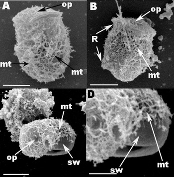
The fixative used for the samples was suboptimal for electron microscopy; nonetheless, it was adequate to permit some observation of the spore wall and its ornamentation. The spores of the rock oyster parasite appeared to have a network of branching microtubule-like structures (Fig. 4) covering the entire spore including the opercula lid. The microtubule-like structures varied in size but were approximately 15–30 nm (mean=20 nm) in diameter (Fig. 4). In some of the spores this network appeared to degrade as the spore aged revealing a smooth spore wall (Fig. 4). A ridge was also present on one side of the spore. It began just below the operculum and ended approximately 2 μm from the aboral end of the spore (Fig. 4). The ridge did not consist of spore wall material but appeared to be composed of the same material as the microtubule-like structures. Indeed, no spore wall-derived ornamentation was observed on any spores.
Scanning electron micrographs indicating the spore ornamentation of Minchinia occulta n. sp. infecting Saccostrea cuccullata. (A) Minchinia occulta spore with a branching network of microtubule-like structures and opercula lid (op). Scale bar=1·5 μm. (B) Spore with a ridge running along the front of the spore (arrow). Scale bar=2·5 μm. (C) Spore with network of branching microtubule–like structures that has been partially removed or degenerated revealing a smooth spore wall (sw) beneath. Some artefact also appears on the spore wall. Scale bar=3·0 μm. (D) Higher magnification view of the degraded microtubule like structures with exposed spore wall beneath. Scale bar=1 μm.

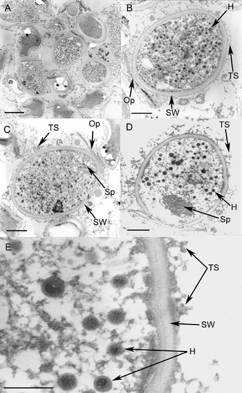
The fixed spores of M. occulta n. sp were elongated 4·5–5·0×3·5–4·1 μm in size (Figs 4 and 5). The spore wall appeared to be 225 nm in thickness and consisted of 3 layers; an inner dense layer of approximately 90 nm, a middle layer of 30 nm and an outer dense layer of 130 nm (Fig. 5). The sporoplasm nucleus was positioned equatorially and usually round in shape and measured 1·5–2·3 μm in diameter (mean=1·9 μm). The operculum was situated in the apical zone of the wall and consisted of a circular lid of 2·5–3·0 μm (mean=2·8 μm) diameter (Fig. 5). The lid of the operculum was about 150 nm thick and was connected to the spore wall by a hinge (Fig. 5).
Transmission electron micrographs (TEM) of spores from Minchinia occulta n. sp. infecting the rock oyster (Saccostrea cuccullata). (A) Group of spores at a variety of different spore stages none of which possess any spore wall ornamentation. Scale bar=2 μm. (B–D) Minchinia occulta spores with operculum (Op), haplosporosomes containing a membrane-bound body (H), microtubular-like structures (TS) and spore wall (SW) and spherule (Sp). Scale bars=1 μm. (E) Higher magnification view of the membrane-bound body within the haplosporosomes (H), the spore wall (SW) and the microtubular-like structures (TS). Scale bar=500 nm.

Round, electron-dense haplosporosomes were present that were 150 nm–180 nm (mean=160 nm) in diameter. A membrane-bound body was also often observed within the haplosporosomes (Fig. 5E). In the apical zone of the endosporoplasm, a spherule approximately 700 nm in diameter was formed by several vesicles (Fig. 5). Dense vesicles (DVs) (diameter=159 nm to 228 nm (mean=200 nm)) were numerous throughout the episporoplasm (Fig. 5). Bundles of microfilaments were also occasionally observed in the episporoplasm as were lipid droplets. A lipid body was often observed adjacent to the middle of the spore (Fig. 5). No spore wall ornamentation or any attached cytoskeletal structures were observed on any spore maturation stages including mature spores (Fig. 5).
Taxonomic summary
Minchinia occulta Bearham, Spiers, Raidal, Jones, Nicholls n. sp.
Description and identifying characteristics
All parasite stages including uni-nucleate and bi-nucleate naked cells are extracellular with uni-nucleate cells approximately 4·5 to 5·8 μm in diameter with a central nucleus. Multi-nucleate plasmodia, possibly associated with sporogenesis, may also be present with between 2 and 24 nuclei. Spore ornamentation consisting of a network of branching microtubule-like structures covering the entire spore including the opercula lid. A ridge is also present on one side of the spore beginning just below the operculum and ending approximately 2 μm from the aboral end of the spore (Fig. 3). The sporoplasm nucleus is positioned equatorially and is usually round.
Type Host
Saccostrea cuccullata Born, 1778 (Mollusca, Bivalva, Ostreidae).
Sites of infection
Minchinia occulta was observed as focal infections in the connective tissue of the gills and as disseminated infections in the mantle, reproductive follicles, and around the digestive diverticulae. Minchinia occulta was also observed in the epithelial cells of the gut but not in the epithelium of the digestive diverticulae with sporulation confined to the connective tissue of the digestive gland.
Type locality
Varanus Island (co-ordinates 20°39′3″ S 115°34′27″ E), Western Australia.
Material deposited
H&E slides in the Marine Invertebrate Zoology Type Collections, Department of Aquatic Zoology, Western Australian Museum, registration number WAM Z27551.
Etymology
Occulta means hidden or concealed and refers to the cryptic nature of the parasite in some H&E sections.
DISCUSSION
The parasite described in this study shows the usual spore morphology, structure and ornamentation of a haplosporidian species. The internal organization of the spore endosporoplasm is similar to other haplosporidian species. This was characterized by the presence of an equatorial nucleus, several electron-dense membrane-bound haplosporosomes and an apical spherule. The existence of an orifice that was covered with an operculum indicates the parasite belongs to Minchinia, Haplosporidium or Bonamia.
The parasite's identity was confirmed as the same parasite species characterized by Bearham et al. (Reference Bearham, Spiers, Raidal, Jones and Nicholls2007) with an in situ hybridization assay utilizing an oligonucleotide ISH probe (SSRDb) on an oyster from the same sample the M. occulta n. sp. spores were obtained. The ISH assay was designed to target the SSU rRNA gene of the Minchinia sp. characterized by Bearham et al. (Reference Bearham, Spiers, Raidal, Jones and Nicholls2007). The assay did not react with any of the other haplosporidian parasites in the assay (Minchinia teredinis, H. costale and H. nelsoni). Individual parasites were also observed within the reproductive tissue of the host and were from the same host species and within the same range as the parasite described by Bearham et al. (Reference Bearham, Spiers, Raidal, Jones and Nicholls2007).
The only previous mention of spore ornamentation in M. occulta n. sp. was by Hine and Thorne (Reference Hine and Thorne2002) in a TEM ultrastructural study of spores. Hine and Thorne (Reference Hine and Thorne2002) noted the presence of surface microtubules present on the wall of mature spores. The spores observed in the current study, utilizing both SEM and TEM, suggested that these microtubules formed a covering of branching microtubules. The size and arrangement of the internal organization of the spores observed here were consistent with those described previously by Hine and Thorne (Reference Hine and Thorne2002).
The ornamentation on the spores of the rock oyster parasite was unique among described haplosporidian species. The spores of the rock oyster parasite did not have spore wall-derived ornamentation and are therefore different from most described Haplosporidium species.
The branching network of microtubules on the spores of M. occulta resembles, at least superficially, the spore ornamentation of Haplosporidium nelsoni and H. costale in Crassostrea virginica (Burreson and Reece, Reference Burreson and Reece2006). These species have ornamentation consisting of a network of branching fibres. However, the composition of the ornamentation differs considerably from the parasite described here. Spore ornamentation in H. nelsoni consists of fibres that are derived from the epispore cytoplasm and are tubular but with considerable internal complexity (Burreson and Reece, Reference Burreson and Reece2006). In addition, the covering on H. nelsoni consists of individual ribbons tightly bound together that separate at the aboral end of the spore and do not cover the opercula lid unlike M. occulta n. sp. (Burreson and Reece, Reference Burreson and Reece2006). The fibres from H. costale while derived from the epispore cytoplasm, are not tubular and have a fine braided structure unlike M. occulta (Perkins, Reference Perkins1969; Burreson and Reece, Reference Burreson and Reece2006). Ornamentation on spores of Haplosporidium louisiana consist of very thin filaments composed of spore-wall material that wrap around the spore (Perkins, Reference Perkins1969). No spore wall-derived ornamentation was observed on the spores of M. occulta n. sp.
Hine and Thorne (Reference Hine and Thorne2002) suggested the microtubule-like structures derived from the episporoplasm in M. occulta appeared similar to microtubules in the epispore cytoplasmic vacuoles of Minchinia species in crabs (Hine and Thorne, Reference Hine and Thorne2002; Perkins, Reference Perkins1975) and similar structures may be seen aligned under the epispore plasma membrane in M. chitonis (Ball, Reference Ball1980; Hine and Thorne, Reference Hine and Thorne2002). Bundles of similar microtubule-like structures also enter the ECE of M. teredinis (Hine and Thorne, Reference Hine and Thorne2002; McGovern and Burreson, Reference McGovern and Burreson1990). Comps and Tige (Reference Comps and Tige1997) also reported conspicuous microtubule-like fibrils of 20 nm in diameter in the cytoplasmic tails of Minchinia sp. infecting the mussel Mytilus galloprovincialis on the Mediterranean coast of France. The microtubule-like structures were also visible in the episporoplasm surrounding the spore wall similar to M. occulta n. sp. except the structures were focally distributed and were aligned (Comps and Tige, Reference Comps and Tige1997). Similar unaligned microtubules have also been reported from Haplosporidium ascidiarum and H. ascidiarum also appears to lack spore wall filaments (Ormieres and de Puytorac, Reference Ormieres and De Puytorac1968; Hine and Thorne, Reference Hine and Thorne2002). Therefore, H. ascidiarum may also be a species of Minchinia.
However, M. occulta n. sp. differs from other Minchinia species since M. tapetis infecting Ruditapes decussatus in Europe has a single posterior epispore cytoplasmic extension (ECE; Azevedo, (Reference Azevedo2001)) while M. teredinis infecting shipworms (Teredo sp.) in the west Atlantic has four ECEs (McGovern and Burreson, Reference McGovern and Burreson1990). Minchinia chitonis, infecting the chiton Lepidochitona cinereus has two opposing ECEs (Ball, Reference Ball1980). While the ornamentation of each of these Minchinia species is composed of epispore cytoplasm, the parasite observed here differs from each of these parasites since it does not appear to possess an ECE(s). Instead, it is entirely covered in branching microtubule-like structures derived from the epispore cytoplasm and possesses a ridge on one side of the spore. The ridge appears to be composed of a similar material as the microtubules and therefore may be a similar ephemeral structure. No evidence of any epispore cytoplasmic extensions (ECEs) or tails were observed on spores of M. occulta n. sp. Because de-paraffinized tissue was used for SEM, the presence of epispore cytoplasm-derived projections typical of other Minchinia species cannot be ruled out. Further analysis needs to be conducted with samples of infected oyster tissue specifically fixed for electron microscopy.
The results presented here suggest the rock oyster parasite should be assigned to Minchinia and not Haplosporidium or Bonamia. The parasite possesses microtubule-like structures similar to other Minchinia species and there was a lack of any spore wall-derived ornamentation that would suggest the parasite should be assigned to Haplosporidium or Bonamia. This is consistent with the results described by Bearham et al. (Reference Bearham, Spiers, Raidal, Jones and Nicholls2007) where the parasite fell within the Minchinia clade in a phylogenetic analysis of the parasite's SSU rRNA gene and is a sister taxon to a clade composed of M. chitonis, M. teredinis and Minchinia sp. of Cyrenoida floridana (Bearham et al. Reference Bearham, Spiers, Raidal, Jones and Nicholls2007).
Hine and Thorne (Reference Hine and Thorne2002) described the development of M. occulta n. sp. including multinucleate and spore-forming stages. However, it is possible that multi-nucleate stages are relatively rare in the life cycle of M. occulta n. sp. At least four subsequent trips to the sites of the original infections failed to detect the parasite. It is possible M. occulta n. sp. was present at the sites but as a uni-nucleate stage not easily recognized in histology. The samples obtained by Hine and Thorne (Reference Hine and Thorne2002) were unusual for the parasite since they were obtained when mortalities were occurring and because subsequent sampling trips failed to detect the parasite (when no mortalities were occurring). Carnegie et al. (Reference Carnegie, Burreson, Hine, Stokes, Audemard, Bishop and Peterson2006) also described spore-forming multi-nucleate stages in Bonamia perspora. It may be that the uni-nucleate stage of the life cycle becomes more dominant in species within the Haplosporidia as those species become more closely related to species of Bonamia.
The definition of Haplosporidium has been confounded by a lack of knowledge of the spore ornamentation of the type species H. scolopi Caullery and Mesnil, 1899. Characterization of spore ornamentation requires both TEM and SEM examination. This has not been achieved for many haplosporidian species and it will probably require DNA sequence analysis of a large number of species in the phylum to elucidate the relationships among the different morphological types. The TEM and SEM analysis presented here confirms the molecular characterization of Bearham et al. (Reference Bearham, Spiers, Raidal, Jones and Nicholls2007) of a Minchinia parasite infecting rock oysters (Saccostrea cuccullata) on the same coastline.
The authors thank Nancy Stokes and Eugene Burreson at the Virginia Institute of Marine Science for providing unstained H. nelsoni, H. costale and Minchinia teredinis sections as well as sharing their knowledge of molecular techniques and electron microscopy. Our gratitude also goes to Michael Slaven and Gerard Spoelstra (Murdoch University) who undertook the histological preparations in this study and to Peter Fallon who processed micrographs for transmission and scanning electron microscopy. The authors wish to acknowledge Kim Elliott from the Fish Health Unit at the Western Australian Department of Fisheries for processing the samples for transmission electron microscopy and thank the anonymous reviewers for their contribution in improving this manuscript. This work is supported by the Australian Government's Fisheries Research and Development Corporation Project No. 2006/064, Murdoch University and the Pearl Producers Association.