Introduction
Originally described by James Ewing in 1921,Reference Ewing1 Ewing's sarcoma is the second commonest malignant bone tumour in childhood, and has the highest fatality rate.Reference Siegal, Olivier and Reinus2 However, it remains exceptionally rare, and accounts for only 4–6 per cent of all primary malignant bone tumours.Reference Siegal, Olivier and Reinus2
Classically, this small, blue, round cell tumour presents within the mid-shaft or diaphysis of long bones (e.g. the femur), or in the flat bones of the pelvis; this presentation is known as skeletal Ewing's sarcoma.Reference Boor, Jurkovic, Friedmann, Plank and Kocan3 Very rarely, the tumour has been reported to arise from soft tissue rather than bone (e.g. within the paravertebral tissues);Reference Tefft, Vawter and Mitus4 this presentation is termed extra-skeletal Ewing's sarcoma.Reference Boor, Jurkovic, Friedmann, Plank and Kocan3
Ewing's sarcoma presenting within the head and neck accounts for 1 to 7 per centReference Siegal, Olivier and Reinus2, Reference Vaccanti, Forte, de Jong and Taylor5–Reference Aferzon, Wood and Powell7 of total reported cases. Skeletal Ewing's sarcoma has been described within the mandible,Reference Vaccanti, Forte, de Jong and Taylor5 maxilla,Reference Hunsuck8 ethmoid,Reference Howard and Lund9 temporal boneReference Beraud and Fortin10 and periorbital skeleton.Reference Vaccanti, Forte, de Jong and Taylor5 There are very few reports of primary extra-skeletal Ewing's sarcoma arising within the head or neck; those we identified included presentations in the scalpReference Chao, Chang and Sheen6 and nose.Reference Boor, Jurkovic, Friedmann, Plank and Kocan3, Reference Howard and Lund9, Reference Csokonai, Liktor, Arato and Helffrich11
To our knowledge, the presented case represents the first report of isolated extra-skeletal metastasis within the sinonasal cavity, arising from a primary skeletal Ewing's sarcoma.
Case report
A 21-year-old man presented to our department with severe, right-sided epistaxis. He was otherwise fit and well. He was a non-smoker and took no medications. He had a past medical history of Ewing's sarcoma of the right proximal femur at the age of 10 years, treated with surgical resection followed by a course of chemotherapy. At the age of 12 years, he had been found to have recurrent Ewing's sarcoma in his left scapula and left lung, for which he had received a further cycle of chemotherapy and high dose radiotherapy.
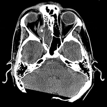
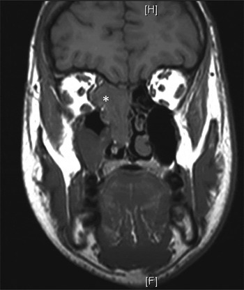
A computed tomography (CT) scan revealed a mass in the right posterior ethmoids, eroding into the orbit and right sphenoid sinus, with a small defect noted within the floor of the anterior fossa; however, there was no evidence of intracranial extension (Figure 1). Magnetic resonance imaging of the paranasal sinuses confirmed a 35 × 25 mm mass arising from the right posterior ethmoid and bulging into the orbit (Figure 2). No evidence of direct infiltration of extraconal fat was seen. A component of the tumour was found to be extending into the sphenoid sinus, superior nasal space and middle meatus. A staging CT showed no evidence of further disease in the chest, abdomen or pelvis.
Axial computer tomography scan showing a poorly enhancing mass 25 mm in diameter (asterisk) arising within the right posterior ethmoid, abutting the lamina papyracea and bulging into the orbit, displacing the medial rectus muscle.

Coronal, T1-weighted magnetic resonance imagining scan showing a mass arising within the right posterior ethmoid and bulging into the right orbit (asterisk). Inferiorly, the mass is shown to pass into the superior nasal space, around the middle turbinate and into the middle meatus.

In view of the radiological evidence, it was decided at the sarcoma multidisciplinary meeting that a tissue diagnosis was required. We thus performed an endoscopic examination of the nasal cavity under general anaesthetic, and took biopsies of the mass. Intra-operatively, frozen section examinations of biopsy specimens were all reported as necrotic tissue. (In retrospect, this could have been secondary to an infarctive effect from the Foley catheter balloon, compressing the tumour.) Frozen section sampling was abandoned, and a further selection of biopsy specimens was sent for full histopathological analysis.
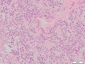
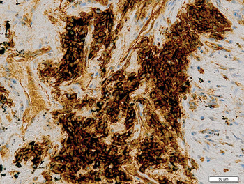
Histological examination showed closely packeted, slightly diffuse, atypical cells with round, hyperchromatic nuclei, scant cytoplasm and occasional mitotic figures, arranged in a sheet-like pattern (Figure 3). Immunohistochemical analysis showed positive staining of the atypical cells for cluster of differentiation 99 glycoprotein only (Figure 4). The appearances were suggestive of a small, round, blue cell tumour with morphological features suspicious of Ewing's sarcoma.
Medium power photomicrograph showing small, round, blue cells with a discohesive, sheet-like growth pattern. (H&E; ×20)

Immunohistochemically stained photomicrograph showing tumour cell positivity for cluster of differentiation 99 glycoprotein (expressing MIC-2 gene). (×20)

In order to consolidate and confirm this diagnosis, samples were sent to the Wessex Regional Genetics Laboratory. There, fluorescent in situ hybridisation was performed, using the Abbot break-apart probe for the Ewing's sarcoma gene. Red-green split signals were seen in the samples, confirming the diagnosis of Ewing's sarcoma.
The patient's histopathological and radiological results were discussed at length at a further sarcoma multidisciplinary meeting. The decision was taken to proceed with macroscopic clearance of the tumour endoscopically, followed by a course of radiotherapy.
Transnasal endoscopic resection of the right sinonasal tumour was performed. The operative findings suggested that the tumour was localised in the posterior ethmoid region, extending along an intact lamina papyracea, eroding the anterior face of the sphenoid and extending approximately 2 mm along the planum sphenoidale. There was no suggestion of any breach of the dura or orbital periosteum. Full macroscopic clearance of the tumour was achieved, leaving the lamina papyracea intact.
Post-operatively, high dose radiotherapy was delivered to the upper nasal cavity, the ethmoid sinus, the sphenoid sinus and the medial part of the right posterior orbit. The patient received 55.8 Gy in 31 fractions delivered over six and half weeks.
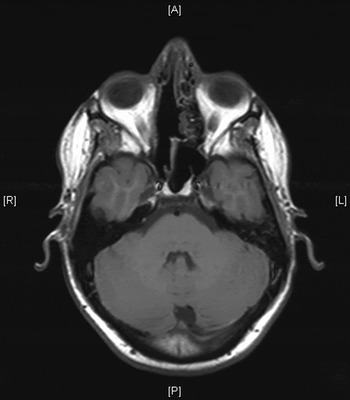
Ten months post-operatively, the patient remained disease-free (Figure 5).
Axial, T1-weighted magnetic resonance imaging scan of the paranasal sinuses and anterior skull base, taken 10 months after initial diagnosis, showing no evidence of residual or recurrent disease. A = anterior; R = right; L = left; P = posterior

Discussion
In his 1921 paper,Reference Ewing1 James Ewing originally described Ewing's sarcoma as a diffuse endothelial myeloma. However, more recently this tumour has been shown to originate from primitive neuroectodermal cells.Reference Csokonai, Liktor, Arato and Helffrich11 As they are separated only by their level of differentiation, Ewing's sarcoma and primitive neuroectodermal tumours are now considered to fall within the same neuroectodermal tumour class.Reference Csokonai, Liktor, Arato and Helffrich11–Reference Wenig, Prasad, Dulfuerov, Fanburg-Smith, Kapadia, Thompson, Barnes, Eveson, Reichart and Sidransky14
Ewing's sarcoma presenting in the head and neck is very rare, and most reported cases involve primary skeletal Ewing's sarcoma.Reference Vaccanti, Forte, de Jong and Taylor5, Reference Hunsuck8–Reference Beraud and Fortin10 The first reported cases of extra-skeletal Ewing's sarcoma were described in 1969, when four cases of paravertebral soft tissue sarcomas were identified as being histopathologically indistinguishable from skeletal Ewing's sarcoma.Reference Tefft, Vawter and Mitus4 Very few reports of head and neck cases have followed; only three cases of primary extra-skeletal Ewing's sarcoma involving the nasal cavity have been described,Reference Boor, Jurkovic, Friedmann, Plank and Kocan3, Reference Howard and Lund9, Reference Csokonai, Liktor, Arato and Helffrich11 plus one case presenting within the scalp.Reference Chao, Chang and Sheen6 Our case is the first documented description of an isolated, extra-skeletal, sinonasal, metastatic deposit from a primary skeletal Ewing's sarcoma.
Within the head and neck region, skeletal and extra-skeletal Ewing's sarcoma appears to present with symptoms and signs secondary to the mass effect of tumour growth, specific to the anatomical location. Presenting symptoms described include pain, loose teeth, local paraesthesia, diplopia, exophthalmos, ptosis, decreasing visual acuity, nasal obstruction and maxillary swelling.Reference Vaccanti, Forte, de Jong and Taylor5, Reference Lane and Ironside15–Reference Kawabata, Yoshifuku, Sagara and Kurono17 Our patient presented with epistaxis and periorbital cellulitis.
It was our patient's unusual presentation of severe epistaxis and periorbital cellulitis, together with a significant past history of recurrent Ewing's sarcoma, which prompted us to undertake further radiological investigation. Plain radiographs can be useful in skeletal Ewing's sarcoma cases, and have been described as showing circumscribed lesions with cortical erosionsReference Vaccanti, Forte, de Jong and Taylor5 with a honeycomb or ‘sun ray’ appearance.Reference Yalcin, Turoglu and Ozdamar18 However, in patients with extra-skeletal Ewing's sarcoma in the head and neck, CT is the radiological investigation of choice.Reference Yalcin, Turoglu and Ozdamar18
The definitive diagnosis of Ewing's sarcoma is confirmed by a combination of histopathology and cytogenetics.Reference Kawabata, Yoshifuku, Sagara and Kurono17
Ewing's sarcoma is defined as belonging to a group of neoplasms known as the small, blue, round cell tumours of childhood.Reference Vaccanti, Forte, de Jong and Taylor5, Reference Kawabata, Yoshifuku, Sagara and Kurono17 Also included within this family of tumours are neuroblastomas, rhabdomyosarcomas and lymphoblastic lymphomas.Reference Dehner19 It can be difficult to morphologically distinguish Ewing's sarcoma from other small, round cell tumours. Light microscopy classically shows sheet-like, densely packed, small, uniformly rounded cells with spherical or oval nuclei surrounded by a minimal, attenuated rim of simplified cytoplasm.Reference Boor, Jurkovic, Friedmann, Plank and Kocan3, Reference Dehner19
For definitive diagnosis of Ewing's sarcoma, the immunohistochemical identification of cluster of differentiation 99 glycoprotein (expressing the MIC-2 gene) is essential.Reference Boor, Jurkovic, Friedmann, Plank and Kocan3, Reference Kawabata, Yoshifuku, Sagara and Kurono17 However, both lymphoblastic lymphoma and rhabdomyosarcoma also show varying degrees of cluster of differentiation 99 glycoprotein immunoreactivity.Reference Kawabata, Yoshifuku, Sagara and Kurono17 Therefore, negative immunoreactions to leukocyte common antigen (also know as cluster of differentiation 45 glycoprotein), neurofilament protein (S-100), and desmin and actin are informative, as they exclude diagnoses of lymphoblastic lymphoma, neuroblastoma and rhabdomyosarcoma, respectively.Reference Kawabata, Yoshifuku, Sagara and Kurono17
Further confirmation of Ewing's sarcoma diagnosis can be obtained from cytogenetic studies. Polymerase chain reaction and fluorescent in situ hybridisation can identify the specific Ewing's sarcoma gene, which shows a characteristic chromosomal translocation between chromosomes 11 and 22 (i.e. t(11;22)(q24;q12)) in 85 per cent of cases and a variant translocation between chromosomes 21 and 22 (i.e. t(21;22)(q22;q12)) in 10 per cent of cases.Reference Kawabata, Yoshifuku, Sagara and Kurono17
• This paper gives the first description of an isolated extra-skeletal metastasis from a skeletal Ewing's sarcoma primary, arising in the sinonasal cavity
• This case highlights challenges in the diagnosis and management of this disease, and describes a successful treatment plan
• This case reinforces the evolving role of the endoscopic transnasal approach in managing sinonasal cavity and anterior skull base tumours
Due to the rarity of extra-skeletal Ewing's sarcoma presenting in the head and neck, we found little precedent in the literature to guide our patient's management. Most authorities agree that Ewing's sarcoma is best managed using an aggressive combination of chemotherapy, radiotherapy and surgical resection.Reference Boor, Jurkovic, Friedmann, Plank and Kocan3, Reference Howard and Daniels16, Reference Coskun, Cinar, Savk, Basak and Dadas20 Boor et al. Reference Boor, Jurkovic, Friedmann, Plank and Kocan3 successfully treated a case of extra-skeletal Ewing's sarcoma of the nose with surgical resection followed by chemotherapy and radiotherapy,Reference Boor, Jurkovic, Friedmann, Plank and Kocan3 whilst Csokonai et al. Reference Csokonai, Liktor, Arato and Helffrich11 utilised surgical resection followed by radiotherapy alone. It is important to be aware that radiotherapy to the sinonasal cavity presents difficulties due to the high risk of morbidity to the eye, and it must therefore be used with caution.Reference Howard and Lund9
Our patient's successful treatment plan originated from crucial initial discussions within the sarcoma multidisciplinary meeting. The extensive assessment of radiological images, the rhinologists' expert opinion and the sarcoma team's past experience combined to enable us to formulate a management plan involving surgical excision followed by six and a half weeks of high dose radiotherapy. Complete macroscopic clearance of the tumour was achieved through transnasal endoscopic sinus surgery followed by post-operative radiotherapy. Ten months post-operatively, the patient remained free of residual and recurrent disease, both endoscopically and radiologically.
Conclusion
This case report not only represents a unique description of an isolated extra-skeletal sinonasal metastasis from a primary skeletal Ewing's sarcoma, but also documents a successful treatment strategy in the face of little guidance from the literature. Furthermore, this case reinforces the constantly evolving role of the endoscopic transnasal approach in the management of neoplastic disease involving the sinonasal cavity and anterior skull base.