Introduction
During cochlear implantation, an electrode is typically placed in the round window or in the scala tympani by cochleostomy.Reference Eshraghi, Hodges, Telischi, Balkany, Jackler and Brachman1 The cochlear implant electrode is passed through the round window to the scala tympani in order to minimise trauma and maximise neural stimulation. However, programmed cell death inevitably occurs as a result of mechanical trauma associated with electrode placement and the oxidative stress that occurs during surgical manipulation.Reference Boggess, Baker and Balkany2–Reference Eshraghi, Yang and Balkany4
It is very important to preserve residual hearing after cochlear implantation. Some studies have suggested that patients with impaired residual hearing can hear better in noisy environments after cochlear implantation.Reference Gantz, Turner, Gfeller and Lowder5 However, other reports (for example, Li et al.Reference Li, Wang, Northrop, Merchant and Nadol6 and W Gstottner (unpublished data)) suggest that residual hearing becomes impaired or worse for the majority of patients receiving a cochlear implant.
This study aimed to present the histopathological and audiological effects of mechanical trauma and chronic irritation over the cochlea associated with the placement of a model electrode in the scala tympani of rats. We also aimed to investigate the benefit of continuous topical corticosteroid application (via a pump) in terms of preventing histopathological and audiological effects in the rats.
Materials and methods
This study was conducted in the Animal Laboratory of the Experimental Surgery Department of Ege University in accordance with the Helsinki Final Act (1986) guidelines related to animal experimentation.
Twenty-one healthy adult female albino rats were used in this study. The rats' weights ranged from 250 to 330 g, with an average weight of 285 g. Rats were kept in a controlled environment with a temperature of 21–22°C, a 12-hour light/dark cycle, free access to food and water, and a background noise level of less than 50 dB. The 21 experimental animals were divided into 3 groups of 7.
Anaesthesia
Forty-five mg/kg ketamine hydrochloride (Ketalar®) plus 5 mg/kg xylazine (Rompun®) was administered intraperitoneally for general anaesthesia of the animals.
Surgical intervention
In all animals, the area behind the right ear was shaved and cleaned with povidone-iodine. Subcutaneous lidocaine (maximum 1–2 ml) was injected to control bleeding. A right retroauricular skin incision was made in a posteroinferior direction. The muscles were cut by blunt dissection to expose the tympanic bulla. The tympanic bulla was opened with a number 11 scalpel to expose the tympanum. In all three groups of animals, the round window membrane was perforated (1–2 mm), and a model electrode with a diameter of 0.4 mm was placed in the round window at a depth of 2 mm.
The animals in group one received no further treatments. The round window of each animal in this group was closed with muscle tissue, and the incision line was sutured with 4-0 polyglactin.
For group two, in addition to the placement of the electrode, an intrathecal microcatheter compatible with the Alzet® mini-osmotic pump was placed in the round window and fastened by muscle tissue. The pump was used to release normal saline. The Alzet mini-osmotic pump was subcutaneously placed between two scapulae during the former retroauricular incision, and the incision line was sutured with 4-0 polyglactin.
In group three, the animals also received an electrode and an Alzet mini-osmotic pump, but instead of releasing saline, the pump released 400 µg/ml dexamethasone (0.25 µl per hour). The dexamethasone was prepared by diluting 1 cc of 8 mg/2 ml dexamethasone with 9 cc of normal saline, and was put into a pump with a 200 µl reservoir volume. A total of 80 µg dexamethasone was released over 14 days.
Two of the animals from group three and one of the animals from group two died within the first 24 hours; their data were excluded from the study. The cause of death was determined to be bleeding during the operation. Another three experimental animals were included as substitutes for the animals that perished.
Audiological assessment
The right ear of each animal was evaluated by distortion product otoacoustic emission (DPOAE) testing before surgical intervention using a GSI Audera audiometer (Cardinal Health, Madison, Wisconsin, USA) with probes designed especially for these animals. Measurements were taken after confirming that the device was in an appropriate measurement position, and that the device probe indicator and stimulus waveform were in the appropriate configuration.
As we could not obtain a result with primary tone levels of L1 = 65 dB SPL and L2 = 55 dB SPL for some of the rats, DPOAEs were elicited using L1 = 70 and L2 = 70 dB SPL primary tone levels. Only the rats with two normal ears were included in the study. The signal-to-noise ratio values were measured at 2f1-f2 frequency for DPOAEs at 2, 3 and 4 kHz.
Hearing was tested again for each group peri-operatively, immediately following surgery. Hearing in the right ear was re-evaluated with DPOAE testing under intraperitoneal anaesthesia on the 14th post-operative day.
All rats were sacrificed via injection of a high dose of intraperitoneal ketamine hydrochloride whilst under general anaesthesia 14 days after the start of the study. The right temporal bones were dissected and placed into 10 per cent formalin solution for histopathological examination.
Histopathological examination
After being kept in 10 per cent formalin solution for 48 hours, the temporal bone samples from each group were decalcified in ethylenediaminetetra-acetic acid solution (100 ml, 0.1 M dissolved in phosphate buffer; pH 7.1): the solution was changed every 2 days over a period of 4 weeks and maintained at 4°C. After dehydration with increasing alcohol concentrations, samples were made pellucid with xylene and embedded in paraffin. Serial sections (5 µm thick) obtained from paraffin blocks were deparaffinised by putting them in a drying oven at 60°C for 1 night with two changes of xylene. They were then rehydrated by applying decreasing alcohol concentrations. The sections were stained with haematoxylin solution for 5 minutes (01562E; Surgipath, Peterborough, UK). The sections were washed under running water, differentiated by acid alcohol solution and stained with eosin for 3 minutes (01602E; Surgipath). The sections were then exposed to an alcohol series and put into xylene. Sections were kept in xylene for 30 minutes and enclosed with a glass coverslip. They were then examined and photographed under a light microscope (Olympus X-40).
Statistics
The data were analysed using the following statistical tests: Kruskal–Wallis, Mann–Whitney U, Kolmogorov–Smirnov, one-way analysis of variance (ANOVA) and Tamhane's post-hoc.
Results
Audiological findings
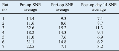
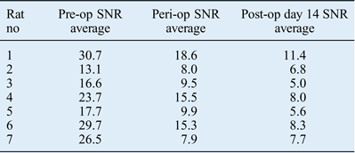
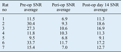
Hearing was assessed in all groups using distortion product otoacoustic emission (DPOAE) testing conducted pre-operatively, peri-operatively and 14th days post-operatively. The arithmetic means of the signal-to-noise ratio values at 2, 3 and 4 kHz are shown in Tables I–III.
Light micrographs of rat cochlear samples, showing haematoxylin and eosin staining of: (a) cochlear stria vascularis (Sv) and scala tympani (ST) (×100), (b) sacala vestibuli (SV) and scala tympani (ST) (×40), (c) stria vascularis (Sv) (×200), (d) stria vascularis (Sv) (×400), (e) spiral ganglion (SG) (×200), and (f) ganglion cells (GC) (×400).

DPOAE test results for group one*
* Electrode only group. DPOAE = distortion product otoacoustic emission; no = number; pre-op = pre-operative; SNR = signal-to-noise ratio; peri-op = peri-operative; post-op = post-operative
DPOAE test results for group two*
* Electrode plus saline pump group. DPOAE = distortion product otoacoustic emission; no = number; pre-op = pre-operative; SNR = signal-to-noise ratio; peri-op = peri-operative; post-op = post-operative
DPOAE test results for group three*
* Electrode plus dexamethasone pump group. DPOAE = distortion product otoacoustic emission; no = number; pre-op = pre-operative; SNR = signal-to-noise ratio; peri-op = peri-operative; post-op = post-operative
Differences between the pre-operative, peri-operative and 14th day post-operative DPOAE values for each group were investigated using the Kruskal–Wallis (non-parametric) test. In this study, p values less than 0.05 were considered to be significant. There were no significant differences between the pre-operative DPOAE values (asymptotic significance = 0.780) or peri-operative DPOAE values (asymptotic significance = 0.252) of the groups. However, there was a significant difference in the post-operative DPOAE values (asymptotic significance = 0.006).
Groups were analysed pairwise with a Mann–Whitney U test. We ran three tests, and considered p = 0.017 as the level of significance. The difference in post-operative values was not significant between group one (electrode only) and group two (electrode plus saline pump) (p = 0.848). However, the difference was significant between group one and group three (electrode plus dexamethasone pump) (p = 0.006), and between group two and group three (p = 0.006).
For group one (electrode only), at 4 kHz, there was a significant difference between the pre-operative and peri-operative test results (p = 0.013), and between the pre-operative and post-operative test results (p = 0.003), but there was no significant difference between the peri-operative and post-operative test results (p = 0.096).
Differences were also observed for group two (electrode plus saline pump) (asymptotic significance = 0.001). The p value was 0.013 for the difference between the pre-operative and peri-operative values (significant), p was 0.002 for the difference between the pre-operative and post-operative values (significant), and p was 0.030 for the difference between the peri-operative and post-operative values (not significant).
There were differences for group three too (electrode plus dexamethasone pump) (asymptotic significance = 0.011). The p value was 0.009 for the difference between the pre-operative and peri-operative values (significant), p was 0.337 for the difference between the pre-operative and post-operative values (not significant), and p was 0.016 for the difference between the peri-operative and post-operative values (significant).
The DPOAE values varied significantly for the three groups in the peri-operative period with respect to the pre-operative values. The post-operative values indicated progressive worsening of hearing impairment for groups one and two. However, the post-operative DPOAE values indicated improvement in the group that received corticosteroids (group three). These values approached pre-operative levels, and there was no significant difference between the pre-operative and post-operative values for this group.
Histopathological findings
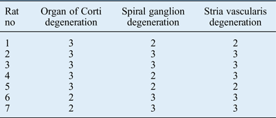
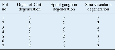
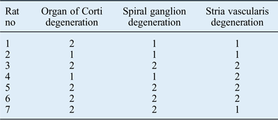
Examination of rat cochlear samples from group one (electrode only) revealed a rupture on the basilar membrane, a fracture on the osseous spiral ligament, chromatolysis on spiral ganglion cells, cytoplasmic and nuclear condensation, a reduction in the number of nuclei and neurons, disorders in the satellite cells, and hydropic and vacuolar degeneration on the stria vascularis (Table IV, Figure 1a). We observed all of the above in the group two (electrode plus saline pump) samples too (Table V, Figure 1b). In the group three (electrode plus dexamethasone pump) samples, histologically the cochlear structures were more intact than in those of the other two groups, although there was: a small distortion in the continuity of the basilar membrane; mild oedema, and cytoplasmic and nuclear condensation on spiral ganglion cells; and mild hydropic and vacuolar degeneration on the stria vascularis (Table VI, Figure 1c).
Cochlear structure histopathological findings for group one*
0 = No degeneration, 1 = mild degeneration, 2 = moderate degeneration, 3 = severe degeneration.
* Electrode only group. No = number
Cochlear structure histopathological findings for group two*
0 = No degeneration, 1 = mild degeneration, 2 = moderate degeneration, 3 = severe degeneration.
* Electrode plus saline pump group. No = number
Cochlear structure histopathological findings for group three*
0 = No degeneration, 1 = mild degeneration, 2 = moderate degeneration, 3 = severe degeneration.
* Electrode plus dexamethasone pump group. No = number
Haematoxylin and eosin staining of cochlear samples from each group were evaluated under a light microscope and scored based on changes in the organ of Corti (hydropic and vacuolar degeneration, and loss of outer and inner hair cells), spiral ganglion (cytoplasmic and nuclear condensation, and nucleus and neuron loss) and stria vascularis (hydropic and vacuolar degeneration), wherein 0 = no degeneration, 1 = mild degeneration, 2 = moderate degeneration and 3 = severe degeneration.
Statistical analysis of histopathological findings
The normality of the distribution of the degeneration scores obtained from the histopathological evaluation was determined using the Kolmogorov–Smirnov test. The distribution of the findings was normal.
The significance of differences between the degeneration scores of the three groups was investigated using a one-way ANOVA. There were differences among the groups for three parameters (organ of Corti, spiral ganglion and stria vascularis) (p < 0.05).
The group findings were compared with each other using Tamhane's post-hoc test. The extent of degeneration in group three (electrode plus dexamethasone pump) was significantly lower than in the other two groups. When group three was compared with group one (electrode only), statistically significant differences were detected (organ of Corti p = 0.007, spiral ganglion p = 0.004 and stria vascularis p = 0.013). When group three was compared with group two (electrode plus saline pump), statistically significant differences were again detected (organ of Corti p = 0.007, spiral ganglion p = 0.013 and stria vascularis p = 0.033). When group one was compared with group two, there were no statistically significant differences (organ of Corti p = 1, spiral ganglion p = 0.941 and stria vascularis p = 0.948).
Discussion
During a cochlear implantation, which is a very important procedure for restoring the hearing of children and adults with severe and very severe hearing loss, an electrode is placed in the round window or in the scala tympani via cochleostomy. The risk of residual hearing loss during the placement of an electrode in the internal ear ranges from 50–70 per cent, although atraumatic techniques have been reported to reduce the risk to 20 per cent (W Gstottner, unpublished data). There has been an increase in surgical procedures on the ear in recent years, and it is important to preserve residual hearing, especially in patients with residual hearing at low frequencies.Reference Eshraghi, Hodges, Telischi, Balkany, Jackler and Brachman1, Reference Boggess, Baker and Balkany2, Reference Gomaa, Rubinstein, Lowder, Tyler and Gantz7 Studies have shown that there is no single reason for residual hearing loss; it can be due to a combination of mechanical causes, such as fibrous tissue on the cochlea, ostosis and electrical stimulation associated with surgery.Reference Eshraghi, Hodges, Telischi, Balkany, Jackler and Brachman1–Reference Mazurek, Amarjargal, Haupt, Fuchs, Olze and Machulik3
Trauma that occurs after the placement of an electrode in the internal ear can be either acute or progressive. Acute trauma occurs from physical damage that is caused by the electrode on the cochlea, especially on hair cells. Progressive trauma occurs as a result of reactive oxygen species in the environment, which are caused by oxidative stress. Tumour necrosis factor α (TNF-α) is a proinflammatory cytokine that plays a role in the pathophysiology of many chronic inflammatory diseases. It is produced in response to oxidative stress and inflammatory mediators, and leads to programmed cell death within neural structures.Reference Wang, Van De Water, Bonny, de Ribaupierre, Puel and Zine8–Reference Yildirim, Coban, Satar, Yetiser and Kunt10 Studies have shown that dexamethasone has an anti-inflammatory effect. In addition, it prevents the ototoxic effects of TNF-α, prevents cell apoptosis by inhibiting mitogen-activated protein kinase (MAPK) and c-jun N-terminal kinase (JNK) pathways, and prevents the loss of hair cells by suppressing TNF-α expression.Reference Yildirim, Coban, Satar, Yetiser and Kunt10–Reference Van De Water, Abi Hachem, Dinh, Bas, Haake and Hoosien13
Electrode placement in the scala tympani offers an alternative to placement on the scala vestibuli; the former causes less trauma to very fragile structures such as the osseous spiral lamina, basilar membrane and modiolus.Reference Eshraghi, Hodges, Telischi, Balkany, Jackler and Brachman1 In experimental animals, the placement of an electrode in the scala tympani mostly causes the loss of hair cells on the cochlea, which results in hearing loss.Reference Kirkegaard, Murai, Risling, Suneson, Järlebark and Ulfendahl14 Auditory brainstem response testing is frequently used to evaluate hearing loss, and distortion product otoacoustic emission (DPOAE) testing (2f1-f2) can be used to diagnose hearing loss originating from the scala tympani.
In this study, the early and late effects of electrode trauma on hearing were measured by DPOAE testing, and evaluated based on the averages of signal-to-noise ratios at 2, 3 and 4 kHz. The DPOAE measurement and noise threshold value can vary, as they are two different measurements that are conducted at different times. The signal-to-noise ratio is more reliable than DPOAE amplitudes and can be used to evaluate DPOAE results.Reference Dagli15 There were significant differences between the pre-operative and peri-operative DPOAE scores for the three groups. The post-operative scores indicated further worsening in hearing impairment for group one (electrode only) and group two (electrode plus saline pump). For group three (electrode plus dexamethasone pump), the 14th day DPOAE values improved, and even approached pre-operative levels. There was no significant difference between the pre-operative and 14th day post-operative values for group three. We suggest that the dexamethasone infusion following implantation of the intracochlear model electrode was more effective than the administration of just one dose of dexamethasone for preventing hearing loss. These findings are consistent with studies showing the protective effect of corticosteroids on hearing.Reference Takemura, Komeda, Yagi, Himeno, İzumikava and Doi16–Reference Parham18
In a study conducted by Eshraghi and colleagues on human temporal bones, rupture on the basilar membrane due to electrode trauma was observed, as well as a fracture on the osseous spiral ligament.Reference Eshraghi, Yang and Balkany4 Histopathological evaluation of the rats' cochleas in our study revealed that the histological structures in group three (electrode plus dexamethasone pump) were more intact than those in group one (electrode only) and group two (electrode plus saline pump). However, a small distortion in the continuity of the basilar membrane, oedema on the spiral ganglion cells, and some hydropic and vacuolar degeneration on the stria vascularis were observed in group three too. Nevertheless, the extent of degeneration on the organ of Corti, spiral ganglion and stria vascularis was significantly lower in group three when compared with the other two groups.
• The study findings show that continuous dexamethasone infusion is beneficial for preventing the loss of hair cells and neurons associated with early and late periods of intracochlear electrode trauma
• Developing technologies that can provide continuous corticosteroid infusion after cochlear implantation is important for protecting residual hearing and improving the quality of hearing after implantation
Topical corticosteroids are increasingly being used to prevent mechanical or acoustic trauma (which initiates apoptosis at the cellular level) associated with the placement of an electrode on the scala tympani.Reference Eshraghi, Adil, He, Graves, Balkany and Van De Water9–Reference Van De Water, Abi Hachem, Dinh, Bas, Haake and Hoosien13, Reference Eshraghi, Polak, He, Telischi, Balkany and Van De Water19 Our findings show that continuous dexamethasone infusion is beneficial for preventing the loss of hair cells and neurons associated with early and late periods of intracochlear electrode trauma. If these findings are supported by clinical investigations, the application of a mini-osmotic pump may be considered as an alternative treatment following cochlear implantation. Developing technologies that can provide continuous corticosteroid infusion after cochlear implantation is important for protecting residual hearing and improving the quality of hearing after implantation.