A 10-year-old boy was admitted because a lateral view of chest X-ray obtained at a regular follow-up visit suggested cardiac strangulation by an epicardial pacemaker lead. He underwent dual-chamber pacemaker implantation via epicardial leads at the age of 3 months because of congenitally complete atrioventricular block. However, he visited the emergency room because of respiratory distress at 6 months after pacemaker implantation. Chest X-ray showed cardiomegaly with a cardiothoracic ratio of 0.73, and echocardiography showed a decrease in the left ventricular ejection fraction (approximately 10%). Thereby, he was diagnosed with pacemaker-induced cardiomyopathy, and subsequently, a ventricular lead was revised from the right ventricular anterior wall to the apical left ventricle; meanwhile, the ventricular lead initially implanted on the right ventricle was not removed.
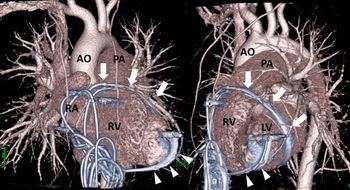
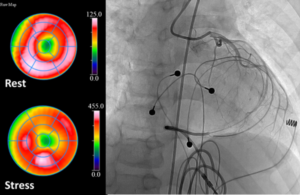
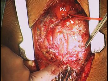
On admission, he had neither chest pain nor exertion on exercise. Contrasted CT showed that the ventricular lead initially implanted was coursing from the anterior wall of the right ventricle along with the atrioventricular groove towards the posterior wall of the left ventricle, which suggested cardiac strangulation (Fig 1). Tc99m tetrofosmin myocardial perfusion scintigraphy showed hypoperfusion in the apex, and selective left coronary angiography showed that the left anterior descending artery was compressed by the lead (Fig 2 and Supplemental Movie). Then, the strangulating lead was successfully removed on the median sternotomy (Fig 3).
Volume rendering images of contrasted CT are shown (left; anterior–posterior image, right: lateral views). The right ventricular lead is coursing from the anterior wall of the right ventricle along with the atrioventricular groove towards the posterior wall of the left ventricle (arrows). Arrow heads means the revised left ventricular lead.

Tc99m tetrofosmin myocardial perfusion scintigraphy at rest (right upper) and adenosis triphosphate stress test (right lower) shows hypoperfusion in the apex, and selective left coronary angiography (left) shows that the left anterior descending artery is compressed by the strangulating lead.

A picture after the median sternotomy is shown. The epicardial pacemaker lead firmly strangled the heart (arrow head) and was subsequently removed on the cardiopulmonary bypass.

Cardiac strangulation has been concerned during childhood or adolescence in patients who has underwent epicardial pacemaker implantation during neonate or early infancy.Reference Alhuzaimi, Roy and Duncan1,Reference Takeuchi and Tomizawa2 Although a simple lateral view of chest X-ray is crucial to detect cardiac strangulation, it is occasionally missed. Previous reports have shown that cardiac strangulation can result in fatal course.Reference Janík, Hejna, Straka, Krajčovič and Novomeský3 Therefore, we should recognise this rare but devastating complication as early as possible.
Supplementary material
To view supplementary material for this article, please visit https://doi.org/10.1017/S1047951120002395
Financial support
This work received no specific grant from any funding agency.
Conflicts of interest
None.
Ethical standards
Not applicable.