Dilated cardiomyopathy is the most common cardiomyopathy and remains the prevailing cause for cardiac transplantation in children and adults.Reference Richardson, McKenna and Bristow 1 , Reference Towbin, Lowe and Colan 2 The phenotype of dilated cardiomyopathy in children is more heterogeneous than in adults. However, in previous Pediatric Cardiomyopathy Registries, aetiologies could not be determined in over half of cases.Reference Towbin, Lowe and Colan 2 Morbidity and, in particular, mortality remain unacceptably high; 5-year rate of death or paediatric cardiac transplantation is reported to be 40–60%.Reference Towbin, Lowe and Colan 2 , Reference Towbin and Bowles 3 Dilated cardiomyopathy is characterised by left ventricular dilation, systolic dysfunction, and secondary diastolic interference.Reference Towbin and Bowles 3 , Reference Everitt, Sleeper and Lu 4 Left atrial congestion and hypertension occurs as a result of left ventricular backward failure and mainly related to diastolic dysfunction.Reference Richartz, Werner, Ferrari and Figulla 5 Symptoms of tachypnoea and dyspnoea correlate with pulmonary congestion and oedema, negative atrial-ventricular coupling, and atrial tachyarrhythmia.Reference Towbin, Lowe and Colan 2 , Reference Towbin and Bowles 3 , Reference Richartz, Werner, Ferrari and Figulla 5 Diuretic use is largely aimed at reducing ventricular pre-load and treating pulmonary interstitial oedema; it is widely subscribed to as a first line management option, neglecting the side effects.Reference Tsirka, Trinkaus and Chen 6 , Reference Kantor, Abraham, Dipchand, Benson and Redington 7 If pharmacological treatment fails, the patient’s condition is classified as “end-stage”.Reference Schranz, Akintuerk and Voelkel 8 For such patients in affluent countries, referral for transplant may be made, with some requiring the bridge of mechanical support.Reference Tsirka, Trinkaus and Chen 6 , Reference Alexander, Daubeney and Nugent 9 However, the mortality of patients awaiting cardiac transplantation is inversely age-related.Reference Jordan, Ichord and Reinhartz 10 This inverse relationship also holds true for the chance of ventricular regeneration and recovery.Reference Amir, Ma and Reddy 11 , Reference Mollova, Bersell and Walsh 12 We hypothesise in the setting of left atrial and consecutive pulmonary congestion requiring high-dose or long-term diuretic treatment that atrial decompression should be considered, especially before extracorporeal life support becomes necessary.Reference Kantor, Abraham, Dipchand, Benson and Redington 7 Our aim was to evaluate the feasibility and efficacy of percutaneous atrioseptostomy for creating a restrictive atrial septal defect, thus unloading the left atrium and ventricle in dilated cardiomyopathy patients with reduced ejection fraction.
Methods
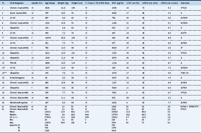
This retrospective study encompasses 27 left atrial decompressions in 22 patients with dilated cardiomyopathy. Patient’s median age was 2 years (24 days to 36.9 years), median body weight 9.9 kg (3–50 kg); nine patients were female. The demographic data are summarised in Table 1. According to the Pediatric Cardiomyopathy Registry,Reference Everitt, Sleeper and Lu 4 dilated cardiomyopathy was defined by depressed left ventricular function with ejection fraction <45% and left ventricular dilatation – left ventricular end-diastolic dimension, z-score >+2. According to the results of histopathological/immunohistological analysis of the obtained endomyocardial biopsies and preferential MRI, 10 patients (45%) revealed dilated cardiomyopathy with inflammation and classified as chronic (n=9) and acute (n=1) myocarditis. Chronic myocarditis is histologically defined as co-existence of already degenerated muscle tissue and fibrosis together with still persistence of infiltration of interstitial tissues. The others were classified as idiopathic (n=5); left ventricular noncompaction (n=4); or associated with mitochondriopathy, arrhythmias, and pacemaker (n=3). NYHA/Ross function was classified as class IV (n=16), III (n=5), and II for the patient with pacemaker-induced cardiomyopathy. Mean left ventricular ejection fraction was 21.5±9.7%; the left ventricular end-diastolic dimension was 49±10 (z-score 5.3±1.6); median serum brain natriuretic peptide was 1576 pg/ml in the range 188–7991 pg/ml. Percutaneous trans-septal decompression was performed 8.4 (±11.3) days after admission. In 11 patients, concomitant myocardial biopsies were taken. Institutional Review Board approval was obtained for analysing all retrospective data. Informed written consent was obtained before the procedures from all patients or their parents.
Characteristics, presentation at admission, and outcome of patients diagnosed with several causes of dilated cardiomyopathy.

A=alive; A*=lost to follow-up; BNP=brain natriuretic peptide; bPAB=bilateral pulmonary banding; D=dead; DCM=dilated cardiomyopathy; EF=ejection fraction; HTX=heart transplantation; LVEDD= left ventricular end-diastolic dimension; LV=left ventricular; LV-NC=left ventricular non-compaction; PAB=pulmonary artery banding; PAB+=pulmonary artery banding + open heart surgery; Potts+=reverse Potts-Shunt
* One
** Two
*** Three additional interventions
Statistical data were calculated with SPSS (IBM). Descriptive data are presented as mean±SD or median and range, as appropriate. Significance was calculated with the Student’s t-test for paired samples. A p-value ⩽0.05 was considered statistically significant. Continuous variables are presented as mean±SD or median and range as appropriate. The calculated z-score is given for the age-dependent left ventricular end-diastolic dimension; a z-value of +2.5 was defined as dilated cardiomyopathy. Specific parameters were analysed only if both pre- and post-intervention data were available.
Interventional technique
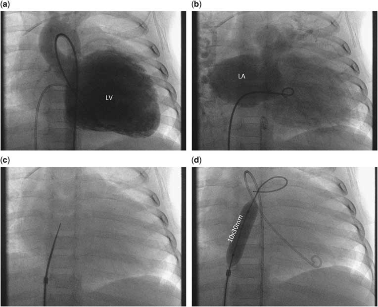
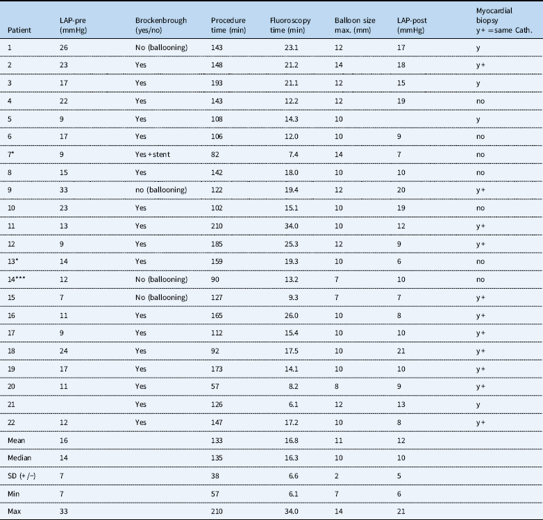
All procedures were performed under fluoroscopy guidance and only by local anaesthesia or in young children with analgo-sedation (Fig 1a–d). No patient underwent general anaesthesia; all received local anaesthetic. In stable patients a complete haemodynamic assessment was performed before intervention. In critically unwell patients, the intervention was performed almost in isolation. In n=18, MRI was performed before or as a hybrid imaging procedure with subsequent heart catheterisation. In younger patients, especially infants, this was carried out under a single episode of sedation; the total procedure time is presented in Table 2. Trans-septal puncture was performed in 18 patients using a Brockenbrough technique (Table 2) with a needle length of 56 or 71 cm (Cook-Medical). In children, the needle was loaded in a 6F, 48 cm-long Check-Flow performer® Introducer (Cook-Medical); in adolescents and adults an 8F Mullins sheath (Cook-Medical) was used. To ensure safety during manipulation the long sheath was advanced through the already placed 2F larger short sheath. The needle-sheath ensemble was pulled back from the upper caval vein in the right atrium during continuous application of contrast medium through the needle. For interatrial septum puncture, the tip of the puncture ensemble was directed posteriorly on the 90° lateral view and between a 3 and 4 o’clock positions on anterior–posterior view. With contact of the fossa ovalis the needle was advanced to inject contrast medium into the atrial septum, then advanced to the left atrium by further continuous injection of contrast medium. After entering the left atrium, the Cook sheath was advanced across the septum and the needle replaced with a 0.035 in. guidewire. Balloon dilatation of the atrial septum was then performed with high-pressure balloons; in most, depending on the patient’s size, Powerflex® (Cordis, Switzerland) or Atlas® (Bard, United States of America) balloons with a length of 20–40 mm were used. The maximal balloon diameter was chosen depending on the desired size of the atrial communication. In all patients, the goal was to achieve a restrictive atrial communication; therefore, a somatic size-dependent communication between almost 4 and 8 mm was created. Based on our long-term experience,Reference Schranz, Akintuerk and Voelkel 8 static ballooning of the intact atrial septum was in part performed by sequential dilatation using balloon diameters up to a maximum of 14 mm resulting in an arterial communication of up to 8 mm. Powerflex® 12×30 mm balloons or, in adolescents and adult patients, Atlas® balloons (14×20 mm) were predominantly used as the final balloon diameter with an average balloon diameter of 11 mm (min 6 mm, max 14 mm) (Table 2). Indentation by the atrial septum on the waist of the inflated balloon, followed by the disappearance of the waist after full inflation was identified in all procedures (Fig 1a–d). If recoil was observed during deflation, repetition of inflation up to the rated burst pressures (8–18 atm.) was performed. At the end of the procedure, full or only partial haemodynamic data were obtained and the created atrial communication was evaluated by transthoracic echocardiography. Persistent foramen ovale dilatation without trans-septal needle puncture was performed in four patients. The same balloon material and technique was used as described. In one patient, an Andra-stent XL was placed into the intact atrial septum, mounted on a 12×30 mm balloon and inflated to a residual diabolo shape, with a waist diameter of 8 mm. Routine stenting of the atrial septum was avoided, to reduce additional procedural risks and to prevent negative aspects of long-term stent placement considering also the chance for recovering. Expecting clinical and haemodynamic improvement after creation of an atrial defect, intended right ventricular myocardial biopsies were always performed after generation of the restrictive septum defect. Procedural success was defined as a successful creation of a patent, but restrictive atrial communication. Restriction was defined by echocardiography diameters between 4 and 10 mm and/or residual left/right atrial pressure gradient of >3–5 mmHg. Gradual balloon dilatation of the intact atrial septum was favoured in some patients, to take care and attention not to create an unrestrictive atrial communication. The decision for re-dilatation of the atrial septum during the later follow-up was made depending on clinical symptoms of pulmonary congestion and echocardiographic evidence of recoil of the atrial septum causing an unwanted degree of restriction at the created atrial communication.
( a – d ) Left ventricular (LV) dilated cardiomyopathy of patient no. 17 at day 445, when a restrictive atrial communication was created. On ( a ) the left ventricular angiography with an extreme dilated LV is depicted (MRI; z-score of left ventricular end-diastolic dimension was 6.5). ( b ) Angiographically obtained left atrial dimension before the atrial septum puncture. ( c ) Trans-septal needle advanced through a 6F Cook sheath in anterior–posterior plane; before the atrial septum is punctured the correct posterior direction to the left atrium has additionally to be demonstrated in the 90° lateral frame (see text); ( d ) Final Powerflex® balloon fully inflated to a diameter of 10 mm within the atrial septum. A diagnostic Pigtail catheter is still re-advanced in the LV cavity.

Procedure related data including the left atrial pressure (LAP) before and after balloon dilatation of the atrial septum, in 18 patients after trans-septal puncture by Brockenbrough technique and 11 additional myocardial biopsies during the same heart catheterisation (y+, cath.).

Cath.=catheterisation; LAP=left atrial pressure
Results
Creation of a restrictive atrial communication was successful in all patients. There were no procedural death or complications. Dilated cardiomyopathy remained idiopathic in 23% of the patients. The average mean procedural length, including MRI, haemodynamic assessments, and myocardial biopsies, was 133±38 minutes; median fluoroscopy time 16.8 minutes (range 6.1 and 34 minutes) (Table 2). Mean left atrial pressure pre-intervention decreased from 16±7 before atrioseptostomy to 12±5 mmHg post-intervention (p=0.005). The left/right atrial pressure gradient decreased from 9.6±5.6 to 5±3.5 mmHg (p=0.005). Left atrial and left ventricular end-diastolic pressure was recorded in seven patients; in this group, both pressures decreased by a mean of 2±1 mmHg. The left ventricular ejection fraction at discharge (n=18) increased significantly from 21.5±9.7 to 29.2±8% (p=0.01). This correlated with immediate improvement of NYHA/Ross functional class from a mean of 3.73 (0.55±) to 2.95 (±0.84). Serum brain natriuretic peptide (n=18) decreased from median 1576 pg/ml (range: 188–7991 pg/ml) to 418 pg/ml (range: 89–4349 pg/ml) (p=0.05) between admission and discharge.
Follow-up
The median in-hospital followed-up time of all treated patients was 42 (range 1–253) days; outcome (n=22) was followed over a median time of 528 (2–2262) days after the procedure. Following the primary intervention, three patients received a second intervention 27, 337, and 645 days later. In patient number 14, the atrial septum was manipulated four times within 71 days. Re-interventions were clinically and echocardiographically indicated for re-dilatation of patient, but recoiling and increasingly restrictive atrial communication. After creation of a restrictive atrial communication all patients improved; three patients with the need for re-dilation only transient. Patient 14 requiring four atrial manipulations and additional pulmonary artery banding died finally 225 days after the first intervention; the family had declined cardiac transplantation. Six patients, who remained listed for cardiac transplantation, were successfully transplanted; two of them died 7 and 13 months after transplantation because of acute graft failure, one associated with a relapse of a fulminant cytomegalovirus and liver failure; the other patient related to probable parental compliance problems.
One patient survived pulmonary artery banding procedure, but without functional cardiac improvement until the end of the observation period; all others showed functional recovery and regeneration with normalisation of left ventricular end-diastolic dimension, six patients, though one recovered after additional pulmonary artery banding placement. Three patients received pulmonary artery banding after open-heart surgery before weaning from cardiopulmonary bypass. Re-shifting of the interventricular septum to the left was related to a consecutive change of the left ventricular cavum from a globated to an ellipsoid form. The decision for pulmonary artery banding was made according to our previously published inclusion criteria,Reference Schranz, Rupp and Müller 13 which provides a normal right heart function with still normal right atrial and end-diastolic pressures and a pulmonary artery pressure of less than half of the systemic blood pressure level. The final functional class of all, but one survivor was class I in n=10 patients, II (n=7), and III (n=1), respectively.
Discussion
This retrospective study reports the impact of transcatheter creation of a restrictive atrial communication in heterogeneous causes of patients with dilated cardiomyopathy. Preliminary human studies have proposed left atrial dysfunction in heart failure with preserved ejection fraction; creation of an interatrial communication was described as a novel strategy.Reference Feldman, Komtebedde and Burkhoff 14 , Reference Burkhoff, Maurer and Joseph 15 The majority of reports are focussed on left atrial decompression during “extra-corporal life support”.Reference Johnston, Jaggers, McGovern and O’Laughlin 16 – Reference Lin, Chen, Wang, Hung, Chang and Lo 18 Creation of an interatrial communication in patients with reduced left ventricular ejection fraction is currently not reported. However, the here-described trans-septal needle technique followed by static balloon dilation of the atrial septum is safe and effective regardless of patient’s age, clinical condition, or cause of cardiomyopathy. At our institution, atrioseptostomy with the described technique is established since newborns with hypoplastic left heart syndrome are treated by “Giessen hybrid” approach.Reference Schranz, Bauer and Reich 19 , Reference Rupp, Michel-Behnke, Valeske, Akintürk and Schranz 20 Based on our long-term experience of atrioseptostomy, the difference of a fully unrestricted atrial communication and the pathophysiological consequences of an overly restrictive atrial septum defect on the haemodynamics and pre-load-dependent performance of the systemic ventricle are previously described.Reference Yerebakan, Murray and Valeske 21 In newborns and infants with borderline left heart structures, the importance of adequate pre-load on left heart growth properties is evident.Reference Yerebakan, Murray and Valeske 21 Considering the pathophysiological features of congestive heart failure, it is natural that restrictive ventricular filling carries prognostic implications in dilated cardiomyopathy patients.Reference Richartz, Werner, Ferrari and Figulla 5 , Reference Pinamonti, Di Lenarda, Sinagra and Camerini 22 We hypothesised that atrial and consecutive pulmonary congestion can be effectively influenced if a left ventricular pre-load is optimised to reduce congestive symptoms, but still permits an adequate left ventricular filling. We note that all the reported patients in this study benefitted from creation of a restrictive atrial communication in both echocardiographic and, importantly, clinical findings. However, haemodynamic and clinical improvements did not necessarily correlate with a change of shape and form of the affected systemic ventricle. Therefore, in whom the criteria for pulmonary artery banding, such as age, right ventricular affection, pulmonary arterial hypertension, written parental consent, with or without an additional open-heart surgery were not fulfilled or spontaneous improvement could not be observed, the decision to remain the patients listed for transplant was based on missing alternatives forcing cardiac regeneration or because of progressive heart failure despite generated restrictive atrial septum defect. Naturally, the most severely impaired patients with acute or chronic left atrial hypertension and consecutive pulmonary congestion showed the most beneficial effect. Patient 14 can exemplarily best demonstrate this; following the declination of cardiac transplantation by the family, three repeat atrial septum interventions were performed during the follow-up, each time the infant’s clinical condition improved and discharge from hospital became possible. Contemporaneously, this case also shows the weakness of exclusive static balloon dilatation for creating an atrial communication. Considering the variable anatomical structure of the atrial septum, the long-term efficacy of the created communication solely performed by balloon dilatation is unpredictable. However, stent placement was avoided as a primary approach as mentioned earlier; flow reduced devices might have a further option, if they become available. In addition, it should be remarked that carefully performed gradual atrial septum balloon dilatation, using sequentially increased balloon diameters, avoids creation of unrestricted atrial septum defects. The function of a restrictive “pop-off valve” does not lead to right ventricular volume overload, but adequate unloading for the failing left ventricle, which could be demonstrated by the patient’s clinical functional class at rest and exercise. Empirically and according to our previous institutional experienceReference Schranz, Akintuerk and Voelkel 8 , Reference Yerebakan, Murray and Valeske 21 as well as based on computer modelling,Reference Kaye, Shah and Borlaug 23 an atrial communication of 4–10 mm remains – depending on the patient’s somatic-size – restrictive and serves as an acute or chronic “pop-off valve” reducing cardiac congestive symptoms. Left atrial decompression with a physiologic pressure gradient between the right and a left atrium of about 3–5 mmHg provides adequate left atrial decompression such that an immediate anti-congestive effect was observed in this cohort of patients with reduced left ventricular ejection fraction and increased left ventricular end-diastolic dimension. Concomitant with a significant decrease of the left atrial pressure, the left atrioventricular coupling improved, which could be demonstrated in those seven patients in whom complete pre- and post-intervention haemodynamic data were obtained. Left ventricular end-diastolic pressure decreased in line with reduction of left atrial pressure. Theoretically, decreased left atrial pre-load with subsequent decrease in left ventricular filling pressure causes a shift within the Frank-Starling curve, which might support favourable remodelling processes, as seen in some of our especially young dilated cardiomyopathy patients. Further prospective studies must clarify whether creation of a restrictive atrial communication contributes really to left ventricular functional recovery though the theoretical advantages are reduced left ventricular wall stress with secondary improvement in sub-endocardial perfusion and reduced myocardial oxygen consumption. Considering our heterogeneous causes of patients with dilated cardiomyopathy, including a high variability of age, it seems that not the disease itself but the consecutive clinical condition and pathophysiology should determine the indication for generation of a restrictive atrial communication. Pathological haemodynamics, including left atrial size and function with the need of diuretic treatment, are the main consideration of establishing a restrictive atrial communication. Based on our further interventional and surgical experience dealing with all faceted congenital and acquired heart failure, we propose that the presence of a restrictive atrial communication is almost always of haemodynamic and clinical advantage even in patients with reduced ejection fraction. Therefore, maintaining or creating of a de novo restrictive atrial communication belongs to our holistic strategy in the management of patients with atrial and pulmonary congestion based also on a reduced left ventricular ejection fraction with increased end-diastolic dimension.Reference Schranz, Akintuerk and Voelkel 8 Despite the limitations of this retrospective analysis, the reported results are promising. Bridging to recovery or transplantation with a less incidence for need of mechanical assist devices are realistic scenarios as well as improved quality of life corresponding with improved haemodynamic parameters and biomarkers. However, the results need to be confirmed further by prospective multi-centre studies. But ascertainable is that infants and children have the best potential for recovery and remodelling, if novel interventional and surgical procedures are combined with adequate age-dependent medical heart failure therapy.Reference Kaye, Shah and Borlaug 23 , Reference Schranz, Akintuerk and Bailey 24 The current report demonstrates a sustained and relevant clinical benefit of a restrictive atrial septum defect as a part of a holistic therapeutic approach. In addition to the efficacy, there was no need for closure of the created atrial defects during the further follow-up.
Study limitations
The study is limited by its retrospective design and partially incomplete data, in particular considering concomitant diastolic dysfunctional data. Furthermore, follow-up cardiac MRI data was not available to better describe the impact of intervention. We were unable to present all detailed haemodynamic measurements and close follow-up. The described cohort is small and heterogeneous in respect to age, body size, and underlying causes of dilated cardiomyopathy. Moreover, there were several different surgical interventions to several patients during the follow-up period. Furthermore, we cannot offer evidence of all underlying pathophysiological or molecular mechanisms that may underpin the clinical findings. Missing data may affect bias and thus, affect the validity of the statistical analysis.
Conclusion
Our experience in creation of a restrictive atrial communication in a small cohort of children and young adults with symptomatic dilated cardiomyopathy is encouraging and might be the basis for prospective multi-centre studies. In patients with reduced left ventricular ejection fraction, decompression of the left atrium by creation of a restrictive atrial communication should be considered as a safe, cost-effective palliation, when symptoms of pulmonary congestion or intrinsic left atrial hypertension become obvious. We are convinced that unloading of the left atrium should be considered, when high dose or long-term diuretic treatment with negative anti-remodelling effects becomes necessary.Reference Ellison and Felker 25 , Reference Schranz 26 In our experience thus far, creation of a restrictive atrial septum defect in patients with left ventricular ejection fraction with increased end-diastolic dimension is both, safe and effective. Indications for the procedure must not be limited to clinicians with access to inter-atrial flow regulator devices;Reference Rossignol and Zannad 27 rather, this important intervention must be made available to all patients who may benefit.
Acknowledgements
This article is part of the thesis of A.B.
Financial support
This research received no specific grant from any funding agency, or from commercial or not-for-profit sectors.
Conflicts of interest
None of the authors have anything to disclosure.