Introduction
Microsporidia, previously classified as protozoa but now suggested to share a common origin with fungi (Vossbrinck et al., Reference Vossbrink, Maddox, Friedman, DebrunnerVossbrinck and Woese1987; Keeling, Reference Keeling2003; Vossbrinck and Debrunner-Vossbrinck, Reference Vossbrinck and Debrunner-Vossbrinck2005), are a group of obligate and intracellular parasites infecting invertebrates and all classes of vertebrates (Canning and Lom, Reference Canning and Lom1986), including humans. The group is ubiquitous in nature and includes approximately 143 genera and over 1200 species (Wittner and Weiss, Reference Wittner and Weiss1999). Presently, about half of the described genera of microsporidia infect insects (Becnel and Andreadis, Reference Becnel and Andreadis1999). Nearly all insect orders are infected by microsporidia, with Lepidoptera and Diptera accounting for half of these hosts. In insects, microsporidia have been reported to infect the gut, fat body, reproductive organs, muscles, Malpighian tubules, and nervous and silk gland tissues (Becnel and Andreadis, Reference Becnel and Andreadis1999; Solter and Becnel, Reference Solter and Becnel2000). In humans, microsporidia are emerging as opportunistic pathogens in immunocompromised patients, especially those with acquired immunodeficiency syndrome (Orenstein, Reference Orenstein1991; Curry and Canning, Reference Curry and Canning1993). They are also important as pathogens of beneficial insects such as honeybees and silkworms (Becnel and Andreadis, Reference Becnel and Andreadis1999; Rao et al., Reference Rao, Muthulakshmi, Kanginakudru and Nagaraju2004). The microsporidium Nosema bombycis causing ‘pebrine disease’ in silkworms is the type species of the genus (Sprague, Reference Sprague1981) and the first described microsporidium.
Several microsporidian isolates from different genera, including Nosema, Pleistophora and Thelohania, have been isolated from the silkworm Bombyx mori L. (Lepidoptera: Bombycidae) (Fujiwara, Reference Fujiwara1980, Reference Fujiwara1984, Reference Fujiwara1985; Jolly, Reference Jolly1986; Ananthalakshmi et al., Reference Ananthalakshmi, Fujiwara and Dutta1994). These microsporidia differ in spore morphology, sites of infection and virulence. The study of the life cycle of any parasite is important, as it provides information about different developmental stages that could lead to the identification of stages that are susceptible to control methods. In this paper, the developmental cycle of five microsporidian isolates infecting B. mori collected from different locations in West Bengal, and their impact on the host midgut, were studied. Since the mid-gut tissue was the first to be infected and, subsequently, severely parasitized, it was designated as the primary site of infection among the tissues.
Materials and methods
Sample collection
Five microsporidian isolates, coded M1, M2, M3, M4 and M5, infecting B. mori were collected from different locations in West Bengal and evaluated. Each of the purified microsporidian isolates was maintained in vivo in isolation by oral inoculation of healthy silkworm larvae of the strain Nistari (M). The spores were isolated from fifth-instar larvae by macerating and suspending them in 0.85% NaCl, followed by filtration through a cheesecloth, and centrifugation at 3000 rpm for 10 min. The spore pellet was purified by Percoll® gradient centrifugation as described by Sato and Watanabe (Reference Sato and Watanabe1980).
Life-cycle study (smear method)
Spore suspensions (1 × 10⁶/ml) of each isolate were separately smeared on mulberry leaf discs (22.26 cm2 each) and allowed to dry at room temperature for 10–15 min. Five groups of larvae, one for each of the five microsporidian isolates, were selected at 1-day post-moult to second instar, and then starved for 30 min and fed with the inoculated mulberry leaf discs. Each larva was fed one leaf disc and only larvae that consumed the entire disc were evaluated. Twenty larvae per microsporidian isolate were treated and maintained separately on mulberry leaves in small perforated plastic boxes at ambient temperature (26 ± 2 °C), relative humidity (74 ± 6%), and light regime (14 h light–10 h dark).
The various intermediary stages of the developmental cycle of each isolate were studied by dissecting one to two larvae every 24 h following the initial inoculation, through the completion of the life cycle. A portion of the mid-gut tissue from each infected larva was excised and placed in a drop of physiological saline on a glass slide and smeared on a clean slide with the help of another slide. The smear was air dried, fixed in absolute methanol for 2 min and stained with Giemsa's staining solution (Sigma) for 15–20 min. The staining solution was prepared by diluting stock Giemsa stain with phosphate buffer (pH 7.4, 1:9, v/v). The slides were then rinsed in running water for 5 min, air-dried and observed under a Leitz Diaplan Phase Contrast microscope (1000 × ). Dimensions of the various intermediary stages were measured using an ocular micrometer. All measurements are presented in μm as mean values followed parenthetically by range values and standard deviations.
Life cycle study (histological method)
Larvae for histological studies were prepared as described above but the mid-gut tissues were excised and fixed in alcoholic Bouin's solution for 10 h, dehydrated in ascending alcoholic grades, cleared in xylol, mounted in paraffin wax (60 °C), sectioned at 6 μm and stained with Delafield's haematoxylin. Life cycle stages were observed and evaluated.
Results
Life cycle study
Merogony
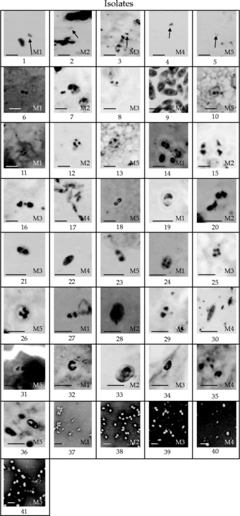
The first vegetative form of the parasite, the sporoplasms, was observed within 24 h post-inoculation (p.i.) (Fig. 1). Meronts were characterized by two round and compact nuclei, stained deep red by Giemsa's solution and a cytoplasm that was very faintly stained. Sporoplasms gradually increased in size and by 24–48 h developed into meronts. Meronts were characterized by their deep blue-stained cytoplasms and were initially ovocylindrical in shape, and then gradually became elongated. The M4 isolate was more elongated than the other isolates. The growth of meronts was followed by a nuclear division, resulting in tetranucleated meronts. Tetranucleated meronts were observed only in the M1, M2 and M5 isolates. However, octanucleated or 12 nucleated meronts were not observed in any of the isolates. The tetranucleated forms eventually divided to form diplokaryotic meronts. Diplokaryotic, tetranucleated and dividing meronts were observed between 24 and 48 h p.i.
Giemsa-stained light micrograph of the merogonic and sporogonic stages of the five microsporidian isolates of Bombyx mori L. (1000 × ). 1–5, sporoplasm; 6–10, diplokaryotic meronts; 11–13, tetranucleated meronts; 14–18, dividing meronts; 19–23, sporont; 24–26, tetranucleated sporont; 27–31, sporoblast; 32–36, immature spores; 37–41, mature spores. Scale bar = 5 μm.

Sporogony
After division, the meronts differentiated into binucleated sporonts. They were characterized by deeply stained compact nuclei, which occupied a central position in the cells. Binucleated sporonts were oval to slightly elliptical in shape, and further nuclear divisions gave rise to tetranucleated sporonts. Sporonts were first observed around 96 h p.i. Tetranucleated sporonts were observed only in the M1, M3 and M5 isolates and were notably absent in M2 and M4. It was significant that neither the tetranucleated meronts nor the tetranucleated sporonts were observed in the M4 isolate.
The tetranucleated sporont completed a single division, and normally produced two sporoblasts, which were observed between 96 and 120 h p.i. The sporoblast was characterized by a faintly stained cytoplasm and intensely stained diplokaryon. Each sporoblast gradually differentiated into an immature spore and then into a mature spore. At the time the spores matured, secondary infective forms resembling the sporoplasm in shape and size were detected. The cytoplasm was stained deep blue with Giemsa. They caused fresh infection of the host cells. These secondary infected forms developed into meronts and repeatedly divided. However, spore dimorphism was not observed in any of the isolates. No short polar tube-type spore was observed. The length (μm) of the polar tube ranged between 67.14 ± 8.93 (M3) and 92.4 ± 3.78 (M2). In the other isolates, it was recorded as follows: 84.09 ± 8.31 in M1, 76.61 ± 5.59 in M4 and 69.87 ± 5.76 in M5.
The diplokaryon was apparent in immature spores while in the mature spores, nuclei were not distinguishable. The entire life cycle of the parasite was completed in 6–8 days.
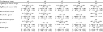
Considerable differences with respect to the shape and size of the different intermediary stages of all the isolates were observed (Table 1). The morphometric observation of the intermediary stages of the parasite through histological methods confirmed the findings obtained from the mid-gut smear method.
Mean values of the different developmental stages of the five microsporidian isolates (μm), as revealed by the study of tissue smears and histology

L, length; B, breadth.
Values indicate mean diameter or as otherwise indicated; values in parentheses indicate the range of values, followed by standard deviation.
Histopathology of the mid-gut
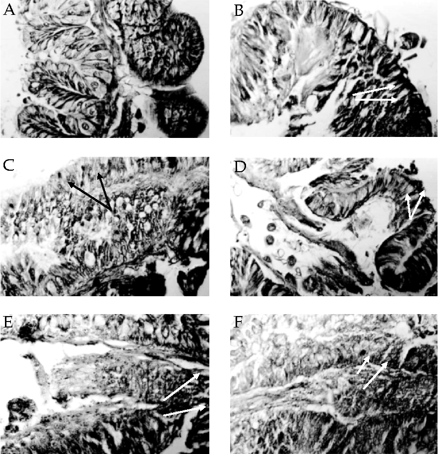
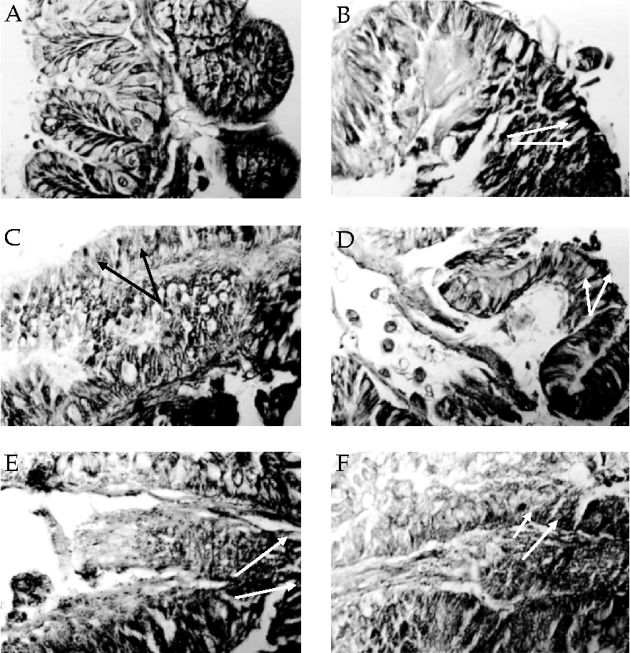
As revealed in Fig. 2, M5 isolate infections in the epithelial cells of the silkworm midgut were most intense, followed by M3, M4, M2 and M1. The nuclei of the spores stained dark purplish and those of the host tissues were lightly coloured. Goblet cells were more severely infected than the columnar cells and were darkish in colour in comparison with those of the uninfected host. In severely infected cells, the cytoplasmic membrane and the nucleus fragmented.
Histological sections of the silkworm mid-gut (400 × ). A, healthy silkworm mid-gut tissue; B–F, silkworm mid-gut tissue infected with M1, M2, M3, M4 and M5 microsporidian isolates, respectively. The arrows indicate the site of infection. The rupture of the mid-gut epithelium is observed in all the isolates. Note the maximum disintegration of the tissue in those infected with the M5 isolate.
Discussion
Studies of the morphological and morphometric differences among the different life cycle stages of microsporidian isolates can substantiate the differences among the isolates based on, for example, spore morphometrics, pathogenicity, and molecular studies. Microsporidia isolated from silkworms are obligatory intracellular parasites, multiplying in host cells in the form of meronts and sporonts (Ishihara, Reference Ishihara1969). Mature spores were found in the faecal material of the infected larvae and are the means of secondary contamination via the food source. The life cycle of entomopathogenic microsporidia ranges from relatively simple to extremely complex. Microsporidia having a simple life cycle are usually characterized by a single sporulation sequence involving only one host individual (Andreadis, Reference Andreadis2007). Nosema bombycis has a relatively simple life cycle in which two types of spores, internal (short polar tube type) and external (long polar tube type) spores, are formed in the same host (Kawarabata and Ishihara, Reference Kawarabata and Ishihara1984; Iwano and Ishihara, Reference Iwano and Ishihara1991). Although the only microsporidia for which intermediate hosts have been identified are from mosquitoes (Amblyospora and Parathelohania) (Becnel and Andreadis, Reference Becnel and Andreadis1999), this situation might be common. Nosema necatrix and Thelohania diazoma were long considered to be dual infections in caterpillars (Kramer, Reference Kramer1965). They were finally proven to be two types of one species, Vairimirpha necatrix, developing in their own way and their own time, in the same host (Fowler and Reeves, Reference Fowler and Reeves1975). Amblyospora californica is an example of a complex developmental cycle in which two generations of mosquitoes and a copepod intermediate host is needed to complete it (Becnel, Reference Becnel1992).
In the present study, spores of all the five microsporidian isolates appeared to be apansporoblastic as the spores were in direct contact with the host cell cytoplasm and possessed no sporophorous vesicle (Gupta et al., Reference Gupta, Hossain, Mohanan, Mitra, Mandal and Bajpai2011). All developmental stages were observed to be diplokaryotic. Binucleate spores germinating in the gut lumen deposit the sporoplasm directly into the mid-gut epithelial cells by means of an extruding polar filament. Each binucleate sporoplasm grew in size and matured into a meront. Meronts divided and differentiated into sporonts, and each sporont further gave rise to two sporoblasts. Sprague and Vernick (Reference Sprague and Vernick1971) characterized Nosema as having sporonts that divide into two sporoblasts and distinguished Nosema from the genera Glugea and Encephalitozoon by their nuclear arrangement. Our observations show all the isolates to be very similar to N. bombycis in its development, as described by Ishihara (Reference Ishihara1969). A similar developmental sequence has been reported in Nosema apis (Fries, Reference Fries1993; De Graaf et al., Reference De Graaf, Raes and Jacob1994). Most Nosema spp. have two types of spores, primary and environmental, e.g. N. portugal and N. chrysorrhoae (Hylis et al., Reference Hylis, Pilarska, Obornik, Vavra, Solter, Weiser, Linde and McManus2006). The dimorphic development of N. bombycis spores in the gut epithelium of B. mori larvae was studied (Iwano and Ishihara, Reference Iwano and Ishihara1991). These authors have identified two types of spores, internal and external. A thin spore wall and a short polar tube that germinates spontaneously within the cytoplasm of epithelial cells characterizes the internal spores. It is believed that the autoinfection serves to spread the parasites to the adjacent epithelial cells. The external spores are thick walled, with a long polar tube, and are produced in large numbers. The infected epithelial cells are filled with spores and finally rupture, releasing the spores into the gut lumen. Ananthalakshmi et al. (Reference Ananthalakshmi, Fujiwara and Dutta1994) reported that the pathogen infects almost all tissues and the mid-gut was designated as one of the primary sites of infection.
In the present study, larvae were maintained at ambient temperatures (26 ± 2 °C) and mature spores were observed in the gut epithelial cells 6–8 days after inoculation in all the five microsporidian isolates. Kawarabata and Ishihara (Reference Kawarabata and Ishihara1984) also reported a similar developmental time after studying the infection and development of N. bombycis in the Antheraea eucalypti (Scott) (Lepidoptera: Saturniidae) cell line. Iwano and Ishihara (Reference Iwano and Ishihara1991), however, observed short polar tube-type spores of N. bombycis in the gut epithelial cells of B. mori at 60 h p.i. and long polar tube-type spores at 72 h p.i. No short polar tube-type spore was observed in the present study.
Significant differences in the dimensions of the various intermediary stages of the five isolates were noted. In M4, tetranucleated meronts and sporonts were not observed. Similarly, tetranucleated meronts and sporonts were not observed in M3 and M2, respectively. The length/breadth ratio of the late-stage meronts in M4 was smaller in comparison with the other isolates, giving it a spindle shape. Octanucleated or 12 nucleated meronts/sporonts were not observed in any of the isolates. In an electron microscopic study of these same five microsporidian isolates, Gupta et al. (Reference Gupta, Hossain, Mohanan, Mitra, Mandal and Bajpai2011) reported variations in the number of polar tube coils among the isolates. They have reported 11 coils in M3, 12 each in M1, M4 and M5 and 14 in M2.
The mid-gut epithelial cells showed pathological changes due to parasitic infection, and these changes were of varying degrees in all the five isolates (Fig. 2). Among the five isolates, the greatest damage to the mid-gut tissue was observed for the M5 isolate and the least for M1 infection. In the mid-gut epithelium, goblet cells were more severely infected than the columnar cells. The present observations corroborate those of Fujiwara (Reference Fujiwara1980, Reference Fujiwara1984) and but no internal spores were observed as reported by Iwano and Ishihara (Reference Iwano and Ishihara1991).
Conclusion
As revealed in our histological studies, the five microsporidian isolates differed with respect to the dimensions of their developmental stages and all caused damage in the mid-gut tissues of B. mori. The M5 isolate was most damaging, followed by M3, M4, M2 and M1.
Acknowledgements
The authors are grateful to the Central Silk Board, Ministry of Textiles, Government of India for the financial support given to the Project ARP 3351 under which this work was carried out.