Introduction
Reproductive cloning or somatic cell nuclear transfer (SCNT) produces an animal with the same genetic material as that of a currently or previously existing animal. Nucleus transfer is a valuable tool for embryological studies in mammals and a method for the multiplication of elite animals. Improved NT methods have enabled the achievement of blastocyst development rates superior to those with in vitro fertilization (IVF) and have increased the rate of production of cloned offspring. However, its overall success rate is low in terms of cloned offspring born due to aberrant epigenetic reprogramming (Bourc’his et al., Reference Bourc’his, Le Bourhis, Patin, Niveleau, Comizzoli, Renard and Viegas-Péquignot2001; Boiani et al., Reference Boiani, Eckardt, Schöler and McLaughlin2002; Xue et al., Reference Xue, Tian, Du, Kubota, Taneja, Dinnyes, Dai, Levine, Pereira and Yang2002; Ogawa et al., Reference Ogawa, Ono, Shimozawa, Sotomaru, Katsuzawa, Hiura, Ito and Kono2003).
Epigenetic reprogramming is a process of resetting the gene expression of a differentiated donor cell to an early embryonic totipotent state. The cloning technique involves reprogramming the genetic elements of a somatic cell to transform it into a totipotent embryonic cell. The epigenetic marks accumulated during the differentiation of the embryo are erased and re-established to new ones after SCNT. The embryonic marks, which are established first, are followed by the establishment of tissue-specific marks. This process is complex and sometimes not fully complete in all instances. This abnormal epigenetic reprogramming may lead to faulty or differential gene expression including persistent expression of genes specific to the donor nucleus and deficient expression of genes involved in pluripotency.
The incomplete dysfunctional reprogramming is responsible for the morbidity and mortality of clones during pregnancy and soon after birth. The epigenetic defects in cloned embryos can be broadly classified into three categories, i.e. hyper-methylation of genes, histone acetylation and methylation, and errors in X chromosome inactivation (Kang et al., Reference Kang, Koo, Park, Choi, Chung, Lee and Han2001). X chromosome inactivation (XCI) is a dosage compensation mechanism that operates in the early embryo and allows the silencing of one X chromosome in female mammals. Genes located on the X chromosome are present in two copies in females and one in males. To overcome the potential unequal expression of genes resulting from unequal copy number of chromosomes, X-inactivation, the transcriptional silencing of a single X chromosome during female embryogenesis, ensures equal dosage of X-linked genes in male (XY) and female (XX) (Lyon, Reference Lyon1961). But such inactivation is not proper in cloned females, due to which their development rate is low compared with males.
SCNT embryos and offspring have been shown to exhibit aberrations in XCI (Nolen et al., Reference Nolen, Gao, Han, Mann, Gie Chung, Otte, Bartolomei and Latham2005). SCNT embryos can reactivate, count, and inactivate X-chromosomes; they are not able to regulate XCI consistently. Significant numbers of abnormalities were observed at all stages of development, including the lack of an inactive X chromosome in some cells of SCNT blastocysts and the absence of imprinted XCI in blastocysts and late-stage placenta (Nolen et al., Reference Nolen, Gao, Han, Mann, Gie Chung, Otte, Bartolomei and Latham2005). The pattern of X-inactivation in aborted bovine SCNT fetuses and dead newborn calves was found to be altered (Xue et al., Reference Xue, Tian, Du, Kubota, Taneja, Dinnyes, Dai, Levine, Pereira and Yang2002).
Most of the X-linked genes (∼90%) are upregulated in the bovine female embryos, suggesting that, in the bovine blastocyst, XCI is far from completion (Bermejo-Alvarez et al., Reference Bermejo-Alvarez, Rizos, Rath, Lonergan and Gutierrez-Adan2010). As XCI is an epigenetic phenomenon and involves histone acetylation and methylation, DNA demethylating agents such as 5-aza-2′-deoxycytidine and S-adenosyl homocysteine have been used on donor cells to chemically reactivate the Xi prior to NT. These treatments partially reactivated the Xi, as evidenced by the re-expression of silenced alleles and earlier chromosome replication (Haaf et al., Reference Haaf, Ott and Schimd1988; Sasaki et al., Reference Sasaki, Hansen and Gartler1992; Lee and Park, Reference Lee, Park, Yang, Jung, Byun, Lee, Yoo, Yeom and Seo2004). Deacetylation and methylation of histones play a key role in the inactivation of the X chromosome as it contains repressive markers such as H3K9me2, H3K4me3 and H3K27me3.
These epigenetic modifiers demethylate histones which enable transcription factors to have access to the genes present on the inactive X chromosome and therefore partially reactivate the inactive X chromosome. Reprogramming in somatic cells containing partially reactivated chromosomes was better compared with the control cells, as significantly more female embryos reached the blastocyst stage compared with control ones. Both approaches have resulted in the improvement of the rate of development of the SCNT embryos to the blastocyst stage (Enright et al., Reference Enright, Sung, Chang, Yang and Tian2005; Jeon et al., Reference Jeon, Coppola, Perrault, Rho, Betts and King2008). Handmade cloning (HMC) is a simple method of SCNT, as there is no need for expensive micromanipulators and skilled expertise (Verma et al., Reference Verma, Arora, Sethi, Mukhopadhyay and Verma2015). In our study, both donor cells, as well as reconstructed HMC-derived cloned buffalo embryos, were treated with 50 nM trichostatin-A (TSA) and 7.5 nM 5-aza-2′-deoxycytidine (5-aza-dC).
The concentration used was decided based on previous experiments in our laboratory (Saini et al., Reference Saini, Selokar, Agrawal, Singla, Chauhan, Manik and Palta2017). In view of this, the present study was carried out to study the effects of an epigenetic modifier on the developmental competence and quantitative expression of the above-mentioned genes in male and female cloned embryos. The rate of blastocyst formation was taken as a measure of the developmental potential, whereas the total cell number (TCN) and the apoptosis level were measured using the terminal deoxynucleotidyl transferase dUTP nick end labelling (TUNEL) assay and were used as parameters for determining the quality of embryos. Epigenetic status was also compared between male (untreated, control) and male blastocysts treated with an epigenetic modifier and female (untreated, control) and female blastocysts treated with an epigenetic modifier in terms of the global levels of histone modifications H3K18ac and H3K9me2.
In addition, the relative mRNA abundance of some X chromosome (XIST, G6PD, HPRT and PGK), apoptosis-related (BCL-XL and CASPASE3), development-related (IGF1R and IFN-tau), pluripotency (OCT4), and epigenetic status-related genes (DNMT1) was also compared between male and female NT blastocysts (treated with epigenetic modifier) using their untreated cloned male and female blastocysts counterparts as controls.
Materials and methods
All chemicals were obtained from Sigma Chemical Co., St. Louis, MO, USA, the media were purchased from GIBCO (Grand Island, NY, USA) and the disposable plasticware was purchased from Nunc (Roskilde, Denmark) unless otherwise stated. Fetal bovine serum (FBS) was obtained from HyClone, Logan, UT, USA. Four adult male (Mu-5710, Mu-5926, Mu-6044, and Mu-6136) and an equal number of adult female Murrah buffaloes (Mu-5579, Mu-5924, Mu-5517, and Mu-5365) available at Livestock Research Centre of NDRI, Karnal, were used to obtain donor cells.
Establishment of somatic cells
Skin biopsies from the ear pinna of the male (Mu-5710) and female (Mu-5579) animals were collected aseptically with the help of an ear notcher in sterile Ca2+- and Mg2+-containing Dulbecco’s phosphate-buffered saline (DPBS) supplemented with 50 mg/ml gentamicin sulfate. The tissues were then washed thoroughly with Ca2+- and Mg2+-free DPBS. The tissue was cut finely into 1–2-mm size pieces, which were cultured in 10 ml of DMEM/F12 medium (1:1 ratio), supplemented with 20% FBS, 0.68 mM l-glutamine and 50 mg/ml gentamicin sulfate in a CO2 incubator in T-25 culture flasks. After 12 h, 3 ml of fresh medium was added to the flask to keep the attached explants submerged. The cells from the outgrowth were removed by trypsinization after they reached confluency, which usually took 5–7 days. The cells were subcultured and grown in T-25 flasks until they attained confluency, following which they were passaged up to 10 times. Aliquots of cells from the early passages (passage 2 or 3) were cryopreserved in DMEM/F12 containing 10% dimethyl sulphoxide (DMSO) and 20% FBS, and were stored in liquid nitrogen for future use. The survival of the cells was evaluated by thawing after 7 days of freezing and culturing the frozen–thawed cells in DMEM supplemented with 10% FBS. The quantitative survivability of the cells was determined by staining the frozen–thawed cells with trypan blue (0.4%), whereas the qualitative survivability was evaluated by allowing the frozen–thawed cells to grow in vitro.
TSA and 5-aza-dC treatment to donor cells
For all experiments, the donor cells were seeded at a concentration of 1 × 10⁴ cells/well in six-well culture plates and were cultured in DMEM supplemented with 0.68 mM l-glutamine, 10% FBS and 50 µg/ml gentamicin for 24 h. The cells were then cultured in medium containing 50 nM TSA and 7.5 nM 5-aza-dC for 24 h before handmade cloning. The cells used in all the experiments were between passages 5–10.
Production of embryos by IVF
Cumulus–oocyte complexes (COCs) collected from abattoir buffalo ovaries were subjected to IVM and IVF as described earlier (Sharma et al., Reference Sharma, George, Kamble, Singh, Chauhan, Singla, Manik and Palta2011). For IVC, the presumed zygotes were washed several times with Research Vitro Cleave medium (K-RVCL-50, Cook®, Queensland, Australia) supplemented with 1% fatty acid-free BSA and were cultured in this medium for up to 8 days post insemination in a CO2 incubator at 38.5°C.
Handmade cloning (HMC)
Buffalo ovaries were collected from a Delhi slaughterhouse immediately after slaughter. Oocytes were collected by aspiration of surface follicles (2–8 mm diameter) with an 18-gauge needle attached to a 10-ml syringe containing the aspiration medium (TCM-199 and 0.3% BSA, 0.68 mM l-glutamine, 50 μg/ml gentamycin sulfate). The contents of the syringe, which included the aspirated oocytes, follicular fluid, granulosa cells and other debris, were poured into 100 mm × 100 mm square Petri dishes with a 13-mm grid. The oocytes were searched under a zoom stereomicroscope at approximately ×20 magnification. The oocytes were then shifted to 35-mm Petri dishes containing the washing medium (TCM-199 and 10% FBS, 0.81 mM sodium pyruvate, 0.68 mM l-glutamine and 50 μg/ml gentamycin sulfate). COCs of usable quality were used for IVM, groups of 18–20 COCs were placed in 100-μl droplets of the IVM medium, overlaid with sterile mineral oil in 35 mm Petri dishes and cultured for 21 h in an humidified CO2 incubator (5% CO2 in air) at 38.5°C. Primary cell cultures of skin cells derived from the ear biopsy of an adult Murrah buffalo were established and donor cell preparation was performed for HMC as reported previously (Selokar et al., Reference Selokar, Shah, Saha, Muzaffar, Saini, Chauhan, Manik, Palta and Singla2012). HMC was performed as described previously (Sandhu et al., Reference Sandhu, Mohapatra, Agrawal, Singh, Palta, Singla, Chauhan and Manik2016).
TUNEL assay
The level of apoptosis was determined using TUNEL staining to assess the quality of blastocysts as described previously (Mohapatra et al., Reference Mohapatra, Sandhu, Singh, Singla, Chauhan, Manik and Palta2015a). Cell counting was performed from the digital images obtained on an inverted Nikon fluorescence microscope. Each experiment was repeated at least three times. Apoptotic index (AI) = (number of TUNEL positive nuclei in the blastocyst/total number of nuclei counted in that blastocyst) × 100.
Immunofluorescence staining
Immunofluorescence staining was performed as described previously (Mohapatra et al., Reference Mohapatra, Sandhu, Neerukattu, Singh, Selokar, Singla, Chauhan, Manik and Palta2015b). To examine the global level of H3K18ac and H3K9me2 in cloned embryos using immunofluorescence staining, the blastocysts were fixed in 4% paraformaldehyde for 1 h at 37°C, washed three times with 0.3% PVA solution (polyvinyl alcohol solution made in Ca2+- and Mg2+-free DPBS) and then permeabilized in 0.5% Triton X-100 for 20 min at 37°C. The blastocysts were blocked for 1 h in 3% BSA and then incubated overnight at 4°C with the respective rabbit primary antibody. Primary antibodies, which included anti-H3K18ac (1:1500, Millipore) and anti-H3K9me2 (1:150, Millipore), were diluted in 3% BSA.
After washing five times with PVA containing 0.1% Triton X-100 (DPBST), the blastocysts were incubated with FITC-conjugated goat anti-rabbit secondary antibody (Sigma) diluted 1:700 in DPBS. After five washings with DPBST, the nuclei were counterstained with H33342 (10 µg/ml) and rinsed in DPBST. The blastocysts were then mounted on slides in mounting medium (2.5% DABCO in glycerol). The slides were observed under a fluorescence microscope, and the images were captured keeping the same optical conditions. NIS-element basic research image processing software (Nikon, Tokyo, Japan) supplied with the microscope was used for image acquisition and quantitative measurements of the mean pixel intensity emitted by each individual nucleus. The images were merged using NIS-Elements BR 3.0 software (Nikon, Tokyo, Japan). At least 10 images (200 nuclei from each image) were analyzed for each epigenetic marker.
Quantitative real-time PCR (qRT-PCR)
RNA was isolated from pools of 10 blastocysts each using an RNAqueous micro kit (Ambion, Austin, TX, USA) as per the manufacturer’s protocol. Following DNase treatment, an RT reaction was performed for cDNA preparation using superscript reverse transcriptase III (Invitrogen). Quantification of mRNA was carried out using qRT-PCR on a CFX 96 I Cycler (Bio-Rad) as described previously by Singh et al., Reference Singh, Mohapatra, Kaushik, Singh, Palta, Singla, Manik and Chauhan2021. The reaction mixture (10 µl) contained 5 µl SYBR Green Mastermix (Maxima SYBR Green Mastermix, Thermo Scientific), 0.2 µl of 10 µM of each primer and 2× diluted cDNA. Thermal cycling conditions consisted of initial denaturation at 95°C for 5 min, followed by 40 cycles of 15 s at 95°C, 15 s at the corresponding annealing temperature and 15 s at 72°C followed by 95°C for 10 s (Supporting information Table S1). All the primer pairs used were confirmed for their PCR efficiency, and specific products were checked using melt curve analysis and for the appropriate size by 2% agarose gel electrophoresis. Primer sequences are provided in the supplementary data (Table S1). The expression data were normalized to the expression of GAPDH and were analyzed using CFX Manager software (Bio-Rad). In all experiments, three trials were carried out, each in duplicate.
Experimental design
Evaluation of the effects of the treatment with epigenetic modifiers on the developmental competence and quality of buffalo cloned embryos:
-
Experiment 1: Based on the results of earlier studies carried out in our laboratory (Saini et al., Reference Saini, Selokar, Agrawal, Singla, Chauhan, Manik and Palta2017), both the donor cells and the fused embryos were treated with 50 nM TSA and 7.5 nM 5-aza-dC. The effect of this treatment on the cleavage and blastocyst rate, and embryo quality was examined by determining the TCN and the apoptotic index using the TUNEL assay.
-
Experiment 2: Evaluation of the epigenetic status of epigenetic modifier-treated male and female embryos. The global levels of H3K18ac and H3K9me2 in cloned embryos produced were examined using immunofluorescence staining.
-
Experiment 3: Measurement of quantitative expression of genes by qPCR in epigenetic modifier-treated male and female embryos. The relative mRNA abundance of genes linked to the X chromosome (XIST, HPRT, PGK and G6PD) and of those related to growth and development (IGF-1R and INF-tau), pluripotency (OCT4) and epigenetic status (DNMT1) were compared between epigenetic-modifier-treated male and female cloned embryos using untreated male and female embryos respectively as controls.
Statistical analysis
Statistical analysis was carried out using Sigma Stat version 3.1 (Aspire Software International, VA, USA). The datasets were analyzed using one-way analysis of variance (ANOVA) followed by the Holm–Sidak test. Percentage values were subjected to arcsine transformation prior to analysis. The differences were considered to be statistically significant at a P-value < 0.05. Data were presented as mean ± standard error of the mean (SEM)
Results
Experiment 1: Evaluation of the effects of the treatment with epigenetic modifiers on the developmental competence and quality of male and female cloned embryos
Based on the results of previous studies carried out in our laboratory, both the donor cells and the fused embryos were treated with 50 nM TSA and 7.5 nM 5-aza-dC. Developmental competence was assessed by determining the cleavage and blastocyst rates at day 8. The cleavage (93.9% ± 0.76) and blastocyst (40.1% ± 2.05) rates of treated male embryos were significantly lower (P < 0.05) than that of control male (95.7% ± 1.05, 49.7% ± 3.80, respectively) embryos (Table 1). Similarly, the cleavage (90.4% ± 1.24) and blastocyst (37.0% ± 0.83) rates of treated female embryos were significantly lower (P < 0.05) than that of control (93.7% ± 1.03, 47.2% ± 2.44, respectively) female embryos (Table 2).
Developmental competence following treatment of donor cells and male embryos with a combination of epigenetic modifiers (50 nM trichostatin-A and 7.5 nM 5-aza-2′-deoxycytidine)

Data from 12 trials.
Data are mean ± standard error of the mean (SEM).
a,b Values with different superscripts within the same column differ significantly (P < 0.05).
Developmental competence following treatment of donor cells and female embryos with a combination of epigenetic modifiers (50 nM trichostatin-A and 7.5 nM 5-aza-2′-deoxycytidine)

Data from 12 trials.
Data are mean ± standard error of the mean (SEM).
a,b Values with different superscripts within the same column differ significantly (P < 0.05).
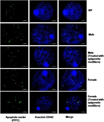
TUNEL assay (Figure 1) revealed that there was no significant difference in the TCN and apoptotic index between cloned blastocysts produced following treatment of donor cells and male embryos (322 ± 23.0 and 4.96 ± 0.31, respectively) with a combination of epigenetic modifiers and the non-treated control blastocysts (360 ± 21.5 and 5.90 ± 0.48, respectively) (Table 3). However, the TCN of both these groups of cloned blastocysts was significantly higher (P < 0.05) than that of IVF blastocysts (191 ± 13.3). The apoptotic index of IVF blastocysts (4.28 ± 0.53) was significantly lower (P < 0.05) than that of untreated control cloned male blastocysts, which was not significantly different from that of cloned blastocysts of the epigenetic modifier-treated group.
TUNEL assay for detection of apoptosis in male and female blastocysts produced using hand-guided cloning following treatment with epigenetic modifiers (trichostatin-A and 5-aza-2′-deoxycytidine) and in blastocysts produced by in vitro fertilization (IVF).

Level of apoptosis treatment of donor cells and male embryos with a combination of epigenetic modifiers (50 nM trichostatin-A and 7.5 nM 5-aza-2′-deoxycytidine)

Data are mean ± standard error of the mean (SEM).
a,b Values with different superscripts within the same column differ significantly (P < 0.05).
Just as for male blastocysts, the TCN of cloned blastocysts (217 ± 9.0) produced following treatment of donor cells and female embryos with a combination of epigenetic modifiers and the non-treated control blastocysts (248 ± 18.9), which was not significantly different, was higher (P < 0.05) than that of IVF blastocysts (191 ± 13.3) (Table 4). The apoptotic index of cloned blastocysts (6.65 ± 0.43) of the epigenetic modifier-treated group was significantly higher (P < 0.05) than that of the control (8.11 ± 0.67) group that, in turn, was higher (P < 0.05) than that of IVF blastocysts (4.28 ± 0.53) (Table 4).
Level of apoptosis treatment of donor cells and female embryos with a combination of epigenetic modifiers (50 nM trichostatin-A and 7.5 nM 5-aza-2′-deoxycytidine)

Data are mean ± standard error of the mean (SEM).
a,b,c Values with different superscripts within the same column differ significantly (P < 0.05).
Experiment 2: Evaluation of the epigenetic status of epigenetic modifier-treated male and female embryos
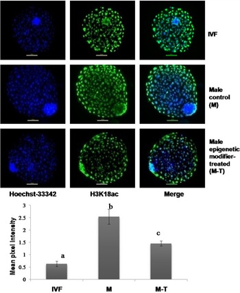
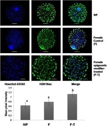
As indicated by mean pixel intensity after immunofluorescence staining, the global level of H3K18ac of male blastocysts from the control group was significantly higher (P < 0.05) than that of male blastocysts of the epigenetic modifier-treated group that, in turn, was higher (P < 0.05) than that of IVF blastocysts (Figure 2). The global level of H3K18ac of the female blastocysts of the control group and that of IVF blastocysts was not significantly different but was lower (P < 0.05) than that of female blastocysts of the epigenetic modifier-treated group (Figure 3).
(Upper panel) Mean pixel intensity of H3K18ac examined using immunofluorescence staining in male control and male blastocysts produced using hand-guided cloning following treatment with epigenetic modifiers (trichostatin-A and 5-aza-2′-deoxycytidine) and in blastocysts produced by in vitro fertilization (IVF). (Lower panel) Bars with different superscripts differ significantly (P < 0.05).

(Upper panel) Mean pixel intensity of H3K18ac examined by immunofluorescence staining in female control and female blastocysts produced using hand-guided cloning following treatment with epigenetic modifiers (trichostatin-A and 5-aza-2′-deoxycytidine) and in blastocysts produced by in vitro fertilization (IVF). (Lower panel) Bars with different superscripts differ significantly (P < 0.05).

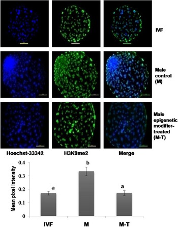
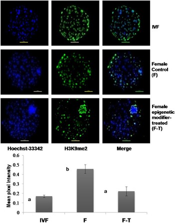
For both male (Figure 4) and female (Figure 5) embryos, the global level of H3K9me2, which was not significantly different between the cloned blastocysts of the epigenetic modifier-treated group and IVF blastocysts, was significantly lower (P < 0.05) than that of the control cloned blastocysts.
(Upper panel) Mean pixel intensity of H3K9me2 examined using immunofluorescence staining in male control and male blastocysts produced using hand-guided cloning following treatment with epigenetic modifiers (trichostatin-A and 5-aza-2′-deoxycytidine) and in blastocysts produced by in vitro fertilization (IVF). (Lower panel) Bars with different superscripts differ significantly (P < 0.05).

(Upper panel) Mean pixel intensity of H3K9me2 examined using immunofluorescence staining in female control and female blastocysts produced using hand-guided cloning following treatment with epigenetic modifiers (trichostatin-A and 5-aza-2′-deoxycytidine) and in blastocysts produced by in vitro fertilization (IVF). (Lower panel) Bars with different superscripts differ significantly (P < 0.05).

Experiment 3: Measurement of quantitative expression of genes by qPCR in epigenetic modifier-treated male and female embryos
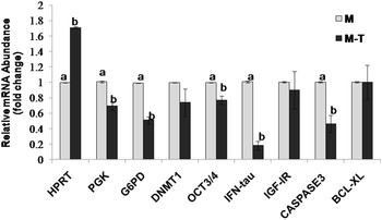
A comparison of gene expression levels between the cloned blastocysts of the epigenetic modifier-treated male and the control groups revealed that the relative mRNA abundance of HPRT was higher (P < 0.05) and that of PGK, G6PD, OCT 4, IFN-tau and CASPASE3 was significantly lower (P < 0.05) in the former compared with that in the latter, and the expression levels of DNMT1, IGF1R and BCL-XL were not significantly different between the two groups (Figure 6).
Relative mRNA abundance of X-linked and development-related genes in male blastocysts produced using hand-guided cloning following treatment with epigenetic modifiers (trichostatin-A and 5-aza-2′-deoxycytidine). M-T: male blastocysts treated with epigenetic modifiers; M: Controls. Bars with different superscripts differ significantly (P < 0.05).

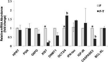
In the female embryos, the relative mRNA abundance of OCT4 was significantly higher (P < 0.05) and that of XIST and CASPASE3 was significantly lower (P < 0.05) in the cloned blastocysts of the epigenetic modifier-treated group compared with that of the control group, whereas the expression levels of HPRT, PGK, G6PD, DNMT1, IFN-tau, IGF1R and BCL-XL were not significantly different between the two groups (Figure 7).
Relative mRNA abundance of X-linked and development-related genes in female blastocysts produced using hand-guided cloning following treatment with epigenetic modifiers (trichostatin-A and 5-aza-2′-deoxycytidine). F-T: Female blastocysts treated with epigenetic modifiers; F: Controls. Bars with different superscripts differ significantly (P < 0.05).

Discussion
We studied the effects of the treatment of male and female embryos with epigenetic modifiers on their developmental competence and quality. Based on the results of previous studies carried out in our laboratory (Saini et al., Reference Saini, Selokar, Agrawal, Singla, Chauhan, Manik and Palta2017), both the donor cells and the fused embryos were treated with 50 nM TSA and 7.5 nM 5-aza-dC.
This is the first report on the effect of the sex of the cloned embryos in response to treatment with epigenetic modifiers. TSA and 5-aza-dC are well known epigenetic modulating agents that have been used to improve reprogramming in various species such as mouse (Kishigami et al., Reference Kishigami, Mizutani, Ohta, Hikichi, Thuan, Wakayama, Bui and Wakayama2006a), rabbit (Meng et al., Reference Meng, Polgar, Liu and Dinnyes2009), pig (Cervera et al., Reference Cervera, Martí-Gutiérrez, Escorihuela, Moreno and Stojkovic2009) and cattle (Enright et al., Reference Enright, Kubota, Yang and Tian2003, Reference Enright, Sung, Chang, Yang and Tian2005; Ding et al., Reference Ding, Wang, Zhang, Wang, Guo and Zhang2008; Iager et al., Reference Iager, Ragina, Ross, Beyhan, Cunniff, Rodriguez and Cibelli2008; Cui et al., Reference Cui, Xu, Shen, Zhang, Zhang and Kim2011; Wang et al., Reference Wang, Su, Wang, Xu, Quan, Liu and Zhang2011a, Reference Wang, Xiong, An, Wang, Liu, Quan, Hua and Zhang2011b).
TSA is a potent histone deacetylase inhibitor that enhances the pool of acetylated histones and DNA demethylation (Yoshida et al., Reference Yoshida, Kijima, Akita and Beppu1990; Hattori et al., Reference Hattori, Nishino, Ko, Hattori, Ohgane, Tanaka and Shiota2004), and can reduce abnormal DNA hypermethylation (Kishigami et al., Reference Kishigami, Van Thuan, Hikichi, Ohta, Wakayama, Mizutani and Wakayama2006b). TSA has been shown to increase the blastocyst rate and improve their quality by inducing hyperacetylation in SCNT bovine embryos (Ding et al., Reference Ding, Wang, Zhang, Wang, Guo and Zhang2008; Cui et al., Reference Cui, Xu, Shen, Zhang, Zhang and Kim2011).
5-Aza-dC, which is a DNA methyltransferase inhibitor, has been shown to improve the epigenetic status of cloned bovine embryos by decreasing their DNA methylation (Enright et al., Reference Enright, Kubota, Yang and Tian2003, Reference Enright, Sung, Chang, Yang and Tian2005). TSA or 5-aza-dC or both have been used to treat activated reconstructed bovine embryos (Iager et al., Reference Iager, Ragina, Ross, Beyhan, Cunniff, Rodriguez and Cibelli2008; Cui et al., Reference Cui, Xu, Shen, Zhang, Zhang and Kim2011) to improve their nuclear reprogramming. Another approach, which has been used in cattle, is to expose the donor cells to either one or both of these drugs (Enright et al., Reference Enright, Kubota, Yang and Tian2003, Reference Enright, Sung, Chang, Yang and Tian2005; Ding et al., Reference Ding, Wang, Zhang, Wang, Guo and Zhang2008; Wang et al., Reference Wang, Su, Wang, Xu, Quan, Liu and Zhang2011a, Reference Wang, Xiong, An, Wang, Liu, Quan, Hua and Zhang2011b). On giving treatment to both the donor cells and the fused embryos with 50 nM TSA and 7.5 nM5-aza-dC, we found that the cleavage and blastocyst rates of treated male cloned embryos were significantly lower (P < 0.05) than in control male embryos.
Similarly the cleavage and blastocyst rates of treated female cloned embryos were significantly lower (P < 0.05) than in control female embryos. These findings agree with an earlier study in cattle (Sangalli et al., Reference Sangalli, De Bem, Perecin, Chiaratti, Oliveira, de Araújo, Valim Pimentel, Smith and Meirelles2012), but are in contrast with others (Ding et al., Reference Ding, Wang, Zhang, Wang, Guo and Zhang2008; Wang et al., Reference Wang, Su, Wang, Xu, Quan, Liu and Zhang2011a) and might be explained by the differences in breed and age of the animals used to derive nuclear donors cells.
Male and female embryos responded in a similar way in relation to blastocyst rate but when their apoptotic index was determined compared with untreated embryos, we found that there was no significant difference between male control and treated embryos, but the apoptotic index of treated female embryos was significantly lower (P < 0.05) than for control female embryos. The lower apoptotic index in treated female embryos may be due to lower expression levels of the XIST gene. It has been reported that activation of XIST expression is correlated with a dramatic increase in apoptotic bodies, suggesting that XIST-mediated XCI may result in cell death and contribute to embryonic lethality (Panning and Jaenisch, Reference Panning and Jaenisch1996; Sandhu et al., Reference Sandhu, Mohapatra, Agrawal, Singh, Palta, Singla, Chauhan and Manik2016). The relative expression level of the XIST gene was significantly lower (P < 0.05) in treated female embryos and this might be the cause of the lower apoptotic index compared with untreated groups.
We found that, as expected, both treated male and female embryos showed significantly lower (P < 0.05) global levels of H3K9me2 than untreated male and female embryos, which agrees with the previous reports, as both 5-aza-dC and TSA are known to induce global DNA and histone demethylation (Xiong et al., Reference Xiong, Dowdy, Podratz, Jin, Attewell, Eberhardt and Jiang2005; Li et al., Reference Li, Kato, Tsuji and Tsunoda2008). As the XIST gene involves enrichment for H3K9me2 during inactivation of the X chromosome (Heard et al., Reference Heard, Rougeulle, Arnaud, Avner, Allis and Spector2001; Mermoud et al., Reference Mermoud, Popova, Peters, Jenuwein and Brockdorff2002; Peters et al., Reference Peters, Mermoud, O’Carroll, Pagani, Schweizer, Brockdorff and Jenuwein2002), reduction in the global level of H3K9me2 can be correlated with lower expression of the XIST gene in treated female embryos. Again, as expected, the global level of H3K18ac in treated female embryos was significantly higher (P < 0.05) than in the untreated control, as TSA has been shown to increase the blastocyst rates and improve their quality by inducing hyperacetylation in SCNT bovine embryos (Ding et al., Reference Ding, Wang, Zhang, Wang, Guo and Zhang2008; Cui et al., Reference Cui, Xu, Shen, Zhang, Zhang and Kim2011).
In contrast, we found that the global levels of H3K18ac in treated male embryos were significantly lower (P < 0.05) than in control male embryos. However, it is difficult to interpret these results as we did not study the expression levels of many other important epigenetics-related genes that are part of epigenetic regulation. Both treated male and female embryos showed significantly higher (P < 0.05) levels of H3K18ac than IVF embryos and there was no significant difference in the global levels of H3K9me2 between treated male and female cloned embryos and IVF embryos, suggesting that epigenetic modifier treatment improved the epigenetic status of cloned embryos.
The effect of treatment with the epigenetic modifier on the male and female embryos was further studied at the gene level. We found that both male and female embryos reacted completely differently to treatment with epigenetic modifiers. In the female embryos, we found no significant difference in the expression levels of X-linked genes HPRT, PGK, and G6PD compared with untreated female embryos, which agreed with an earlier report on porcine (Park et al., Reference Park, Jeong, Jeong, Lee, Jeong, Shin, Kim, Jeung, Hyun, Lee, Lee and Hwang2012). Similarly no significant difference was observed in the expression levels of DNMT1, IFN-tau, IGF1R and BCL-XL between female untreated and epigenetic modifier-treated cloned embryos, although the expression levels of XIST and CASPASE3 were significantly lower (P < 0.05) in treated female embryos than in the untreated control. This could be correlated with the low apoptotic index in treated female embryos compared with the control. In male treated embryos, the expression levels of PGK, G6PD, OCT 4, IFN-tau and CASPASE3 were significantly lower (P < 0.05), whereas HPRT abundance was significantly higher (P < 0.05), although there was no significant difference between the expression levels of DNMT1, IGF1R and BCL-XL and there was no expression of XIST in male treated and control embryos. Therefore in both male and female embryos similar effects of treatment were observed on genes related to growth and development, but the effect on expression of X-linked genes varied. In conclusion, these results indicated that not all X-linked genes in the cloned embryos responded to TSA and 5-aza-dC treatment in the same way. Moreover, the effectiveness of this treatment during the SCNT process may be limited to a few genes. However, it is difficult to interpret these results as we only investigated one male and female cell line and results may vary between different cell lines due to differences in genotype.
Supplementary material
To view supplementary material for this article, please visit https://doi.org/10.1017/S0967199422000600.
Acknowledgements
Funding provided by the Department of Biotechnology, Ministry of Science and Technology, Government of India, and National Agriculture Innovative Project (NAIP) grants to SKS (C 2-1-(5)/2007) and MSC (C-2067 and 075) for the present work are gratefully acknowledged. AS is a recipient of an CSIR-SRF.
Author contributions
RSM, AS and MKS conceived and designed the experiments. SKS and MSC provided the funds. AS and SKM performed the experiments. SKM analyzed the data. SKM and AS prepared the manuscript. All authors contributed to the article and approved the submitted version.
Author disclosure statement
The authors declare that there are no conflicting financial interests.
Ethical approval
As the buffalo ovary samples were collected at an abattoir, ethical approval was not required. Approval of the Animal Ethics Committee of NDRI, Karnal, was obtained before carrying out animal experiments.