INTRODUCTION
Oriental theileriosis caused by the members of the Theileria orientalis complex, is an emerging tick-borne disease of bovines in the Asia-Pacific region. Recently, a number of outbreaks of the disease occurred in cattle in Australia and New Zealand (e.g. Izzo et al. Reference Izzo, Poe, Horadagoda, De Vos and House2010; Islam et al. Reference Islam, Jabbar, Campbell, Cantacessi and Gasser2011; McFadden et al. Reference McFadden, Rawdon, Meyer, Makin, Clough, Tham, Mullner and Geysen2011). Currently, based on the sequence analysis of the major piroplasm surface protein (MPSP) gene, at least 11 distinct genotypes of the T. orientalis (types 1–8 and N1–N3) have been recognized globally. Of those, only type 1 (chitose) and type 2 (ikeda) are considered to be pathogenic and known to be associated with clinical form of oriental theileriosis (reviewed by Sivakumar et al. Reference Sivakumar, Hayashida, Sugimoto and Yokoyama2014), which is characterized by high fever, anaemia, decreased milk production, abortions and/or mortality in dairy and beef cattle (Izzo et al. Reference Izzo, Poe, Horadagoda, De Vos and House2010; Islam et al. Reference Islam, Jabbar, Campbell, Cantacessi and Gasser2011; McFadden et al. Reference McFadden, Rawdon, Meyer, Makin, Clough, Tham, Mullner and Geysen2011; Perera et al. Reference Perera, Gasser, Anderson, Jeffers, Bell and Jabbar2013, Reference Perera, Gasser, Firestone, Anderson, Malmo, Davis, Beggs and Jabbar2014).
Although known to have a cosmopolitan distribution (reviewed by Sivakumar et al. Reference Sivakumar, Hayashida, Sugimoto and Yokoyama2014), oriental theileriosis has not been reported from bovines in many countries, and the genetic characterization of T. orientalis is lacking on a global scale (reviewed by Sivakumar et al. Reference Sivakumar, Hayashida, Sugimoto and Yokoyama2014). One of the main possible reasons for the spread of pathogenic genotypes of T. orientalis from one country to another and/or interstate within a country could be associated with the transportation of cattle infected with T. orientalis and/or infested with vectors responsible for the transmission of the parasite, without appropriate quarantine measures (Stewart et al. Reference Stewart, Standfast, Baldock, Reid and de Vos1992; Kim et al. Reference Kim, Tsuji, Kubota, Wei, Lee, Ishihara and Onuma1998; Gebrekidan et al. Reference Gebrekidan, Gasser, Perera, McGrath, McGrath, Stevenson and Jabbar2015). For example, in Australia, oriental theileriosis is known to be introduced from Japan following an importation of cattle infested with Haemaphysalis ticks (Stewart et al. Reference Stewart, Standfast, Baldock, Reid and de Vos1992). Recently, movement of cattle among different states of Australia has been found to play an important role in an emergence of oriental theileriosis (e.g. Islam et al. Reference Islam, Jabbar, Campbell, Cantacessi and Gasser2011; Gebrekidan et al. Reference Gebrekidan, Gasser, Perera, McGrath, McGrath, Stevenson and Jabbar2015).
Australia is one of the world's largest exporters of live cattle, particularly to Asian and Middle Eastern countries (ABARES, 2014). Every year, hundreds of thousand beef and dairy cattle are exported, thereby contributing hundreds of million dollars into the Australian economy. In 2014, the largest number (730 257) of Australian cattle was exported to Indonesia followed by Vietnam (181 542) and China (117 906) (www.livecorp.com.au). Owing to the strict quarantine laws, Australian cattle are free from many infectious diseases, thus offering less risk of spreading diseases associated with cattle movement.
Australia has been exporting live cattle for many years to several countries around the world, and no issues had been raised and/or reported from importing countries regarding any infectious diseases, including oriental theileriosis. However, more recently, pregnant dairy cattle were exported from Australia to Vietnam, and some of them aborted and died on their arrival in Vietnam. It was suspected that these abortions and deaths in imported cattle were due to oriental theileriosis. Therefore, the objective of this study was to determine whether abortions and/ or deaths in cattle imported from Australia to Vietnam were associated with T. orientalis.
MATERIALS AND METHODS
Case history
Twenty one hundred Holstein-Friesian, pregnant (~4–8 months) heifers were exported from Australia to Vietnam in October 2015. Cattle were mainly sourced from dairy farms located in three states in Australia, including New South Wales, South Australia and Victoria, and kept in a quarantine facility for almost 1 month in Victoria. Blood tests (for enzootic bovine leukosis and Johne's disease), vaccinations (for tick fever vaccine against Anaplasma marginale, and Babesia bovis and B. bigemina), leptospirosis and clostridial diseases and infectious bovine rhinotracheitis) as well as anthelmintic treatment (pour-on preparation of ivermectin) were performed during a 4-week quarantine period. A livestock carrier ship left Australia on 12 September 2015 and arrived Vietnam on 3 October 2015.
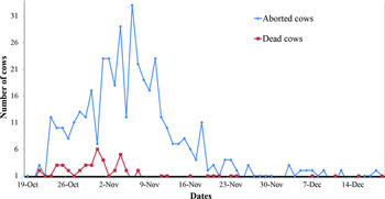
On 5 October 2015, all animals were blood tested (for tuberculosis) and vaccinated against foot-and-mouth (FMD) disease and haemorrhagic septicaemia after their arrival on the destined dairy farm. Over the course of the next 2 weeks, each animal was given a booster dose of the FMD vaccine and also tested for the stage of pregnancy. On 23 October 2015, a number of heifers exhibited clinical signs including lethargy, pyrexia (40 °C), emaciation, anorexia, depression, extremely pale mucous membranes, jaundice, abortion and even death of some animals with or without abortion. The number of animals showing these signs increased in a week, and 439 heifers aborted and 58 died by mid December 2015 (Fig. 1). Based on the physiological status of heifers and clinical signs consistent with oriental theileriosis (Izzo et al. Reference Izzo, Poe, Horadagoda, De Vos and House2010; Islam et al. Reference Islam, Jabbar, Campbell, Cantacessi and Gasser2011; McFadden et al. Reference McFadden, Rawdon, Meyer, Makin, Clough, Tham, Mullner and Geysen2011; Perera et al. Reference Perera, Gasser, Anderson, Jeffers, Bell and Jabbar2013, Reference Perera, Gasser, Firestone, Anderson, Malmo, Davis, Beggs and Jabbar2014), a tentative diagnosis of oriental theileriosis was made, and the affected animals were first treated with Liquamycin LA-200 (Oxytetracycline dihydarate 200 mg mL−1, Intravenous, Zoetis, USA) and Flunixin (Flunixin meglumine 50 mg mL−1, Intravenous, Norbrook, UK) in late October 2015. As the abortions and deaths of heifers continued to occur (Fig. 1), to prevent further losses associated with oriental theileriosis, 1000 heifers were treated with Butalex (Buparvaquone 50 mg, Intramuscular, Sarabhai Zydus Animal Health Limited, India) on 27 November 2015, and abortions and deaths of animals markedly decreased and stopped by mid December 2015.
Incidence of abortions and deaths due to oriental theileriosis in cows imported to Vietnam from Australia.

Pathological investigations
Following abortions and deaths of pregnant heifers, a number of laboratory tests were conducted to confirm oriental theileriosis and rule out other conditions/diseases presenting similar syndrome in pregnant cattle. Haematological examination of 25 aborted heifers revealed a regenerative anaemia, with a packed cell volume (PCV) ranging from 0·12 to 0·24 (reference range for PCV in cattle: 0·24–0·46). Similarly, the examination of stained (DiffQuik staining method) blood smears showed the presence of Theileria piroplasms (Allison and Meinkoth, Reference Allison, Meinkoth, Weiss and Wardrop2010), with no evidence for the presence of other tick-borne pathogens, including Anaplasma and Babesia. Additional tests were performed for diseases, including other forms of bovine theileriosis, brucellosis, leptospirosis and neosporosis, which possibly could cause abortions and deaths in near-term cattle. An examination of various tissues from 10 aborted heifers revealed that they were test-negative for Brucella abortus, Leptospira spp., Neospora caninum (tested using ELISA), Theileria annulata and Theileria parva (tested using blood smear examination).
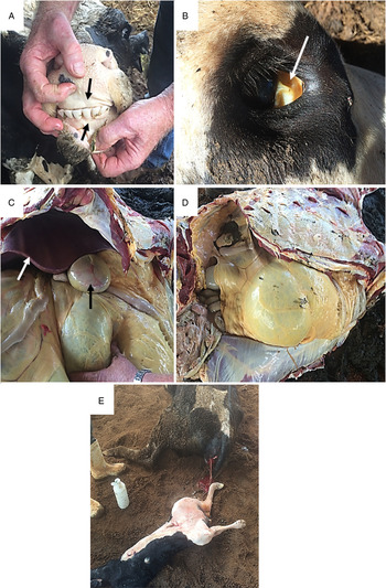
Furthermore, post-mortem examination was conducted on some dead heifers (n = 10) and almost all of them had similar gross lesions, including marked pale mucous membranes, jaundice, enlarged livers, enlarged gall bladder and necrotic fetal membranes (Fig. 2), all of which were consistent with post-mortem findings of oriental theileriosis (Izzo et al. Reference Izzo, Poe, Horadagoda, De Vos and House2010).
Lesions in a cow affected by oriental theileriosis. (A, B) Extremely pale mucous membranes (black arrow); (C) rounded liver (black arrow) and enlarged gall bladder (white arrow); (D) pale discoloration of carcass due to icterus; (E) an aborted cow with dead fetus.

Molecular diagnosis of T. orientalis by multiplexed tandem PCR (MT-PCR)
To determine and quantitate the pathogenic genotypes (ikeda and chitose) of T. orientalis, we tested individual blood samples from a total of 112 cattle using an established molecular diagnostic tool, MT-PCR (Perera et al. Reference Perera, Gasser, Firestone, Smith, Roeber and Jabbar2015a ). Based on the disease and shipment status of heifers, these blood samples were divided into three groups. Group 1 (n = 49) were from heifers, which aborted and/or showed severe clinical signs of oriental theileriosis in Vietnam, group 2 (n = 23) were pre-shipment bloods (kept in Australia) collected from the same heifers, which aborted and/or showed clinical signs of oriental theileriosis in Vietnam, while group 3 were from cattle which were from the same original batch of heifers which were sent to Vietnam but sent to Taiwan. Blood samples were collected from cows from the coccygeal vein into EDTA tubes by registered, practicing veterinarians.
DNA was extracted from individual blood samples (200 µL) using the DNeasy Blood and Tissue Kit (Cat. no. 69506, Qiagen, USA), following the manufacturer's protocol, and eluted in 100 µL buffer, and then tested by MT-PCR. This assay was conducted in the Easy-Plex platform (AusDiagnostics Pty Ltd., Australia) as described previously by Perera et al. (Reference Perera, Gasser, Firestone, Smith, Roeber and Jabbar2015a ) using primers (Cat. no. 38170R; AusDiagnostics) designed specifically to the major piroplasm surface protein gene (MPSP) gene (genotype chitose) and the first internal transcribed spacer (ITS-1) of nuclear ribosomal DNA (genotype ikeda). Following primary and secondary amplifications, the peak high-resolution melting temperature of each amplicon was compared with those of pre-determined reference temperatures representing individual pathogenic genotypes: chitose (82·1 ± 1·5 °C) and ikeda (87·4 ± 1·5 °C) (Perera et al. Reference Perera, Gasser, Firestone, Smith, Roeber and Jabbar2015a ). The relative intensity of infection of each of these two pathogenic genotypes was expressed as a DNA copy number (Perera et al. Reference Perera, Gasser, Firestone, Smith, Roeber and Jabbar2015a ). All amplicons had the peak melting temperatures within respective reference values; randomly selected amplicons representing each genotype were subjected (with positive and negative controls) to single-strand conformation polymorphism analysis and targeted sequencing (Cufos et al. Reference Cufos, Jabbar, de Carvalho and Gasser2012; Perera et al. Reference Perera, Gasser, Anderson, Jeffers, Bell and Jabbar2013).
Molecular-phylogenetic analysis
In order to demonstrate that the genotypes associated in clinical disease in Vietnam were originated from Australia using a molecular-phylogenetic approach, all samples were also tested using conventional PCR employing primers MPSP-F and MPSP-R for the amplification of ~776 bp region of the MPSP gene as described previously (Ota et al. Reference Ota, Mizuno, Kuboki, Igarashi, Nakamura, Yamashina, Hanzaike, Fujii, Onoe, Hata, Kondo, Matsui, Koga, Matsumoto, Inokuma and Yokoyama2009). PCR products were then treated with shrimp alkaline phosphatase and exonuclease I (Germentas Inc., USA), and subjected to bi-directional, automated sequencing (BigDye® Terminator v.3·1, Applied Biosystems, CA, USA) using (separately) the same primers employed in PCR. The quality of nucleotide sequences was appraised using the program Geneious Pro 5·6·5 (Biomatters Ltd., Auckland, New Zealand).
In order to determine the relationship of the MPSP sequences of T. orientalis determined herein and those available in GenBank from previously published studies, mainly from Australia (Kawazu et al. Reference Kawazu, Sugimoto, Kamio and Fujisaki1992; Gubbels et al. Reference Gubbels, Hong, van der Weide, Qi, Nijman, Guangyuan and Jongejan2000; Islam et al. Reference Islam, Jabbar, Campbell, Cantacessi and Gasser2011; Kamau et al. Reference Kamau, de Vos, Playford, Salim, Kinyanjui and Sugimoto2011; Cufos et al. Reference Cufos, Jabbar, de Carvalho and Gasser2012; Perera et al. Reference Perera, Gasser, Anderson, Jeffers, Bell and Jabbar2013; Bogema et al. Reference Bogema, Deutscher, Fell, Collins, Eamens and Jenkins2015) and Vietnam (Kawazu et al. Reference Kawazu, Kamio, Kakuda, Terada, Sugimoto and Fujisaki1999; Altangerel et al. Reference Altangerel, Lan, Long, Ueno, Li, Luo, Macedo, Matsumoto, Inokuma, Kawazu, Igarashi, Xuan and Yokoyama2011a ) as well as from other countries, including Japan (Kubota et al. Reference Kubota, Sugimoto and Onuma1996; Zakimi et al. Reference Zakimi, Kim, Oshiro, Hayashida, Fujisaki and Sugimoto2006), Korea (Kim et al. Reference Kim, Tsuji, Kubota, Wei, Lee, Ishihara and Onuma1998), USA (Kakuda et al. Reference Kakuda, Shiki, Kubota, Sugimoto, Brown, Kosum, Nopporn and Onuma1998), China (Gubbels et al. Reference Gubbels, Hong, van der Weide, Qi, Nijman, Guangyuan and Jongejan2000) and Thailand (Altangerel et al. Reference Altangerel, Sivakumar, Inpankaew, Jittapalapong, Terkawi, Ueno, Xuan, Igarashi and Yokoyama2011b ) were compiled. Theileria annulata (Shiels et al. Reference Shiels, d'Oliveira, McKellar, Ben-Miled, Kawazu and Hide1995) was used as an outgroup and all sequences were aligned over 695 bp. Phylogenetic analyses were performed using Bayesian Inference (BI) and Neighbour-Joining (NJ) methods as previously described by Gebrekidan et al. (Reference Gebrekidan, Gasser, Baneth, Yasur-Landau, Nachum-Biala and Hailu2016). The Generalised Time Reversible model of evolution, with gamma distribution and a proportion of invariable sites (GTR + Γ + I), was utilized for the analyses.
Statistical analyses
Pearson's chi-square contingency table analysis, Fisher's Exact Test and descriptive statistics were used to analyse the prevalence of genotypes determined by the MT-PCR and conventional PCR assays. DNA copy numbers (a measure of the intensity of infection) for the two pathogenic genotypes (chitose and ikeda) of T. orientalis were log-transformed (log10) and analysed by ANOVA (one-way analysis of variance) and Tukey's Honestly Significant Difference test and/or paired-sample t-test. The SPSS Statistics 22 package (IBM) was used for statistical analyses, and a P-value of <0·05 was considered as statistically significant.
RESULTS
Of 112 cattle blood samples tested, 81 (72·3%) were test-positive for T. orientalis using MT-PCR. Apparent prevalences of cattle in groups 1, 2 and 3 were 77·6% (38/49), 86·9% (20/23) and 57·5% (23/40), respectively, and the difference in prevalence was statistically significant (P < 0·0001) between groups 1 and 3 (Table 1).
Prevalence of the two pathogenic genotypes of Theileria orientalis detected using MT-PCR from cattle

Different superscript letters (a, b, c, d) denote significantly different (P < 0·05) prevalence of T. orientalis.
Superscript letters within columns indicate difference in overall prevalence and prevalence of each genotype among the three groups of cattle, while those within rows show difference in genotypes of T. orientalis within each group separately.
Group 1, heifers which aborted and/or showed severe clinical signs of oriental theileriosis in Vietnam, group 2 pre-shipment bloods from heifers, which aborted and/or showed clinical signs of oriental theileriosis in Vietnam and group 3 cattle from the same batch of heifers, which were sent to Vietnam but sent to Taiwan.
Overall, the majority of cattle were infected with monogenotypic infections of either ikeda 44·6% (50/112) or chitose 6·3% (7/112), followed by mixed infections with these genotypes (21·4%; 24/112) (Table 1). In group 1, genotype ikeda had the highest prevalence (57·2%; 28/49) followed by chitose (10·2%; 5/49) and mixed infections (10·2%; 5/49) (Table 1), and the prevalence of single infection with ikeda was significantly higher than those of chitose and mixed infections with chitose and ikeda (P < 0·0001) (Table 1). In group 2, the highest prevalence was recorded for mixed infections with prevalence of chitose and ikeda (43·5%; 10/23) followed by monogenotypic infections with ikeda (39·1%; 9/23) and chitose (4·3%; 1/23), respectively (Table 1). Prevalences of monogenotypic infection with ikeda and of mixed infections were significantly higher than that of single infection with chitose (P < 0·0001) (Table 1). Similarly, in group 3, a significantly higher monogenotypic infection with ikeda (32·5%; 13/40) and mixed infections (chitose and ikeda) (22·5%; 9/40) were detected compared to that of single infection with chitose (2·5%; 1/40) (P < 0·0001) (Table 1).
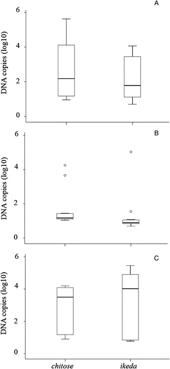
The relative intensities of infection (DNA copies) of genotypes chitose and ikeda varied among the different groups. The infection intensities of chitose and ikeda were higher in groups 1 (57 721 and 33 709, respectively) and 3 (5897 and 61 766, respectively) than those in group 2 (2071 and 6331, respectively) (Table 2). Furthermore, the infection intensities of genotype ikeda were significantly different among three groups of cattle tested (P < 0·05) (Table 2). Overall, in group 1, the average DNA copies of genotype chitose was relatively higher than those of genotype ikeda (Table 2; Fig. 3A). However, in groups 2 and 3, the average DNA copies of genotype ikeda were relatively higher than those of genotype chitose (Table 2; Fig. 3B and C). In group 2, very low average DNA copies of both chitose and ikeda were recorded compared with those in groups 1 and 3 (Table 2; Fig. 3B).
Box plot diagrams indicating the number of DNA copies (log10-transformed) of the two pathogenic genotypes (chitose and ikeda) estimated by MT-PCR from blood samples of three groups (A, B, C) of cattle.

Infection intensities of the two pathogenic genotypes of T. orientalis estimated using MT-PCR in cattle

Different superscript letters (a, b, c, d) denote significantly different (P < 0·05) prevalence of T. orientalis.
Superscript letters within columns indicate difference in DNA copy numbers of each genotype among the three groups of cattle.
Group 1, heifers which aborted and/or showed severe clinical signs of oriental theileriosis in Vietnam, group 2 pre-shipment bloods from heifers which aborted and/or showed clinical signs of oriental theileriosis in Vietnam and group 3 cattle from the same batch of heifers which were sent to Vietnam but sent to Taiwan.
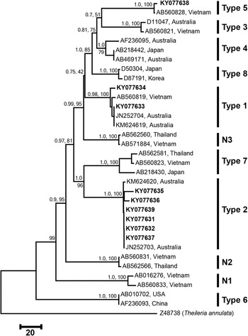
To further characterize T. orientalis, blood samples were tested using conventional PCR targeting the MPSP gene, and 34·8% (39/112) of them were test-positive for T. orientalis. Apparent prevalences of cattle in groups 1, 2 and 3 were 44·9% (22/49), 17·4% (4/23) and 32·5% (13/40), respectively, and the difference in prevalence was statistically significant (P < 0·05) among each group. DNA sequencing of PCR amplicons revealed the presence of six variants of ikeda (GenBank accession nos. KY077631, KY077632, KY077635-KY077637, KY077639; with a pairwise difference of 0·3–1·9%), two variants of chitose (GenBank accession nos. KY077633-KY077634; with a pairwise difference of 1·2%) and one sequence of type 5 (GenBank accession no. KY077638) genotypes of T. orientalis. Pairwise comparisons of MPSP sequences of chitose and ikeda determined herein and those previously reported from Australia (chitose: JN252704 and KM624619; ikeda: JN252703 and KM624620) revealed nucleotide differences of 0·7–1·3 and 0·4–1·0%, respectively. Furthermore, phylogenetic analyses revealed that MPSP sequences from this study grouped, with strong statistical support, with previously published ikeda, chitose and type 5 genotypes of T. orientalis genotypes to the exclusion of all other currently known genotypes of T. orientalis globally (Fig. 4). In addition, genotypes chitose and ikeda grouped with respective types previously reported from Australian cattle (Fig. 4).
Genetic relationships of Theileria orientalis genotypes from cattle imported to Vietnam from Australia (bold) with reference genotypes selected from previous studies. Each genotype is represented by a unique GenBank accession number followed by the country of origin. The relationships were inferred based on the phylogenetic analysis of major piroplasm surface protein sequences using Neighbour-Joining (NJ) and Bayesian inference (BI) methods, and Theileria annulata was used as an outgroup. Nodal support values are indicated: posterior probability for BI (first/above) and bootstrap for NJ (second/below). The scale bar indicates distance.

DISCUSSION
This is the first report of oriental theileriosis leading to abortion and sudden deaths in dairy cattle imported to Vietnam from Australia. We detected two pathogenic genotypes chitose and ikeda of T. orientalis from different groups of cattle using the MT-PCR assay. To our best knowledge, this is the first report of genotype ikeda in Vietnam. In addition, a molecular-phylogenetic analysis of the major piroplasm surface protein gene sequences revealed that genotypes chitose and ikeda found in this study were closely related to those previously reported from Australian cattle. Overall, the recorded DNA copies of these genotypes were relatively low compared with DNA copies recorded in cattle herds with and/or without outbreaks of oriental theileriosis in Australia and New Zealand employing the same MT-PCR assay (Gebrekidan et al. Reference Gebrekidan, Gasser, Perera, McGrath, McGrath, Stevenson and Jabbar2015; Perera et al. Reference Perera, Gasser, Firestone, Smith, Roeber and Jabbar2015a , Reference Perera, Gasser, Read, Malmo, Nguyen, Nyein, Cheng, Jex, Rawlin, Spithill and Jabbar b , Reference Perera, Gasser, Pulford, Stevenson, Firestone, McFadden and Jabbar c ).
In the present study, an overall apparent prevalence of T. orientalis was high (72·3%), which is in concordance with cattle on farms experiencing outbreaks of oriental theileriosis in Australia (53·8–75·5%) (Perera et al. Reference Perera, Gasser, Anderson, Jeffers, Bell and Jabbar2013, Reference Perera, Gasser, Firestone, Smith, Roeber and Jabbar2015a , Reference Perera, Gasser, Read, Malmo, Nguyen, Nyein, Cheng, Jex, Rawlin, Spithill and Jabbar b ; Gebrekidan et al. Reference Gebrekidan, Gasser, Perera, McGrath, McGrath, Stevenson and Jabbar2015) and New Zealand (Perera et al. Reference Perera, Gasser, Pulford, Stevenson, Firestone, McFadden and Jabbar2015c ). Interestingly, in all groups of cattle, the prevalence of genotype ikeda was higher (32·5–57·2%) compared with that of genotype chitose (2·5–10·2%) which confirms that ikeda is mainly involved in a clinical form of oriental theileriosis as reported elsewhere (e.g. Ota et al. Reference Ota, Mizuno, Kuboki, Igarashi, Nakamura, Yamashina, Hanzaike, Fujii, Onoe, Hata, Kondo, Matsui, Koga, Matsumoto, Inokuma and Yokoyama2009; Izzo et al. Reference Izzo, Poe, Horadagoda, De Vos and House2010; Islam et al. Reference Islam, Jabbar, Campbell, Cantacessi and Gasser2011; McFadden et al. Reference McFadden, Rawdon, Meyer, Makin, Clough, Tham, Mullner and Geysen2011).
Although in cattle originating from Australia, this study reports a high prevalence of genotype ikeda (type 2) in Vietnam. Based on conventional PCR targeting MPSP (as used in this study), only seven genotypes of T. orientalis (types 1, 3, 5, 7 and N1–N3) were previously reported from domestic animals and ticks in Vietnam (Altangerel et al. Reference Altangerel, Lan, Long, Ueno, Li, Luo, Macedo, Matsumoto, Inokuma, Kawazu, Igarashi, Xuan and Yokoyama2011a ). Prior to our study, there was no report regarding the presence of the most virulent genotype ikeda either from bovids or ticks from Vietnam. Furthermore, all the available studies sampled clinically healthy animals, therefore reported low prevalence of T. orientalis infection. For example, Altangerel et al. (Reference Altangerel, Lan, Long, Ueno, Li, Luo, Macedo, Matsumoto, Inokuma, Kawazu, Igarashi, Xuan and Yokoyama2011a ), reported 14% (34/243) prevalence of T. orientalis infection from domestic animals. An absence of the pathogenic genotype ikeda as well as a low prevalence of chitose and other genotypes of T. orientalis in the country, for instance in the study reported by Altangerel et al. (Reference Altangerel, Lan, Long, Ueno, Li, Luo, Macedo, Matsumoto, Inokuma, Kawazu, Igarashi, Xuan and Yokoyama2011a ) may explain why clinical oriental theileriosis has not been a commonplace in bovines from Vietnam. Furthermore, a higher (72·3%) prevalence of T. orientalis reported herein than the previous study (14%) by Altangerel et al. (Reference Altangerel, Lan, Long, Ueno, Li, Luo, Macedo, Matsumoto, Inokuma, Kawazu, Igarashi, Xuan and Yokoyama2011a ) could be due to the use of different diagnostic methods between the two studies, as the MT-PCR assay is reported to be more (~1000 times) sensitive than conventional PCR (cf. Perera et al. Reference Perera, Gasser, Pulford, Stevenson, Firestone, McFadden and Jabbar2015c ). In addition, the differences in prevalence could also be due to variation in the host susceptibility, immunity and stress as well as geographic variability.
The molecular-phylogenetic analysis of MPSP sequences determined herein and those previously reported suggested that chitose and ikeda genotypes of T. orientalis involved in outbreak of oriental theileriosis in imported cattle to Vietnam were very similar to genotypes chitose (pairwise difference: 0·7–1·3%) and ikeda (pairwise difference: 0·4–1·0%) reported from Australian cattle. In addition, both genotypes chitose and ikeda grouped, with strong statistical support, with respective types previously reported from Australia (see Fig. 4). This high genetic relationship of genotypes chitose and ikeda determined in this study and those previously reported from Australia suggests that these pathogenic genotypes of T. orientalis might have been introduced to Vietnam from Australia via infected cattle as the outbreak of the disease started immediately upon arrival of the cattle to Vietnam.
In this study, we found that the average DNA copies of pathogenic genotypes (chitose and ikeda) of T. orientalis in cattle showing clinical signs and/or aborting ascribed to oriental theileriosis were low compared with those of the same genotypes recorded in cattle herds with and/or without outbreaks of oriental theileriosis in Australia and New Zealand employing the same MT-PCR assay. For instance, in three different Australian studies, the average DNA copies of chitose varied from 91 414 to 270 679, whereas those of ikeda ranged from 184 759 to 329 775 (Gebrekidan et al. Reference Gebrekidan, Gasser, Perera, McGrath, McGrath, Stevenson and Jabbar2015; Perera et al. Reference Perera, Gasser, Firestone, Smith, Roeber and Jabbar2015a , Reference Perera, Gasser, Read, Malmo, Nguyen, Nyein, Cheng, Jex, Rawlin, Spithill and Jabbar b ). Similarly, a study by Perera et al. (Reference Perera, Gasser, Pulford, Stevenson, Firestone, McFadden and Jabbar2015c ) reported higher average DNA copy numbers of chitose (70 684) and ikeda (349 271) in cattle from New Zealand than those of both genotypes recorded in this study. This information raises a question as to why dairy cattle with low average DNA copies (groups 1 and 2; see Table 2 DNA copy numbers) imported to Vietnam from Australia suffered from clinical oriental theileriosis associated with abortions and deaths, whereas other cattle (group 3; see Table 2 DNA copy numbers) of the same origin but sent to Taiwan at the same time did not suffer from a clinical disease. Following a systematic investigation of events (undertaken by a veterinarian, L. Nelson) that occurred following the arrival of cattle in Vietnam revealed that cattle might have been stressed due to vaccination (on-board the ship) against FMD, haemorrhagic septicaemia and tuberculosis. In addition, stress levels could have exacerbated when cattle were taken to the destined dairy farm after a 10-h trip by road in a truck, and it even exacerbated further when cattle were blood tested and re-vaccinated after their arrival on farm. It is believed that cattle infected with pathogenic genotypes (ikeda and chitose) of T. orientalis can remain carriers for many years, and they can exhibit clinical form of oriental theileriosis when they are stressed, particularly around calving (Kubota et al. Reference Kubota, Sugimoto and Onuma1996; Gebrekidan et al. Reference Gebrekidan, Gasser, Perera, McGrath, McGrath, Stevenson and Jabbar2015). Therefore, based on the findings of the present study, we suspected that the pregnant heifers were asymptomatically infected with ikeda and chitose when they left Australia, but showed clinical signs along with abortions and deaths associated with oriental theileriosis when they arrived in Vietnam due to a sustained level of stress, poor nutritional status and husbandry.
To date, limited information is available on bovine theileriosis in Taiwan. Previously, Wang et al. (Reference Wang, Kubota, Kakuda, Kuo, Hsu and Onuma1998) used a variety of diagnostic methods, including microscopy, conventional PCR, allele-specific PCR and Southern blotting and reported buffeli and chitose genotypes of T. orientalis from cattle in Taiwan. Similarly, Kim et al. (Reference Kim, Tsuji, Kubota, Wei, Lee, Ishihara and Onuma1998) reported genotypes buffeli and chitose in cattle using PCR restriction length fragment polymorphism analysis. It is not clear that why cattle exported to Taiwan from Australia did not experience clinical oriental theileriosis as they were also infected with T. orientalis (see group 3 in Tables 1 and 2, and Fig. 3). Based on the comparison of management and husbandry practices between the farms in Taiwan and Vietnam, it is hypothesized that better on-farm handling and overall management in Taiwan helped to further reduce stress on pregnant heifers after a voyage, thereby avoiding an outbreak of oriental theileriosis in this country.
The documentation of a pathogenic genotype (ikeda) of T. orientalis in imported cattle to Vietnam is not surprising, as import/export of cattle had been found to be associated with the introduction of infectious diseases from endemic to non-endemic countries globally. For instance, East Coast fever caused by T. parva was first identified in Zimbabwe at the end of the 19th century, and later on, it rapidly spread into other surrounding 11 African countries (Burundi, Democratic Republic of Congo, Kenya, Malawi, Mozambique, Rwanda, Tanzania, Uganda, South Africa, South Sudan and Zambia) following introductions of cattle from Zimbabwe (reviewed by Uilenberg, Reference Uilenberg1999). This situation warrants an application of proper prevention and quarantine measures for pre-departure and post-arrival procedures for exporting and importing countries, respectively, to prevent the introduction of potential pathogenic organisms or their strains.
In conclusion, this study reports the first outbreak of oriental theileriosis in dairy cattle imported to Vietnam from Australia. The two pathogenic genotypes (chitose and ikeda) of T. orientalis were detected using the MT-PCR and conventional PCR assays, and it appears that low infection intensities (DNA copies) of pathogenic genotypes can also cause clinical oriental theileriosis when pregnant cattle experience a period of sustained stress. Pre-shipment testing of cattle, and quarantine and prevention programmes in the importing countries should be strengthened to control the spread of T. orientalis Gebrekidan et al. (Reference Gebrekidan, Abbas, Wajid, Ali, Gasser and Jabbar2017) as well as other infectious agents during import and export of cattle globally.
ACKNOWLEDGEMENTS
Authors would like to thank Natalie Courtman and Kathryn Jenkins for their assistance in microscopic examination of stained blood smears.
FINANCIAL SUPPORT
This project was supported by Frontier International Agri, Australia. H.G. is a grateful recipient of the Melbourne International Research Scholarships (MIRS) and Melbourne International Fee Remission Scholarships (MIFRS) through The University of Melbourne.