Introduction
Premature ventricular complexes are being recognised as a common and often troubling clinical electrocardiographic finding. Reference Ballı, Kanlıoğlu and Akgun1 It is well known that a significant proportion of these arrhythmias usually originates from the outflow tract of either the right or the left ventricle. Premature ventricular complex-induced cardiomyopathy is defined as the development of left ventricular dysfunction caused by a high burden of premature ventricular complexes (usually ≥10% of the total number of QRS complexes). Reference Panizo, Barra and Mellor2,Reference Cronin, Bogun and Maury3
Transcatheter ablation has emerged as a pivotal curative treatment option. In recent years, the integration of three-dimensional mapping and high-density mapping catheters has revolutionised the approach to complex arrhythmias. Reference Steyers, Sodhi and Faddis4 These advanced technologies enable more precise mapping of arrhythmic substrates, thereby significantly enhancing the success rates of ablation procedures. Historically, conventional mapping techniques were utilised; however, the advent of high-density mapping catheters has become increasingly prevalent in modern practice. Reference Martin, Ramos and Padilla5 Despite these advancements, there remains a paucity of studies specifically examining the use of high-density mapping in paediatric right ventricular outflow tract ablations. In this study, we aim to compare the outcomes of right ventricular outflow tract arrhythmias treated with conventional versus high-density mapping catheters, providing new insights into the efficacy and safety of these techniques in children. This research will contribute to the growing body of knowledge and potentially guide future clinical practices in paediatric arrhythmia management.
Materials and methods
Patient population
This study was a retrospective cohort analysis conducted at a single healthcare facility. It included 92 consecutive patients who underwent ablation procedures from January 2015 to June 2024. A comparison was made between 45 patients who underwent mapping with conventional catheters from 2014 to 2019 and 47 patients who underwent mapping with the Advisor HD Grid Catheter (Abbott Technologies, Minneapolis, MN) from 2019 to 2024. Conventional mapping procedures were performed using a tacticath catheter. Ethical clearance was secured from the local ethics committee, and informed written consent was obtained from each patient before the procedure. Clinical data and information related to the procedure were gathered from electronic medical records and the electrophysiology laboratory database.
Inclusion criteria and evaluation
The inclusion criteria for this study identified idiopathic right ventricular arrhythmia as the occurrence of ventricular tachycardia or premature ventricular complexes without any structural heart disease or known predisposing factors. Prior to undergoing the ablation procedure, all patients received a thorough evaluation, which included a physical examination, 12-lead electrocardiography, 24-hour Holter monitoring, exercise testing, and a transthoracic echocardiogram to evaluate left ventricular function. Ventricular systolic functions were assessed by the same operator using the Simpson’s method. Ejection fraction values below 50% were considered as reduced systolic function. A detailed family history concerning sudden death was also obtained. Patients with congenital, structural, or inflammatory heart conditions, such as myocarditis and pericarditis, were not included in the study. Antiarrhythmic drugs were discontinued at least five half-lives before the electrophysiological study.
Ablation indications and procedure
In children with idiopathic left ventricular arrhythmia, indications for ablation included symptomatic episodes of ventricular tachycardia, a high burden of ventricular arrhythmia leading to ventricular dysfunction, unsuccessful antiarrhythmic drug therapy, and arrhythmias induced by exercise or resistant to suppression. In all patients, the Ensite Precision system (Abbott, St Paul, MN, USA) was employed for mapping to facilitate the procedure and minimise or avoid the use of fluoroscopy.
Mapping system overview
This system combines magnetic cardiac geometry creation with an impedance-based platform, resulting in a dual-technology approach. The EnSite Precision mapping system offers features such as AutoMap and TurboMap. AutoMap continuously captures multiple simultaneous points from several electrodes for mapping anatomy, voltage, and activation sequences. TurboMap allows for the rapid replay of any tachycardia episodes recorded during the initial mapping, operating at a speed ten times faster than normal, which helps in creating activation maps without the need for manual remapping. SparkleMap enhances this process by providing a video overlay of the activation on the voltage map within the three-dimensional geometric model. In addition, colour-coded fractionation maps automatically label fractionated electrograms, aiding in the identification of ablation targets. Another key feature is AutoMark, which utilises real-time contact force recordings to offer colour-coded designations for lesion formation.
HD grid catheter
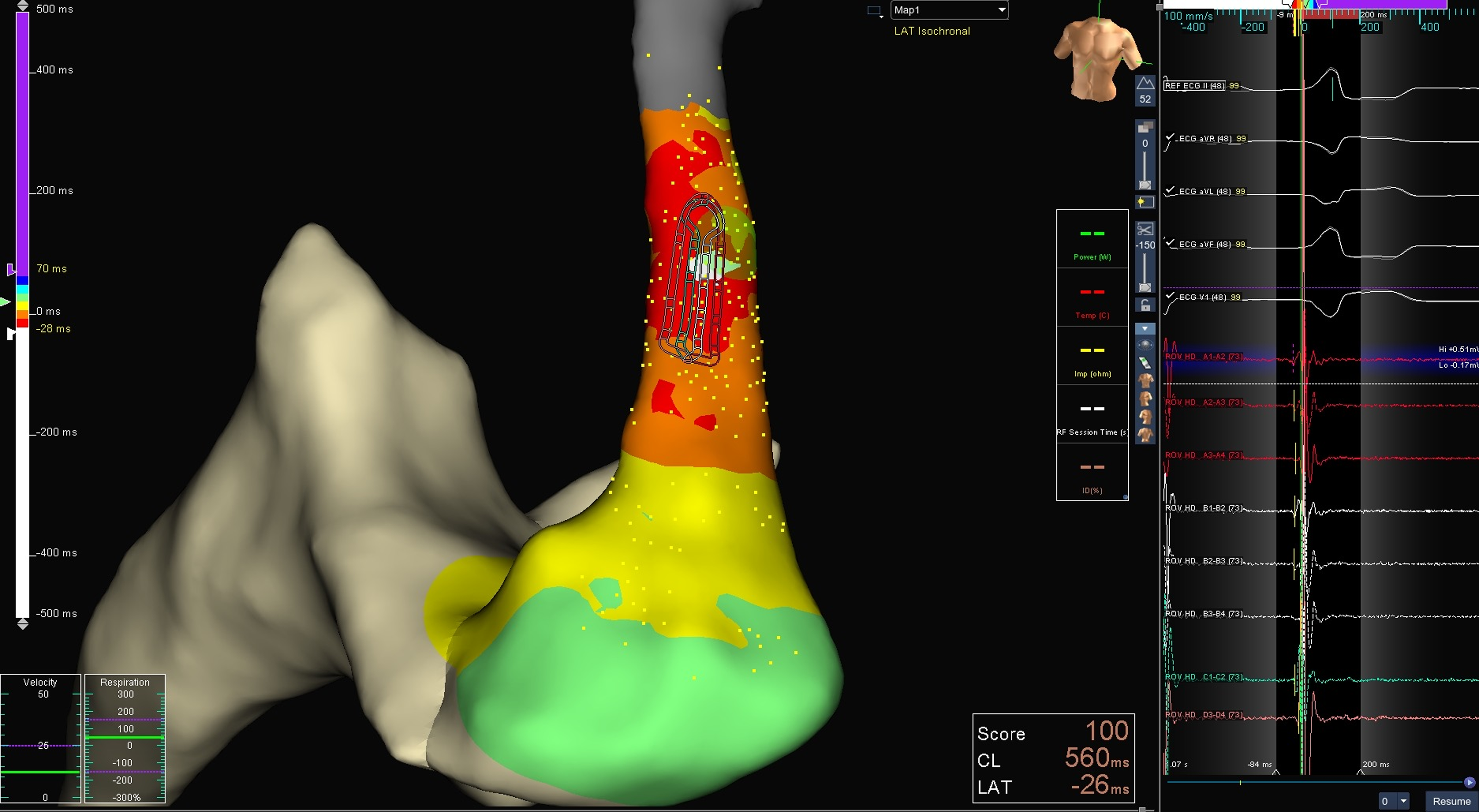
The Advisor™ HD Grid Catheter (Abbott Technologies, Minneapolis, MN) was employed for mapping purposes (Figure 1). This catheter features 16 electrodes with a flexible design and 3 mm equidistant electrode spacing. It can record both parallel and perpendicular to the splines, facilitating rapid assessment of voltage, activation, and conduction directionality, as well as omnipolar electrogram evaluation. The bidirectional capability of the HD Grid catheter allows it to flex up to 35 mm in the D curve and up to 42 mm in the F curve. The catheter is eight French in size and 110 cm in length. Long sheaths (Agilis-Abbott) were utilised to manoeuver the HD Grid Catheter effectively.
Using the Ensite X mapping system, a total of 2560 points acquired in a remarkably brief duration of just 3 minutes with the Advisor HD grid catheter. The white area located at the position of the HD grid catheter indicates the region of early activation. Notably, the earliest area appears to precede a ventricular ectopic beat by approximately 26 milliseconds.

Ablation procedure
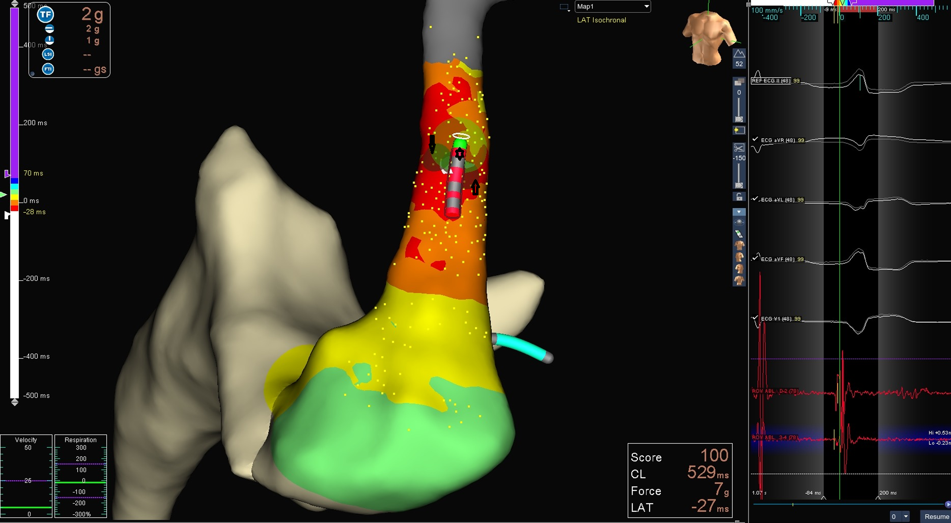
Radiofrequency energy was delivered using the cooled-tip radiofrequency catheter (Medtronic, Inc., Minneapolis, USA). In instances where radiofrequency ablation did not achieve the desired results, an irrigated ablation catheter was used (Figure 2). The radiofrequency energy was initially set at 25 W and gradually increased to 35 W within 30–60 s. During the procedure, the goal was to achieve a 15% drop in impedance. If ventricular tachycardia or premature ventricular complexes persisted 10 s after the ablation began, the procedure was halted. For areas close to the conduction system and coronary arteries, cryoablation was performed using the Freezor Cryocath (Medtronic, Inc., Minneapolis, USA), aiming for a temperature of −75°C for a period of 240 s.
It is shown that an ablation procedure was performed using a Tacticath catheter on the early activation zone identified with a high-density catheter. The ablation points are indicated by arrows, while the Tacticath catheter is represented by a star.

Statistical analysis
All analyses were performed using SPSS version 22 (IBM Corporation, Armonk, New York, United States) statistical software. The results were expressed as the mean ± SD, unless otherwise stated. Differences between outcomes were analysed using unpaired Student’s t test or the Mann–Whitney test. Incidences in groups were tested for significance with the chi-squared test.
Results
Demographic and electrocardiographic features
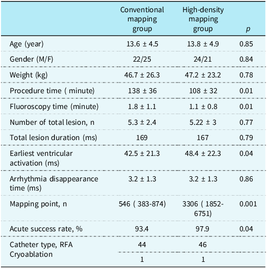
Our study comprised two groups, with a total of 92 patients. Among them, 45 patients underwent conventional three-dimensional mapping, while 47 patients underwent high-density three-dimensional mapping. The demographic characteristics of the patients are detailed in Table 1. There were no significant differences between the groups in terms of age, gender, or weight.
Patient characteristics and procedural results

RFA = radiofrequency ablation catheter; ms = millisecond; n = number.
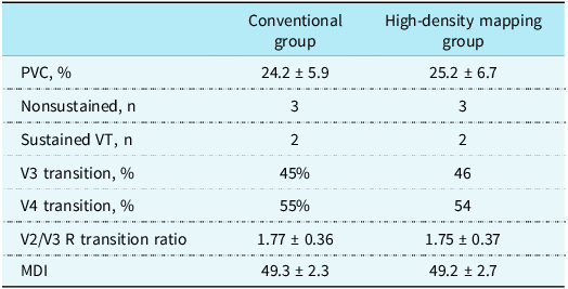
The frequency of ventricular arrhythmias showed no notable variation between the groups, whether the arrhythmias were sustained or nonsustained. There were eight patients with systolic dysfunction in total: four in the high-density mapping group and four in the conventional mapping group. Electrocardiographically, there were no significant differences in the transition zone or the V2/V3 R transition ratio. Additionally, the Maximum Deflection Index was consistent across both groups. The detailed results and statistics regarding procedural details are comprehensively provided in Table 1.
In the high-density mapping group, there was one patient with sustained ventricular tachycardia in whom the procedure was not successful. It was suspected to be epicardial. A 15-year-old patient underwent successful ablation with an irrigated ablation catheter from the great cardiac vein branches in the ventricular summit region 15 days later. In the conventional mapping group, successful ablation was not achieved in three patients.
Procedural features
In the high-density mapping group, the procedure time was significantly shorter. Additionally, there was a notable difference regarding the reduced fluoroscopy time. The total lesion time and number were also significantly lower in the high-density mapping group. The earliest activation time was significantly longer in this group. The procedure success rate was 97.9 % for the high-density mapping group, compared to 93.4% for the conventional mapping group. Successful ablation was performed on one ventricular extrasystole in the parahisian localisation in each group, while a cooled type radiofrequency catheter was used for all other cases. The detailed results and statistics regarding procedural details are comprehensively provided in Table 2.
Electrocardiographic findings

PVC = premature ventrıcular contraction; MDI = Maxımum Deflection Index; n = number.
Follow-up
Follow-up was managed through either inpatient or outpatient visits or through telephone communication without a predetermined follow-up schedule. The average follow-up period was 68.2 ± 24.4 months (conventional mapping group) and 34.4 ± 19.2 months (high-density mapping group). During the 48-hour Holter monitoring after the procedure, transthoracic echocardiography was conducted just before discharge and at three and six months post-ablation for all patients. By the end of the six-month period, all six patients had their systolic function restored to normal. In the conventional mapping group, three patients were unsuccessful due to isolated ventricular extrasystole. The first of these patients was 12 years old and refused a repeat ablation, so we are treating them with antiarrhythmic drugs. The second patient initially did not want a second ablation, but due to being symptomatic despite antiarrhythmic treatment, underwent an ablation two years after the initial procedure. Because we were not using high-density mapping at that time, a successful ablation was performed using conventional mapping. The third patient, who was not successfully treated, began to show polymorphic ventricular extrasystole during follow-up and was diagnosed with arrhythmogenic right ventricular disease. This patient is being monitored with medical treatment.
Discussion
In recent years, the use of electroanatomic mapping and the subsequent development of high-density mapping catheters have become increasingly prevalent in cardiac procedures. Reference Sciacca, Fink and Bergau6 In our study, comparing high-density mapping with conventional mapping methods in right ventricular outflow tract ablations, we observed several significant advantages of high-density mapping. Notably, the procedure time was considerably shorter, allowing for a more efficient workflow and reduced patient discomfort. Additionally, the success rate of the procedures was notably higher, indicating that high-density mapping provides a more accurate and comprehensive understanding of the cardiac anatomy and arrhythmogenic substrates. This enhanced precision likely contributes to the improved outcomes. Furthermore, the recurrence rate post-ablation was notably lower, suggesting that high-density mapping may offer more durable results and decrease the likelihood of patients requiring repeat procedures. These findings highlight the potential of high-density mapping to improve the efficacy and efficiency of ablation procedures, offering substantial benefits over traditional mapping techniques.
In both ischaemia-related and primary cardiomyopathies, high-density mapping catheters are frequently used in activation mapping, especially for showing tachycardia isthmus, as well as in isochronal mapping for observed ventricular tachycardias. Reference Sohinki, Nakagawa and Stavrakis7,Reference Adav, Ramasamy and Theodore8 To the best of our knowledge, there are no studies using high-density mapping in children with focal arrhythmias such as right ventricular outflow tract arrhythmias, and this study is the first to use high-density mapping in right ventricular outflow tract ablations in the paediatric population.
In the study conducted by Yue-E. He and colleagues used conventional mapping, and radiofrequency ablation was successful in 68 out of 75 children with premature ventricular complexes, resulting in a success rate of 90.67%. The recurrence rate during follow-up was reported to be low at 2.94%. Reference He, Xue and Gharbal9 In another study, conducted by Akdeniz et al. using the EnSite Velocity system in children, a total of 35 patients underwent the procedures, which were performed using conventional mapping catheters in conjunction with three-dimensional mapping. During the follow-up period, there was a 20% recurrence rate of ventricular tachycardia. Reference Akdeniz, Gul and Celik10 Conventional mapping catheters often extend procedure times due to their limited pole numbers and rigid structures, which lead to more frequent ventricular premature beats. These frequent premature beats complicate the mapping process and can cause errors in identifying the correct ablation area, especially when premature beats occur near the ablation site. Furthermore, these catheters are inadequate in displaying fractionated signals, which is crucial for accurate mapping. Our results demonstrate that high-density mapping significantly enhances ablation success and reduces recurrence rates.
High-density mapping catheters have revolutionised the approach to cardiac mapping by enabling the acquisition of more data points in a significantly shorter time frame. This efficiency is largely attributed to their design, with many catheters featuring a 16-pole configuration that enhances their data-gathering capabilities. The flexible structure of these catheters allows for softer contact with the cardiac tissue, which not only improves patient comfort but also reduces the incidence of premature beats during the procedure. Studies have highlighted that the ability to gather dense and detailed mapping data facilitates a more accurate identification of arrhythmogenic substrates, thus enhancing the precision of ablations. Reference Balli, Kanlioğlu and Altin11 The combination of these features positions high-density mapping catheters as a superior tool in electrophysiological interventions, offering improved procedural outcomes and reduced complication rates. Although the use of high-density mapping catheters incurs additional costs, we can confidently say that their benefits far outweigh the costs when considering their ability to increase the success rate of the procedure, shorten procedure time, and reduce radiation exposure.
The Orion© catheter is equipped with 64 small electrodes, featuring a compact interelectrode spacing of 2.5 mm, which allows for rapid simultaneous mapping with enhanced near-field signal quality. When used in conjunction with the Rhythmia© mapping system and its automated intelligent annotation module, it offers precise and swift mapping of any premature ventricular complex. However, its size and shape can pose handling challenges in smaller ventricles. On the other hand, the Pentaray© catheter comprises 20 electrodes distributed across five soft, flexible, 3-French branches. When paired with the innovative Carto Prime© system mapping module, it significantly enhances ablation safety and efficacy through features like Parallel mapping©, which enables simultaneous three-dimensional activation mapping of different premature ventricular complex morphologies within the same cardiac chamber, and LAT Hybrid©, which correlates the PVC Local Activation Time information with its normal sinus rhythm location. Reference Dragasis, Vlachos and Frontera12 Lastly, Abbott’s Ensite X© system, featuring AutoMap and TurboMap capabilities, when combined with the Advisor HD Grid© catheter (16 electrodes), facilitates rapid and ultra-high-density mapping of any premature ventricular complex with an optimal signal-to-noise ratio. Multipolar catheters are invaluable in patients with both high and low premature ventricular complex burdens, as they considerably expedite the mapping process and reduce X-ray exposure for both the patient and the operator . Reference Dragasis, Vlachos and Frontera12
The use of high-density mapping system provides reliable data in the ablation of intraatrial re-entrant tachycardia and focal atrial tachycardia in a shorter time by collection of more points during mapping. This facilitates the identification of critical ablation targets. Reference Balli, Kanlioğlu and Altin11,Reference Chieng, Lahiri and Sugumar13
The study conducted by RA Bertels and colleagues highlights that syncope was present in 33% of patients with left ventricular dysfunction at diagnosis, and cardiomyopathy was a critical concern. Reference Bertels, Harteveld and Filippini14 Achieving success in ablation is crucial, especially when considering the potential reversibility of left ventricular dysfunction by reducing the burden of premature ventricular complexes. This underscores the importance of high-density mapping, as it plays a significant role in increasing the success rate of ablation procedures. High-density mapping allows for more precise localisation of arrhythmia sources, improving the chances of a successful ablation and reducing the risk of syncope and progression to cardiomyopathy in paediatric patients.
High-density mapping has been shown to increase the success rate in accessory pathway ablations, with studies indicating even greater success when using the Open Window technique. An example of this is the study conducted by JL Martinez and colleagues, where 33 patients underwent accessory pathway ablation using the Open Window technique with HD mapping. This advanced technique demonstrated reduced mapping time, lower radiofrequency application time, and decreased X-ray exposure, all while achieving comparable success and recurrence rates to conventional methods. Reference Bertels, Harteveld and Filippini14
Ventricular arrhythmias associated with ventricular ischaemia and cardiomyopathy are commonly observed in adult patients, with high-density mapping being widely utilised in this population. In paediatric patients, however, studies primarily focus on its use in atrial or ventricular re-entrant tachycardias related to surgically repaired congenital heart disease. Reference Chuang, Chung and Lee15 High-density mapping has become the preferred technique for catheter ablation of ventricular arrhythmias, surpassing the traditional point-by-point mapping method. Compared to point-by-point mapping, high-density mapping provides significantly enhanced resolution in identifying myocardial scars and delineating conducting channels. This advanced capability allows for a more precise characterisation of arrhythmogenic substrates, leading to reduced procedure durations and improved procedural efficiency. Moreover, the superior mapping accuracy achieved with high-density mapping has been associated with better long-term outcomes, including higher rates of arrhythmia-free survival, making it a critical advancement in the management of ventricular arrhythmias. Reference Yamashita, Cochet and Sacher16–Reference Berte, Relan and Sacher18
Study limitations
This study was limited by its retrospective design. Our study was performed on a small number of patients. More reliable results may be obtained if the study is performed in a larger patient group.
Conclusion
In children with ventricular outflow tract arrhythmias, high-density mapping can be utilised with high success rates and safety. This advanced mapping technique allows for a more detailed and precise visualisation of the cardiac anatomy and arrhythmogenic substrates. By providing improved spatial resolution, high-density mapping enhances the ability to accurately identify the origin of arrhythmias, leading to more effective ablation procedures. Consequently, this approach not only increases the likelihood of successful outcomes but also makes it a reliable option for managing ventricular outflow tract arrhythmias in paediatric patients.
Acknowledgements
None.
Financial support
This research received no specific grant from any funding agency, commercial, or not-for-profit sectors.
Competing interests
None.