INTRODUCTION
Leishmania are pathogenic trypanosomatid protozoans responsible for a diverse spectrum of human diseases. These parasites exhibit a dimorphic life-cycle, consisting of extracellular promastigotes that reside within the midgut of the sandfly vector and intracellular amastigotes that reside within the phagolysosome of host macrophages (Alexander and Russell, Reference Alexander and Russell1992). Chemotherapy relies mainly on pentavalent antimony-containing drugs although miltefosine is appearing to be very useful (Croft and Coombs, Reference Croft and Coombs2003). Unresponsiveness of parasites to antimonials is a serious health problem and resistance has now reached epidemic proportions in India (Sundar, Reference Sundar2001; Sundar and Murray, Reference Sundar and Murray2005). Resistance, as established by in vitro studies on laboratory mutants, and more recently in clinical isolates is multifactorial (Gourbal et al. Reference Gourbal, Sonuc, Bhattacharjee, Legare, Sundar, Ouellette, Rosen and Mukhopadhyay2004; Singh, Reference Singh2006).
DNA microarrays are powerful tools beginning to be employed in the field of parasitology (Rathod et al. Reference Rathod, Ganesan, Hayward, Bozdech and Derisi2002; Diehl et al. Reference Diehl, Diehl, El-Sayed, Clayton and Hoheisel2002; Guimond et al. Reference Guimond, Trudel, Brochu, Marquis, El Fadili, Peytavi, Briand, Richard, Messier, Papadopoulou, Corbeil, Bergeron, Legare and Ouellette2003; Akopyants et al. Reference Akopyants, Matlib, Bukanova, Smeds, Brownstein, Stormo and Beverly2004; Almeida et al. Reference Almeida, Gilmartin, McCann, Norrish, Ivens, Lawson, Levick, Smith, Dyall, Vetrie, Freeman, Coulson, Sampaio, Schneider and Blackwell2004). The utility of microarray technology for studying drug responses and resistance mechanisms has been established in microorganisms and cancer cells (Wilson et al. Reference Wilson, Derisi, Kristensen, Imboden, Rane, Brown and Schoolnik1999; Cheok et al. Reference Cheok, Yang, Pui, Downing, Cheng, Naeve, Relling and Evens2003). Studies have validated the use of microarrays in studying drug resistance in Leishmania tarentolae and have pinpointed new genes over expressed either by gene amplification or by other mechanisms (Diehl et al. Reference Diehl, Diehl, El-Sayed, Clayton and Hoheisel2002). In this study, we made use of cDNA microarray as a rapid screening tool to identify genes in promastigotes of L. donovani clinical isolates associated with antimony resistance. One of the known mechanisms of resistance discovered in laboratory mutants has been drug efflux by ABC transporters (El Fadili et al. Reference El Fadili, Messier, Leprohon, Roy, Guimond, Trudel, Sarvia, Papadopoulou, Legare and Ouellette2005; Haimeur et al. Reference Haimeur, Brochu, Genest, Papadopoulou and Ouellette2000). Since ABC transporters were detected as 2-fold differentially regulated genes in our microarray experiments, their functionality was further validated by flow cytometry.
Although preliminary, this study has, however, established important leads and can be complemented later by studies on amastigotes for further elucidation of the molecular mechanism(s) of antimony resistance in L. donovani clinical isolates.
MATERIALS AND METHODS
Collection of clinical isolates
Two patients were selected from the Kala-azar Medical Research Center of the Institute of Medical Sciences, BHU, Varanasi and also from its affiliated hospital situated at Muzaffarpur, Bihar. The diagnostic criteria for VL were the presence of LD bodies (Leishman Donovan) in splenic aspirations performed and graded as per standard criteria (Chulay and Bryceson, Reference Chulay and Bryceson1983). After diagnosis, the patients were administered a course of sodium antimony gluconate (SAG) (Albert David, Calcutta, India) 20 mg/kg body weight once daily for 30 days. Response to treatment was evaluated by a repeat splenic aspiration on day 30 of treatment. Patients were designated responsive (2001) based on the absence of fever, clinical improvement with reduction in spleen size and absence of parasites in the splenic aspirate while patients who showed presence of parasites in splenic aspirates were labelled as antimonial unresponsive (39). These patients were subsequently treated successfully with amphotericin B.
Culture conditions
Splenic aspirates of responsive (2001) and unresponsive (39) kala-azar (VL) patients obtained after treatment with SAG were inoculated into NNN medium (Gibco BRL, Israel) containing 10% heat inactivated fetal bovine serum (FBS, Gibco BRL, Israel) and passaged every 7th day into a tube containing fresh NNN medium. Promastigotes were then progressively adapted to medium M199 (Sigma, St Louis, USA) supplemented with 10% FBS, 25 mm HEPES, pH 7·4, and cultures were maintained at 26°C.
Assay for drug sensitivity
Assay for drug sensitivity was done as described (Singh, Reference Singh2002). The virulence and level of susceptibility or resistance of these isolates was also confirmed in vivo, by infection in golden hamsters (Dube et al. Reference Dube, Singh, Sundar and Singh2005).
RNA isolation and Northern hybridization
Logarithmic phase promastigote cultures of the sensitive (2001) and resistant (39) isolates were used to isolate total RNA using TRIZOL reagent (Gibco BRL) using standard procedures (Chomezynski and Sacchi, Reference Chomezynski and Sacchi1987). All RNA samples were tested for purity and integrity. The quality of RNA was assessed by examination of purified total RNA by gel electrophoresis, which revealed prominent 18S and 24S alpha and 24S beta ribosomal bands indicating that the RNA was not degraded. Purified total RNA was quantified spectrophotometrically at 260 nm, and 280 nm, and ratios A260 nm/280 nm between 1·9 and 2·1 were considered acceptable.
For Northern hybridizations 10 μg of the total promastigote RNA of the sensitive and resistant isolate was run on 1·5% denaturing agarose gel and transferred to a nylon membrane by typical downward capillary transfer protocol. Primers for the P-glycoprotein like-protein (MDR) (AI034703) were designed based on the L. major sequence in GeneDB (www.genedb.org). Probe was made from PCR amplified 954 bp product from L. donovani nuclear DNA as template using primers: (F-5′ CGCCAT-ATGTCTGCCAACAAT-3′ and R-5′-GGCCT-CGAGTCAATTCTTC-3′). Alpha tubulin gene primers obtained as a kind gift from Marc Ouellette, Quebec, Canada, were used for amplification and probe preparation. Probes were made by labelling 25 ng of the DNAs with [α-32P] dCTP by random priming method (BRIT/BARC, India).
Probes for microarray hybridization
Probes comprised of fluorescent cDNAs synthesized from log phase promastigote total RNA, isolated from drug sensitive (2001) and resistant (39) clinical isolates of L. donovani. CyScribe First-Strand cDNA Labeling Kit (Amersham) was used to incorporate Cy3-dUTP and Cy5-dUTP into the drug sensitive and resistant probes respectively for use in dual colour microarray hybridizations. Purification of labelled cDNA was done using spin column chromatography (CyScribe GFX Purification kit, Amersham). Agarose gel electrophoresis was used to visualize the fluorescence of Cy3 and Cy5 labelled cDNA.
Leishmania major microarray creation and hybridization
The cDNA microarrays used in this study were a kind gift from Jenefer M. Blackwell, UK (Almeida et al. Reference Almeida, Gilmartin, McCann, Norrish, Ivens, Lawson, Levick, Smith, Dyall, Vetrie, Freeman, Coulson, Sampaio, Schneider and Blackwell2004). Briefly, expressed sequence tags (ESTs) were obtained by partial 5′ sequence analysis of 2183 cDNAs from libraries constructed from mRNA harvested from L. major promastigote and lesion-derived amastigotes. The resulting sequences were deposited into dbEST. EST to EST BLASTN hits with probabilities <1·0×10−10 identified 1094 unique (i.e. non redundant) genes amongst the 2183 cDNAs sequenced. Microarrays were then constructed using PCR amplified inserts from the 2183 cDNA EST clones that included all redundant copies of the 1094 unique genes. Accounting for PCR and spotting failures, the arrays contained 1830 cDNA features representing 1001 unique genes. This array was used to study differential gene expression for drug resistance in promastigotes of Leishmania donovani clinical isolates.
The 2 differently labelled probes were combined and put for hybridization essentially according to the protocol described (Almeida et al. Reference Almeida, Gilmartin, McCann, Norrish, Ivens, Lawson, Levick, Smith, Dyall, Vetrie, Freeman, Coulson, Sampaio, Schneider and Blackwell2004) by using Automatic Slide Processor (Amersham Lucidea Module 1). The slides were scanned immediately after washing and drying at both the Cy3 and Cy5 wavelengths (Array Scanner Molecular Dynamics Generation III). First, control hybridizations were performed to account for sample heterogeneity and possible variation due to hybridization. Initially, microarrays were hybridized with fluorescently labelled Cy5 and Cy3 promastigote cDNA, both prepared from the same RNA sample. This theoretically should give a log expression ratio (Cy5/Cy3) of 1 for all the elements arrayed onto the slide. A log10 plot of Cy5 versus Cy3 calibrated fluorescent response showed a tightly packed distribution of most genes along the line of best fit, with a regression correlation coefficient r of 0·99 (data not shown). Once the washing and hybridization conditions were optimized and hybridization signals could be consistently reproduced, the arrays were used for the analysis of gene expression in L. donovani clinical isolates.
Data analysis
The data were extracted and analysed by Array-Vision (Version 6.0) software supplied with the scanner. Local background was subtracted from the intensity value of each spot on the array. Spots were manually examined to assess their quality and those that exhibited poor quality or were saturated were discarded for further analysis. ‘Total intensity normalization’ (Quackenbush, Reference Quackenbush2002), taking into account the internal control genes, was carried out within arrays. In order to compare expression levels between RNAs, the log2-transformed Cy5/Cy3 ratios were calculated from the normalized values. Normalization to account for the systematic dependence of ratio on intensity was performed by locally weighted linear regression (LOWESS) (Cleveland, Reference Cleveland1979). Four replicate arrays were hybridized to look for the differential expression of genes with phenotype of interest i.e. drug resistance, 3 with drug resistant (39) promastigote RNA labelled with Cy5 and drug sensitive (2001) promastigote RNA labelled with Cy3; 1 with drug resistant (39) promastigote RNA labelled with Cy3 and drug sensitive (2001) promastigote RNA labelled with Cy5. The Cy5 signal was plotted against the Cy3 signal on a scatter plot. For each gene represented in the array, the Cy5 fluorescence/Cy3 fluorescence ratio measured at the corresponding array element is a quantitative measurement of the relative abundance of the transcript of that gene in the 2 isolates.
Statistical analysis
A modified Bonferroni's correction for multiple testing (Hochberg, Reference Hochberg2002) was used to estimate Z-score for each transcript, which was then translated into P-values to measure the significance of findings.
Annotation of cDNA sequences onto the Leishmania genome sequence
All annotations were performed using the ARTEMIS software (Rutherford et al. Reference Rutherford, Parkhill, Crook, Horsnell, Rice, Rajandream and Barrel2000). The current L. major codon usage table (obtained from http://www.kazusa.or.jp/codon) was utilized. In order to analyse the putative functions of the statistically significant genes identified, the amino acid sequence predicted from each putative gene were used to carry out a local BLASTP search of the non-redundant protein database and TBLASTN searching of a kinetoplastid specific nucleotide database. These searches were run through ARTEMIS. Generally, hits with BLAST scores of >50 and e-values of <1×e −6 were considered potentially significant. Each protein sequence was then searched against numerous collections of protein motifs and families (SWISS_PROTT Release 39.27, ProDom version 2001.2, PRINTS Release 31.0) as well as being blasted against the database of Clusters of Orthologous Groups of proteins (COGs) and NCBI's Conserved Domain Database and Search Service, version 1.54. These analyses allowed the putative functions to be identified for many of the genes spotted onto these arrays.
Flow cytometry
Flow cytometry was employed to ascertain the functionality of ABC transporters in the promastigotes of resistant (39) and sensitive (2001) isolates. Two substrates, rhodamine 123 and calcein were used. R123 (stock: 3 mm in methanol) was diluted to 100 μm in 0·02 m phosphate-buffered saline, pH 7·2 (PBS), and used at a final concentration of 1 μm. Calcein AM (1 mm in DMSO) was diluted to a final concentration of 1 μm in medium. To identify the nature of the pump, known modulators of MDR and MRP, namely verapamil (Sigma, St Louis, USA, 4 mm in PBS, final concentration 10 μm) and probenecid (Sigma, St Louis, USA, 0·4 m in PBS, pH 8·5, final concentration 4 mm) respectively were used.
Fluorescence labelling of L. donovani promastigotes
Log phase promastigotes (2×10⁶/ml) were washed twice with medium M199 (Sigma, St Louis, USA) supplemented with 10% FBS, 25 mm HEPES pH 7·4 (Medium A) and incubated at 26°C for fluorescence labelling.
Accumulation assays
Promastigotes (2×10⁶/ml) were initially washed twice in HEPES buffered saline referred to as ‘HBS1’ (21 mm HEPES, 137 mm NaCl, 5 mm KCl, 0·7 mm NaH2PO4 and 20 mm glucose, pH 7·4). After addition of R123, fluorescence was measured at 0, 15, 30 and 45 min whereas with calcein AM, fluorescence was measured at 0, 15, 30, 45 and 60 min.
To study the influence of energy depletion upon pump activity, accumulation of both fluorochromes was measured. Promastigotes were washed twice in HEPES-buffered saline referred to as ‘HBS1’ (21 mm HEPES, 137 mm NaCl, 5 mm KCl, 0·7 mm NaH2PO4, 20 mm glucose, pH 7·4). They were transferred to a modified HBS1 where glucose had been specifically excluded and 20 mm NaN3 included, referred to as ‘HBS2’ and incubated in HBS2 at 26°C for 30 min and transferred to PBS containing the fluorescent substrates in the presence or absence of modulators and readings were taken at different time-points (Perez-Victoria et al. Reference Perez-Victoria, Castanys and Gamarro2003).
Retention assays
Promastigotes were pre-loaded with calcein by incubating them with Calcein AM for 1 h at 26°C. Cells were centrifuged at 800 g for 5 min at 4°C, then washed twice with ice-cold Medium A, immediately resuspended in the same medium with or without probenecid, but notably excluding Calcein AM, and placed on ice. Thereafter, the first reading was taken and was considered as the zero min reading. Promastigotes were transferred to 26°C and fluorescence measured at 15, 30, 45, 60 min.
Thiol level detection
To measure intracellular thiol levels, Cell Tracker Green CMFDA (Molecular Probes, stock 10 mm in DMSO; final concentration 10 μm in serum-free medium) was used. Promastigotes were incubated with Cell-Tracker both in normal and energy-depleted conditions and fluorescence measured after 45 min of incubation at 26°C.
Flow cytometric analysis
Monitoring of dye accumulation and retention was carried out on a flow cytometer (FACS Calibur, Becton Dickinson) equipped with an argon-ion laser (15 MW) tuned to 488 nm. Data analysis was carried out with Cell Quest (BD) software. Fluorescence of rhodamine 123 (R123, Sigma, St Louis USA) and calcein (Calcein AM, Molecular Probes) was collected in the photomultiplier tube designated FL1, which is equipped with a 530/30-nm band pass filter. Filter combination and protocols were used to generate scatter-grams and list mode data on forward vs side-scatter and counts vs FL1 height. Samples were analysed at the flow rate of 100–200 cells/sec and a typical analysis was based on examination of 10 000 cells. Drug fluorescence was measured on a log scale while cell counts were on a linear scale. Dead cells or cells with compromised membranes were discriminated by adding propidium iodide (PI, 5 μg/ml) in every tube. Fluorescence of PI was taken at photomultiplier tube designated FL2 equipped with a 585/42-nm band pass filter. Each result is a representative of 4 sets of independent experiments. In order to eliminate the individual variance of basal fluorescence of each sample, we have represented the data as the ratio of fluorescence at any time-point divided by its zero min fluorescence and plotted on the Y-axis.
RESULTS
Drug sensitivity profile of isolates
In our previous study (Singh, Reference Singh2002) we have already confirmed that the clinical isolates used in this study namely, 39 and 2001, are truly resistant and sensitive to SAG respectively. The specific chemotherapeutic response of these isolates was persistent even after repeated passages in cultures as promastigotes and in vivo in experimental models (Dube et al. Reference Dube, Singh, Sundar and Singh2005), which again indicates that these isolates are truly refractory to SAG treatment in field conditions. Although there is stage specificity to antimony susceptibility (Ephros et al. Reference Ephros, Bitnun, Shaked, Waldman and Zilberstein1999) our study (Singh, Reference Singh2002) established the fact that promastigotes are not altogether unresponsive to SAG but do show an intermediate susceptibility phenotype to antimony, thus justifying the use of promastigotes as a preliminary screen to verify research methods and pinpoint potentially important genes using a stage that is easily grown in culture, and not contaminated by host material.
Expression profiling of drug-resistant genes
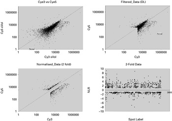
The results from a representative hybridization experiment comparing Cy5-labelled promastigote RNA from resistant isolate (39) versus Cy3 labelled promastigote RNA from sensitive isolate (2001) is shown in Fig. 1. A scatter plot of hybridization intensities showed that most spots fit a predominantly linear relationship, and that a fraction of the points deviated from the line of best fit indicating possible differential expression. The data in Fig. 1 were first transformed to the log2 ratio of the Cy5/Cy3 intensities and then normalized. Most of the spots are tightly grouped along the log2 (ratio)=0 line (no change in expression) and a small number are above the +1 or below the −1 lines (2-fold increase or decrease) indicating differential expression. Similar results were obtained in every experiment performed comparing resistant versus sensitive RNA expression (not shown). For further statistical analysis, we have focused on transcripts showing at least 2-fold differential expression. From more than 1000 genes on the array, 452 genes were identified to be differentially expressed in promastigotes of the resistant isolate 39 in this experiment.
Expression profiling by microarray of resistant (39) vs sensitive (2001) transcripts in log phase promastigotes of Leishmania donovani clinical isolates. Results of a single representative experiment comparing resistant isolate 39 (Cy5, Y-axis) vs sensitive isolate 2001 (Cy3, X-axis). Intensities are shown prior to ‘normalization’ of Cy3 vs Cy5 intensities. Distribution of log2 transformed normalized ratios for a single microarray experiment. The line of best fit is shown in all panels.

Regulated genes identified by statistical and bioinformatic analyses
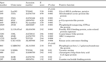
Robust statistical methods were then applied on these 452 genes to further identify genes whose expression changes at the level of mRNA abundance. Z scores for each gene were computed as the ratio of mean difference between the two groups for each gene, divided by standard error for the corresponding gene. To measure the significance of finding, Z scores were translated into P-values under asymptotic normality. To address the multiple comparison issue, the threshold was adjusted for declaring genes differentially expressed using a modified Bonferroni's correction (Hochberg, Reference Hochberg2002). The higher the Z score, the greater is the confidence that the transcript is differentially expressed between the 2 phenotypes. Out of the 452 genes analysed in this experiment, 11 showed a statistically significant (P<0·01) increase in expression (Table 1). Similar analysis was carried out on the results of 3 other hybridizations.
Putative targets from microarray analysis of differential expression between sensitive and resistant Leishmania isolates

In order to analyse the putative functions of the identified genes (Table 1), BLAST search was done. Generally hits with BLAST scores of >50 and e-values of <1×e−6 were considered potentially significant. It was seen that most of the genes identified as differentially expressed had sequences that showed no homology with previously described genes. It will be interesting to study this ‘unclassified category’ of genes later. Encouragingly, a survey of the genes scored as ‘regulated’ in 4/4 experiments revealed many that were expected, based upon prior studies on drug resistance mechanisms in Leishmania, including ABC transporters (Table 1) also showing overlap between slides. On grouping the identified genes by functional category, most remarkably the transport proteins were the most abundant namely, P-glycoprotein-like protein (AI034703), ABC1 transporter (AA741759), phospholipid transporting ATPase-like protein (AI03-4607), mitochondrial carrier protein-like protein (AA741710). Cell surface molecules were abundantly detected namely, phosphoglycan beta 1, 3 galactosyltransferase-like protein, UDP-N-acetyl glucosamine-dolichyl-phosphatase. Modulation of enzymes involved in lipid synthesis and of others possibly involved in cell wall metabolism, may modify access of drug to the plasma membrane. The coherence of this complex strategy for conferring drug resistance in clinical isolates is remarkable and is already under scrutiny by us (Kothari et al. Reference Kothari, Kumar, Sundar and Singh2006). We have lately identified a gene (Accession no. AF273843), localized on the pellicular plasma membrane of the resistant isolate.
Flow cytometry shows differential expression of ABC transporters between resistant and sensitive isolates
Dye accumulation
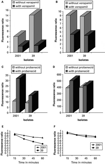
A time kinetic study of the accumulation of Rhodamine 123 showed that a rapid uptake of this dye occurred in promastigotes of both the isolates, being maximal in the antimonial-resistant isolate 39 (Fig. 2A). With the addition of verapamil, a known MDR modulator, a consistent decrease was observed in both the isolates. In order to assess whether ATP influenced this accumulation of Rhodamine 123, cellular ATP was depleted by the addition of NaN3 in the incubation buffer. The removal of ATP did not alter the R123 accumulation as net fluorescence in the presence or absence of ATP was no different (Fig. 2B). Once again, the addition of verapamil resulted in a decrease in R123 accumulation.
(A) Rhodamine accumulation in promastigotes of sensitive (2001) and resistant (39) isolates under normal conditions after 45 min of incubation. (B) Rhodamine accumulation in promastigotes of sensitive (2001) and resistant (39) isolates under ATP depletion after 15 min of incubation. (C) Calcein accumulation in promastigotes of sensitive (2001) and resistant (39) isolates under normal conditions after 60 min of incubation. (D) Calcein accumulation in promastigotes of sensitive (2001) and resistant (39) isolates under ATP depletion after 30 min of incubation. (E) Calcein retention in promastigotes of sensitive (2001) and resistant (39) isolates without probenecid. (F) Calcein retention in promastigotes of sensitive (2001) and resistant (39) isolates with probenecid.

With regard to the accumulation of calcein, both the isolates showed comparable levels of fluorescence, with 2001 having a marginally higher level of fluorescence (Fig. 2C). The addition of probenecid caused a consistent increase in fluorescence in both the isolates, maximal reversal was observed in 2001. Under energy-depleted conditions, calcein accumulation was greatly enhanced in both the isolates 2001 and 39, the fold increase being 36·1, 44·0, respectively in comparison to their levels of accumulation in the presence of ATP. With the addition of probenecid, the accumulation of calcein was marginally amplified in 2001 and 39, being 1·3 and 1·1 respectively (Fig. 2D).
Dye retention
In the retention assay, cells were pre-loaded with Calcein AM, washed and then incubated in Calcein AM-free medium in the presence or absence of probenecid. This allowed us to study the amount of calcein being retained within the parasites, which would indirectly reflect MRP activity. As seen in (Fig. 2E and F) at 60 min, the sensitive isolate 2001 showed maximum retention of calcein as compared to the resistant isolate 39. The resistant strain 39, showed minimum calcein retention at each time-point that was most pronounced at 60 min, indicating maximum efflux activity that was validated by reversal with probenecid. Probenecid enhanced the retention of calcein, indicating the presence of an MRP-like pump.
Thiol levels
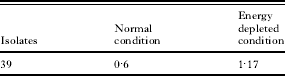
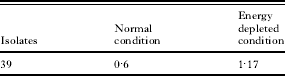
Measurement of total intracellular thiol levels in energy-depleted conditions in Leishmania isolates indicated that 39 had greater thiol levels than 2001 whereas under normal conditions thiol levels were 2001>39 (Table 2).
Fold increase of total thiol levels in promastigotes of SAG resistant isolate (39) with respect to the SAG sensitive isolate (2001) as calculated by the ratio of fluorescence of 39 to 2001
Northern blot analyses
Out of the identified transport proteins through our microarray experimental analysis (Table 1), we chose to look for the mRNA over expression of those genes documented to be involved in antimony resistance in laboratory mutants namely, MDR and MRP (Callahan and Beverley, Reference Callahan and Beverley1991; El Fadili et al. Reference El Fadili, Messier, Leprohon, Roy, Guimond, Trudel, Sarvia, Papadopoulou, Legare and Ouellette2005). Northern analysis performed to detect the transcript for the P-glycoprotein (MDR)-like protein (AI034703) failed to show the transcript under our experimental conditions even after prolonged exposure. Alpha tubulin probe used as control showed transcripts, proving the integrity of the blots (data not shown). This could be due to its very low level of expression as also corroborated by flow cytometric analysis showing functionality of the MRP pump and relative absence of MDR pump in the clinical isolates studied.
DISCUSSION
Having established parasite resistance to drug by observing the relationship between clinical response and SAG sensitivity in promastigotes and amastigotes of 39 and 2001 in vitro, we pursued our microarray analysis on promastigotes of these L. donovani clinical isolates. Microarrays have been used for measuring drug responses and resistance mechanisms in microorganisms and cancer cells (Wilson et al. Reference Wilson, Derisi, Kristensen, Imboden, Rane, Brown and Schoolnik1999; Cheok et al. Reference Cheok, Yang, Pui, Downing, Cheng, Naeve, Relling and Evens2003) and more lately in Leishmania mutants made resistant to drugs (Guimond et al. Reference Guimond, Trudel, Brochu, Marquis, El Fadili, Peytavi, Briand, Richard, Messier, Papadopoulou, Corbeil, Bergeron, Legare and Ouellette2003).
Extensive post-translational modifications are known to be associated with certain Leishmania proteins (Ilgoutz and McConville, Reference Ilgoutz and McConville2001). In gene expression analysis by microarray, the correlation between induced mRNA and induced levels of protein are not always well aligned. Subtle changes in transcript levels that have significant biological impact could be missed. Other limitations of microarray analysis include the impact of alternative splicing during transcript processing, decreased sensitivity of the arrays to the detection of genes with low expression levels (low-abundance genes), the partial genome coverage of the our microarray, and the use of promastigote RNA to investigate the antimonial drug resistance phenotype which is actually only selected in the amastigote stage, all point towards the limitations of this study. The results of our microarray screen proved disappointing as we could mainly confirm the already established determinants of antimonial resistance. The undetectable expression of many more genes can be attributed to the fact that in the clinical condition there is a very modest over expression of resistance, only about 2 to 3-fold, as opposed to many folds higher resistance found in laboratory mutants (Singh et al. Reference Singh, Singh and Sundar2003).
To adjust for unequal fluorescence intensities of the 2 RNA samples and allow comparison from experiment to experiment, various methods of normalization have been devised (Yang et al. Reference Yang, Dudoit, Luu, Lin, Peng, Ngai and Speed2002). The software utilized by us selects the LOWESS (locally weighted linear regression curve fit) normalization method (Cleveland, Reference Cleveland1979). Dye-swap analysis indicated that there was little bias after normalization functions were performed. To assess the overall similarity of the gene expression measurements produced by each of the slides, we examined the distributions of the Z scores of expression measurements. Z score for each gene is computed as the ratio of mean difference between the sensitive and resistant phenotype for each gene, divided by the standard error for the corresponding gene. Thus, analysis of the Z scores permitted direct comparisons of signal distributions and error levels across slides. To measure the significance of findings, Z scores were translated into P-values under asymptotic normality. To address the multiple comparison issue, the threshold is adjusted for declaring genes differentially expressed using a modified Bonferroni's correction (Hochberg, Reference Hochberg2002). The higher the Z score, the greater is the confidence that the transcript is differentially expressed between sensitive and resistant promastigotes. We used very stringent cut- off criteria (P<0·01) for the statistical analysis. The use of rigorous statistical methods rather than simple fold changes is now becoming the standard for analysis of DNA microarray experiments (Newton et al. Reference Newton, Kendziroski, Richmond, Blattner and Tsui2001) since the latter does not take into account the variability of measurements being considered (Pavlidis and Noble, Reference Pavlidis and Noble2001) and can lead to substantial false positives and false negatives (Kerr and Churchill, Reference Kerr and Churchill2001). The use of P-value estimates allowed the identification with high confidence.
Only about 2 overlaps in the lists of genes differentially expressed in resistance between the slides were observed. It has been reported that microarray experiments are subject to considerable variability because of high noise-to signal ratios, differences in hybridization conditions and biological variations between cell populations (Tan et al. Reference Tan, Downey, Spitznagel, Eu, Fu, Dimitroy, Lempicki, Raaka and Cam2003). In addition the efficiency with which Cy3 and Cy5 dyes can bind to a cDNA may vary depending on a number of different factors such as properties of the dye or the sequence of the transcript and dye bias may cause false positives. Also differences arise from the intrinsic properties of the microarray themselves and/or the processing and analytical steps of these microarrays. However, as has been discussed below, when looking for a higher level of comparison such as biological themes on the 11 highest ranking resistant genes, it appeared that there was better concordance between the slides, suggesting that enough genes within distinct GO (genomic organization) categories were detected by each slide to arrive at a common biological theme. Therefore, results were internally consistent because different cDNA clones on the array that represents the same gene, largely grouped in clusters of expression patterns.
Most of the genes identified as differentially expressed had no homology to known proteins. This is because the Leishmania genome sequencing project though complete, however, has a number of genes, which do not match annotated sequence within the sequence database. So the majority of genes belong to the unclassified category, indicating that they have an as yet unknown function. It will be interesting to study these genes as they might encode proteins with functions specific to phenotype of interest and may provide novel targets for therapeutic intervention. Several other clones identified in our study, not represented in geneDB with no assigned putative function, are indicative of the possibility of not being protein-coding sequences.
It is known that transporters play a critical role in resistance (Perez-Victoria et al. Reference Perez-Victoria, Parodi-Talice, Torres, Gamarro and Castanys2001; Chiquero et al. Reference Chiquero, Perez-Victoria, O'valle, Gonzalez-Ros, del Moral, Ferragut, Castanys and Gamarro1998). Studies carried out using laboratory mutants have shown that the parasite evades cytotoxic effects of therapy by enhanced efflux of drugs through over-expressed membrane proteins belonging to the superfamily of ABC (ATP-binding cassette) transporters (Dey et al. Reference Dey, Papadopoulou, Haimeur, Roy, Grondin, Dou, Rosen and Ouellette1994). Since Leishmania relies more heavily upon regulatory mechanisms at the protein level than upon changes in mRNA abundance to control gene regulation (Beverley et al. Reference Beverley, Akopyants, Goyard, Matlib, Gordon, Brownstein, Stormo, Bukanova, Hott, Li, Macmillan, Muo, Schwertman, Smeds and Wang2002), we used flow cytometry, to assay the functional activity of ABC transporters in promastigotes of clinical antimony resistant 39 and sensitive 2001 Leishmania isolates which were found to be differentially expressed in the resistant isolate 39 in this microarray study. It is anticipated that cells showing a resistant phenotype would have a higher level of energy dependent pump activity and therefore show less fluorescence as compared to cells with normal levels of energy dependent pumping activity (sensitive isolate). In the presence of pump modulators, both sensitive and resistant cells are expected to give a similar level of fluorescence. As cells with a compromised cell membrane would record higher fluorescence than healthy cells, it was essential to eliminate this false positivity. For this purpose we discriminated the dead cells by addition of PI (5 μg/ml). The functional activity of drug efflux pumps was evaluated by measuring both cellular dye accumulation and dye retention under both normal (promastigotes suspended and assayed in culture medium) as well as ATP depleted conditions. NaN3 is an established depleter of ATP known to cause 85–90% decrease in cellular ATP concentration within 30 min of incubation (Perez-Victoria et al. Reference Perez-Victoria, Perez-Victoria, Parodi-Talice, Jimenez, Ravelo, Castanys and Gamarro2001).
The fluorescent substrate R123 in its monomeric form is freely permeable across cell membranes and on entering the cell it polymerizes, depending upon mitochondrial membrane potential. The polymerized R123, which is fluorescent, can only be effluxed out of the cell via the MDR pump (Versantvoor et al. Reference Versantvoor, Broxterman, Pinedo, De Vries, Feller, Kuiper and Lankelma1992). Therefore, it can be extrapolated that cells having less MDR activity would be expected to have higher levels of fluorescence. Our data indicate that although a rapid net accumulation of R123 occurred in both isolates irrespective of their chemosensitivity profiles it could not be significantly increased with the addition of verapamil or ATP depletion. This suggests that Leishmania donovani clinical isolates lack an energy-dependent classical MDR efflux system that can be reversed by verapamil. As can be seen from the accumulation assay, there is no hint of classical MDR pump activity, therefore, we did not perform the retention assay of rhodamine. This finding was also corroborated by the results of Northern blot analysis where no transcript could be detected using P-glycoprotein like protein (MDR) probe. Microarray, being more sensitive, showed significant expression of MDR gene in promastigote RNA but was not detected in Northern blots. Two recently published reports (Singh, Reference Singh2006; Singh et al. Reference Singh, Kumar, Ramesh, Negi, Singh and Salotra2006) wherein the origin of the clinical isolates is solely from the same endemic region in India as also used by us, indicate that there is no classical MDR pump activity in promastigotes. Perhaps in the amastigote stage, where the antimony specificity is greatly expressed, a different situation might be encountered.
Calcein AM freely permeates the cell membrane and is converted from a non-fluorescent substrate to fluorescent calcein via intracellular non-specific esterases (Homolya et al. Reference Homolya, Hollo, Germann, Pastan, Gottesman and Sarkadi1993). Calcein is impermeable to the cell membrane and can only be pumped out through an energy dependent efflux pump (mainly MRP1) (Essodaigui et al. Reference Essodaigui, Broxterman and Garnier-Suillerot1998). So cells having less MRP-like activity would therefore show higher fluorescence. Furthermore, this can be corroborated by an increase in fluorescence in the presence of probenecid, a specific modulator of MRP1. Calcein accumulation under normal conditions showed that the sensitive isolate accumulates slightly more calcein than the resistant isolate in the absence of blocker. In the presence of probenecid each isolate showed enhanced fluorescence, the increase being maximal with 2001 and the reversal being minimum with 39. These results indicate that there is a basal level of pumping activity in all clinical isolates tested but the same concentration of probenecid cannot reverse the pump activity to the same level as seen in 39. This indicates that 39 may have some MRP pump activity. In ATP-depleted conditions calcein accumulation increased dramatically in both isolates, indicating that the pump is MRP-like and addition of probenecid resulted in a marginal increase of calcein accumulation of 2001 and 39, indicating that ATP depletion itself is sufficient to block the pump activity. 2001 showed maximum calcein retention while 39 showed minimum retention, thereby, showing maximum efflux, as has been validated by reversal with probenecid, indicative of an MRP-like pump.
The membrane-permeant probe Cell Tracker Green CMFDA (5-chloromethylfluorescein diacetate) contains a chloromethyl group which, after entering the cell, reacts with thiol, while the acetate portion of CMFDA is cleaved by esterases to fluorescent 5-chloromethylfluorescein. Therefore, the fluorescence levels serve as an indirect measure of intracellular thiols. The higher thiol levels of the resistant isolate 39 compared to 2001 indicate towards increased formation of metal thiol adducts and its extrusion by the ATP-coupled MRP pump. It has been documented that antimony forms a conjugate with cellular thiols and is then extruded by MRP (El Fadili et al. Reference El Fadili, Messier, Leprohon, Roy, Guimond, Trudel, Sarvia, Papadopoulou, Legare and Ouellette2005).
Therefore, our studies suggest that the antimonial-resistant clinical isolate 39 has developed resistance by an increased MRP-like pump activity, although the degree of resistance observed is not completely reflected by MRP activity. Taken together, this suggests that MRP is not the sole contributor for antimonial resistance, parasites use various types of mechanism simultaneously.
We are grateful to Jenefer Blackwell, UK for the gift of the microarray slides. The authors thank Jyotsana Dhawan, CCMB, Hyderabad for help in the use of Array Vision software for analysis of data. R. A. acknowledges the financial assistance from the British Medical Research Council. We acknowledge Mr Ali Kausar of the photography section of the Central Drug Research Institute, Lucknow, for editing the photographs and figures. The technical assistance of Mr Vishwakarma in flow cytometry is gratefully acknowledged. H. K. was supported by a grant from the Department of Biotechnology, India No BT/PR3021/Med/12/135/2002. P. K. acknowledges the Council of Scientific and Industrial Research, India for a fellowship.