I. INTRODUCTION
The current progress of micro- and nanotechnology has made a strong impact in the field of biomedical research.Reference Santos, Bimbo, Lehto, Airaksinen, Salonen and Hirvonen1–Reference Godin, Tasciotti, Liu, Serda and Ferrari3 The development of new materials and techniques can help to improve and control targeting and delivering the drug to a specific site or tissue. Nanosystems also provide researchers with new and sophisticated tools for imaging and treatment of dreadful diseases that threaten the mankind. The development of imaging techniques to identify and monitor diseased tissue in vivo allows the visualization of the tissue morphology and cell function in real time for an intact organism, which can reveal physiological and pathological changes that often cannot be studied in isolated cells or tissues.Reference Fass4 Whole-body imaging has also the advantage of being noninvasive, quantitative, and relatively harmless.
Currently, more advanced approaches to image functional changes at tissue and cellular levels are being explored. Combining therapy and diagnostic imaging represents the next generation of personalized care systems.Reference Fass4–Reference Minchin and Martin12 The integration of imaging and therapeutic agents into a single carrier (e.g., theranostic systems) enables detection and diagnosis of the disease, as well as improvement in the treatment of the disease.Reference Kelkar and Reineke9 This strategy can provide insights into the real-time monitoring of cellular phenomena or imaging biological events and therapeutic outcomes in living systems, including patients.Reference Godin, Tasciotti, Liu, Serda and Ferrari3, Reference Weissleder13 The increasing growth of the multifunctional theranostic nanodelivery systems requires new methodologies to study their fate following administration and to provide real-time monitoring, i.e., in vivo access to the critical information at the molecular scale. Two major goals in the development of therapeutic agents or imaging contrast formulations are greater targeting selectivity and better delivery efficiency. Drug development and imaging have become increasingly intertwined in recent years owing to the advantages of using noninvasive and high-throughput imaging studies in drug discovery.Reference Fass4, Reference Kelkar and Reineke9, Reference Massoud and Gambhir14
The noninvasive imaging modalities used for the detection of human cancers include high-sensitivity noninvasive tumor imaging such as: positron emission tomography (PET) and single photon emission computed tomography (SPECT); optical imaging including fluorescence-mediated tomography, and near-infrared fluorescence spectroscopy (NIR); x-ray-based computer-assisted tomography (CT); and magnetic resonance imaging (MRI).Reference Iagaru, Masamed, Keesara and Conti15–Reference Campbell, Arora, Cowell, Garg, Eu, Bhargava and Bansal20 Advances in nanotechnology have also shown the promise of nanoparticles for tumor-targeted drug delivery and noninvasive tumor imaging.Reference Wan, Wang and Liu21 Almost all the imaging modalities can be used in drug studies to provide anatomic, pharmacokinetic, and pharmacodynamic information.Reference Fass4, Reference Kelkar and Reineke9, Reference Massoud and Gambhir14
Tumor imaging plays also a key role in clinical oncology. Nuclear imaging methods such as PET and SPECT offer superior sensitivity compared with CT and MRI. However, CT and MRI can also provide high-resolution images with great anatomical resolution and soft-tissue contrast. Consequently, multimodality imaging is emerging as a powerful combination to provide complementary information for pharmaceutical applications. Before initiating cancer treatment, it is essential to carry out diagnostic imaging to understand the cellular phenotype(s) and heterogeneity of the tumor.Reference Fass4, Reference Kelkar and Reineke9, Reference Massoud and Gambhir14 In addition, the codelivery of an imaging contrast agent and chemotherapeutic drugs can provide real-time validation of the targeting strategy, providing early feedback of therapeutic efficacy. The ultimate goal of a theranostic system is to image and monitor the diseased tissue, delivery kinetics, and drug efficacy and to give feedback to tune the therapy and dose with attainable control. The most promising aspects of utilizing nanoparticles as theranostics are their potential to localize (or be targeted) in a specific manner to the site of disease and to reduce or eliminate the possible numerous untoward side-effects.
Many different kinds of nanoparticles have been used as imaging agents due to their photonic properties originated from quantum confinement and tunable with particle size, such as quantum dots (QDs), gold nanoshells, superparamagnetic iron oxide, nanorods, and other inorganic nanoparticles, offering a direct ability to assess their interaction within the biological systems and provide diagnostic capability.Reference Gao, Cui, Levenson, Chung and Nie22–Reference Huang, Khu and Yeh24 However, metal nanoparticles are not biodegradable, and thus, their tissue accumulation poses toxicity concerns. Moreover, some QDs system may consist of toxic core materials (i.e., cadmium). Nevertheless, the emerging applications of such nanostructures can offer diagnostic and therapeutic combinations suitable for biomedical and even clinical applications.
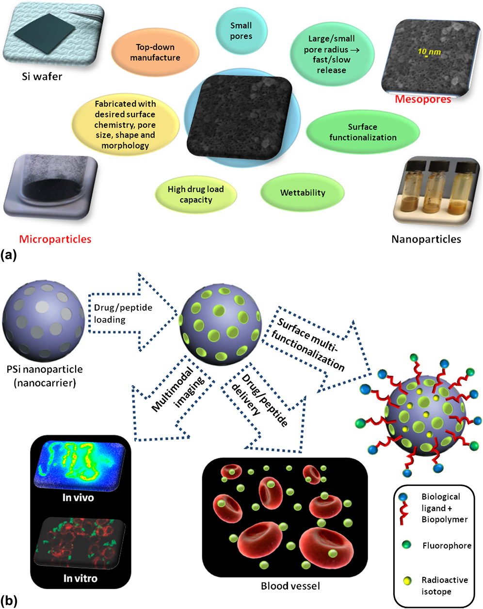
Nanoparticles offer diagnostic and therapeutic capabilities that are not available with small or microscale tools. Therefore, the ideal theranostic nanoparticle system should comprise several features such as: (i) being selective and rapidly accumulating in the diseased tissue; (ii) with capability to elucidate the biochemical and morphological characterization of the area of interest; (iii) having effective delivery of therapeutics; and (iv) being safe and biodegradable with nontoxic by-products. In this respect, porous silicon (PSi) materials have emerged as a promising drug delivery material when loaded with therapeutic agents due to its unique properties (Fig. 1).Reference Santos, Bimbo, Lehto, Airaksinen, Salonen and Hirvonen1, Reference Godin, Tasciotti, Liu, Serda and Ferrari3, Reference Salonen, Kaukonen, Hirvonen and Lehto26–Reference Wu, Park, Park, Segal, Cunin and Sailor36 The PSi materials have been loaded with different drug/protein molecules to modulate their solubility and to control the drug release, showing also their markedly biocompatible characteristics, concurrently serving also as diagnostic imaging tools.Reference Godin, Tasciotti, Liu, Serda and Ferrari3, Reference Tasciotti, Godin, Martinez, Chiappini, Bhavane, Liu and Ferrari37–Reference Canham, Kluczewska, Barley and Varajao50 For example, the PSi quantum sponge structureReference Bisi, Ossicini and Pavesi51 provides tunable photonic properties for biomedical diagnostics,Reference McInnes and Voelcker34, Reference Cunin, Schmedake, Link, Li, Koh, Bhatia and Sailor52 and infrared photoluminescence. This feature has allowed PSi vectors to be exploited to assess their fate on systemic administration.Reference Park, Gu, von Maltzahn, Ruoslahti, Bhatia and Sailor39 Recently, a multistage PSi-based vector has been introduced as a versatile delivery platform for bioactive materials,Reference Tasciotti, Godin, Martinez, Chiappini, Bhavane, Liu and Ferrari37, Reference Tasciotti, Liu, Bhavane, Plant, Leonard, Price, Cheng, Decuzzi, Tour, Robertson and Ferrari38 with the possibility to be used as an in vivo imaging tool when conjugated with a dye onto the surface of PSi. Radiolabeling of the PSi surface has also been demonstrated to be efficient enabling the follow up of the particles during biodistribution studies in rats.Reference Sarparanta, Bimbo, Makila, Salonen, Laaksonen, Helariutta, Linder, Hirvonen, Laaksonen, Santos and Airaksinen41, Reference Bimbo, Sarparanta, Santos, Airaksinen, Makila, Laaksonen, Peltonen, Lehto, Hirvonen and Salonen42
List of the most relevant properties of the nanostructured PSi carriers. (a) Si wafer and PSi powder-based microparticles (left), mesoporous structure (∼10 nm) and PSi solution-based nanoparticles (right). Other properties emphasized are: the tunable pore sizes, yet large or small enough to allow a fast or slow drug release; the possibility to functionalize the surface of PSi materials for targeted therapy; the PSi nanocarriers can be hydrophilic (or hydrophobic) enhancing the wettability properties; considerable amounts of therapeutic compounds can be loaded inside the PSi nanopores; the nanostructured PSi fabrication can be finetuned to produce a certain surface chemistry, pore size and shape, and morphology of the material; due to its top-down manufacture, the nanostructured PSi can be easily scaled-up. (b) Schematic representation of a spherical-shaped nanostructured PSi carrier and its potential in drug delivery, cancer therapy and bioimaging: (i) first the therapeutic compounds (drug/peptide) are loaded into the pores of nanostructured PSi; (ii) the surface of PSi nanocarriers can then be modified (functionalized) with different biological ligands and polymers, labeled with fluorophores and/or radioactive isotopes for noninvasive imaging applications; and (iii) the PSi carriers can then travel in the bloodstream and release the therapeutic compounds in the vicinity of unhealthy cells or tissues and simultaneously provide a real-time monitoring of its actions.Reference Wu, Andrew, Cheng, Freeman, Pearson and Sailor25

In this mini-review, we highlight the recent innovative diagnostic imaging aspects of PSi materials and emphasize their potential as a theranostic platform and tool for the clinic. We first address the existing techniques for imaging in a more clinical-oriented fashion and then outline the multiple biomedical imaging applications of the PSi materials, functionalized with imaging agents, to promote diagnostic function such as fluorescence imaging/optical imaging, NIR, MRI, and nuclear imaging techniques including SPECT, PET, CT, and radiotracers.
II. IMAGING MODALITIES AND PROPERTIES
The emergence of nanosystems is expected to convey substantial benefits to the field of cancer therapy,Reference Ferrari8 among others, as it offers a less invasive alternative when compared with the conventional therapeutic strategies, thereby enhancing both the expectancy and quality of life of the patients. The possibility of loading a therapeutic compound within a labeled nanocarrier, multiplies manifold the benefits obtained as compared with a situation when only one of these strategies is used at a time. Specially designed nanodelivery systems that can overcome the different physiological barriers are actively sought after and are expected to yield substantial benefits for personalized therapy. Although the 21st century has seen tremendous strides in diagnostics and imaging methods, new materials are intensively studied for fulfilling the overwhelming need of a safe, effective, flexible and affordable platform for drug delivery applications.
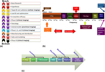
Biomedical imaging is one of the cornerstone activities in comprehensive cancer care and has several advantages including real-time monitoring, accessibility without tissue destruction, minimal or no invasiveness, and functioning over wide ranges of time- and size scales involved in the biological and pathological processes. Diagnostic systems are expected to provide early detection, enhanced signal, targeted and active (detection of biological activity) diagnosis. This is attainable by the conjugation of structural imaging of the morphology or anatomy, functional imaging of the organ and cancerous tissues, along with molecular imaging of the biological processes. Figure 2 presents the in vitro, cellular, preclinical and clinical imaging techniques used in the various phases of drug discovery and the relative frequencies of electromagnetic radiation.Reference Fass4 Imaging systems vary in physical properties including sensitivity, temporal and spatial resolution. For example, most clinical imaging systems are based on the interaction of electromagnetic radiation with body tissues and fluids. High frequency electromagnetic radiation using gamma rays, x-rays or ultraviolet light is ionizing and can cause damage to the human body.
(a) Different imaging steps taking place during pharmaceutical drug discovery (arrows indicate transitions from bench to bedside and vice-versa). (b) Frequency spectrum of electromagnetic radiation imaging techniques. (c) Relative sensitivity of different imaging technologies: x-ray, CT, and ultrasound can provide anatomic imaging; MRI, PET, and other optical techniques can further provide information on physiological and biochemical mechanisms; and nanosystems can further be used for imaging of biological systems. Reprinted and adapted from Ref. Reference Fass4. Copyright © (2008) Elsevier B V.

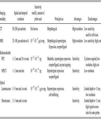
The relative sensitivity and resolution of the different imaging technologies are presented in Table I. PET and nuclear medicine are the most sensitive clinical imaging techniques (sensitivity between nanomol/kg and picomol/kg), whereas MRI and x-ray systems (including CT) have sensitivities of approximately 10 millimol/kg and 1 millimol/kg, respectively.Reference Fass4, Reference Massoud and Gambhir14 When considering cancer research, preclinical fluorescence and bioluminescence-based optical imaging systemsReference D’Hallewin, El Khatib, Leroux, Bezdetnaya and Guillemin53, Reference He, Wang, Hartmann, Cheng and Low54 are routinely used today. However, fluorescent dyes are not a preferred option for clinical imaging due to the problems related to tissue autofluorescence and deep tissue photon penetration and absorption.
Characteristics of some of the in vivo imaging modalities. Adapted from Ref. Reference Massoud and Gambhir14.

In addition, cancer is a multifactorial disease and imaging needs to be able to demonstrate the various mechanisms and phases of pathogenesis. The use of different imaging modalities, various imaging agents and various biomarkers is, therefore, required to achieve a diagnostic procedure as accurately as possible. This also means that the combination of independent and uncorrelated imaging technologies might be needed. The currently performed in vivo imaging modalities differ in both their sensitivity and resolution (Table I). Optical imaging, which includes fluorescence and bioluminescence detection, and radionucleotide-based imaging (PET and SPECT) are both very sensitive, but typically have a resolution greater than several millimeters, which can limit their use especially in small animal models, in which higher resolution is more informative.Reference Fass4, Reference Kelkar and Reineke9, Reference Massoud and Gambhir14 In addition, optical imaging is also restricted to a depth of only several centimeters, and quantification of the signal can be problematic due to the significant tissue absorption of the signal. On the other hand, MRI and CT are much less sensitive with a resolution of less than 1 mm. MRI can image deep tissues and has the added advantage of not exposing the patients to radiation, but data acquisition is slow compared with the other approaches. Combining imaging systems, such as PET/CT, SPECT/CT and in the future the combination of systems using for example, PET and MRI, will indeed improve the diagnostics detection capacity and will allow a better therapeutic response.
With the aim of improving diagnostics for efficient therapy, nanoplatforms have been developed for enhancing tissue contrast or for allowing the identification of specific biological changes. There are several advantages of using nanoparticles as imaging probes due to their unique physicochemical properties and, in some cases, their apparent lack of toxicity. These include the surface area-to-volume ratio and effective surface functionality, which can be used for finetuning the particles for tissue or cell targeting in vivo. Nanoparticles that bind with high affinity to specific receptors can be used to monitor receptor density in real-time. Probes are usually used in molecular imaging and they are required for monitoring the physiological or biochemical changes in vivo. These probes are required to have some property that promotes their accumulation at the site of interest and that allows them to be imaged. The nanotherapeutic and imaging approaches to cancer detection comprise a rational design of nanosystems for their intended action by combining the imaging and therapy in different clinical situations. Therefore, some important aspects have to be addressed regarding the effects of the nanotherapeutic drugs on the pharmacokinetics, biodistribution, and side-effects. When associated or conjugated with a ligand or carrier, one should clarify the effects on the nanoparticle safety profile, on the accumulation and biodegradation after administration, as well as on the side-effects that can result from the ability of the nanoparticles to cross the blood–brain barrier. For being efficient and for overcoming the sensitivity or resolution limitations, the nanoparticles for imaging purposes have to avoid the hostile environments encountered in vivo. These include the interactions with plasma proteins and the recognition by the phagocytic cells and consequent removal from the systemic circulation by the mononuclear phagocyte system (earlier known as the reticuloendothelial system), together with the potential acute and chronic toxicity of the particles themselves.
Multi imaging modalities can facilitate the tracking of nanoparticles both in vitro and in vivo. For monitoring the therapy and response at the cellular level, fluorescent-labeling agents can be used to achieve high-resolution images delineating different organelles inside the cell. Fluorophores are commonly used in the in vitro studies. In vivo, PET or SPECT agents and MRI contrast agents are usually preferred to overcome problems of background fluorescence and photobleaching.Reference Fass4 These in vitro imaging agents are important during the early drug development, as well as in advanced studies that may be promising for clinical trials. However, the imaging modalities above have still several limitations and only with a proper design of the nanoimaging systems can possibly overcome some of the problems faced in the diagnostic detection.
III. PSi AS A DIAGNOSTIC TOOL: PRECLINICAL APPLICATIONS
There is a plethora of recent literature describing the therapeutic applications of PSi materials,Reference Santos, Bimbo, Lehto, Airaksinen, Salonen and Hirvonen1, Reference Godin, Tasciotti, Liu, Serda and Ferrari3, Reference Anglin, Cheng, Freeman and Sailor27, Reference Serda, Godin, Blanco, Chiappini and Ferrari55 and thus, these aspects will not be reviewed herein. The aim of this mini-review is to illustrate some typical cases on the diagnostic potential of the PSi materials for clinically oriented applications.
A. Optical/light imaging
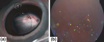
Imaging can be applied to follow the particle-mediated drug delivery and targeting in vivo. The optical properties of PSi have been investigated for numerous applications as a result of its tunable photonic properties.Reference Cunin, Schmedake, Link, Li, Koh, Bhatia and Sailor52 PSi shows a distinctive particle spectrum that can be observed through human tissue,Reference Cheng, Anglin, Cunin, Kim, Sailor, Falkenstein, Tammewar and Freeman40, Reference Li, Cunin, Link, Gao, Betts, Reiver, Chin, Bhatia and Sailor56 which can be used to monitor the loading and release of various organic molecules or biomolecules. Using this approach, Cheng et al. monitored the molecular loading and release of drugs over time in ophthalmic applications and highlighted the intraocular properties of the PSi particles as a platform for acting as a self-reporting drug delivery systems in living animals (Fig. 3).Reference Cheng, Anglin, Cunin, Kim, Sailor, Falkenstein, Tammewar and Freeman40
(a) Photograph of the rabbit eye after intravitreal injection of hydrosilylated PSi particles seen as suspended in the center of the vitreous. (b) Fundus photograph of the particles 3 mo after the injection seen as dispersed in the vitreous, showing a distinctive blue color corresponding to partial degradation and dissolution. Reprinted with permission from Ref. Reference Cheng, Anglin, Cunin, Kim, Sailor, Falkenstein, Tammewar and Freeman40. Copyright © (2008) BMJ Publishing Group Ltd.

B. NIR
Current clinical detection and diagnosis techniques such as PET, x-ray, radiography, CT, and MRI, have insufficient specificity and sensitivity with a potential risk of radioactivity in some cases.Reference Fass4 To overcome this problem, optical imaging has emerged as a promising high-resolution, nonionizing, noninvasive and real-time diagnosis technique, providing molecular information of biological tissues based on scattering, absorption, transmission, fluorescence and reflection properties of the tissue constituents.Reference Choy, Choyke and Libutti57 Among different optical imaging technologies, the development of theranostic agents for simultaneous NIR fluorescence imaging and drug targeting is anticipated to be appealing for imaging of complicated sample matrices. This is expected to considerably affect the future of cancer diagnosis and therapy owing to the high tissue penetration depth of the NIR fluorescence technique.
PSi also exhibits visible photoluminescence at room temperature originating from quantum confinement effects,Reference Canham58 and under excitation the PSi emits in the NIR region.Reference Tasciotti, Godin, Martinez, Chiappini, Bhavane, Liu and Ferrari37, Reference Jurbergs, Rogojina, Mangolini and Kortshagen59, Reference Hong, Lee, Son, Hong and Lee60 This remarkable property has made possible the development of silicon (Si) nanocrystal quantum dots (QDs) capped by functional organic molecules that passivate and protect the Si particles from oxidation, enabling the particles to be used as optical probes in hydrophobic and hydrophilic environments.Reference Tilley and Yamamoto61 This is a crucial prerequisite for in vivo fluorescence imaging, because at lower wave lengths the absorption and autoluminescence of the surrounding tissues can severely limit signal-to-background ratio of the detection.
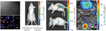
Park et al. took advantage of this PSi property and were the first to demonstrate the imaging of a tumor and other organs using biodegradable PSi nanoparticles in live animals.Reference Park, Gu, von Maltzahn, Ruoslahti, Bhatia and Sailor39 PSi nanoparticles below 200 nm in size with intrinsic luminescent (emitting in the NIR region, 810–875 nm, and excited with red or NIR radiation, 615–665 nm or 710–760 nm, or by two-photon NIR excitation) showed a significant luminescence in HeLa cells using excitation wave lengths of 370, 488, and 750 nm (two-photon excitation) 2 h after incubation. After intravenous, subcutaneous and intramuscular administration of the luminescent PSi nanoparticles, the whole-body fluorescence imaging of nude mice was obtained using a fluorescence mode (green fluorescent protein excitation filter, 445–490 nm and indocyanine green emission filter, 810–875 nm) (Fig. 4). Using a similar approach, Tasciotti et al. have also demonstrated the accuracy of in vivo NIR imaging as a tool for evaluation of PSi nanovector biodistribution.Reference Tasciotti, Godin, Martinez, Chiappini, Bhavane, Liu and Ferrari37
In vitro, in vivo, and ex vivo fluorescence imaging of luminescent PSi nanoparticles. From left to right: HeLa cells treated with PSi for 2 h (red and blue indicate the particles and cell nuclei, respectively; 20 μm); fluorescence image of PSi nanoparticles injected subcutaneously and intramuscularly on each flank of a mouse and after 8 h post intravenous injection without (upper picture) and with (lower picture) surface modification (arrows indicate the spleen); and in vivo image showing the injected PSi particles in the bladder (Bl) and liver (Li), respectively. Reprinted and modified with permission from Ref. Reference Park, Gu, von Maltzahn, Ruoslahti, Bhatia and Sailor39. Copyright © (2009) Macmillan Publishers Ltd.

The fluorescence spectrum of the PSi nanoparticles enabled imaging in the NIR-emission range, which is a convenient window for the in vivo imaging owing to the low levels of NIR autofluorescence of mouse skin excited with visible light.Reference Gao, Cui, Levenson, Chung and Nie22 The whole-body fluorescence imaging showed a different pattern of the PSi nanoparticles’ uptake by the clearance-related organs, enabling a noninvasive monitoring of their biodistribution and degradation in the mice animal as well as the microscopic observation of their localization in the organs. For detecting and imaging of deep-tissue diseases, such as tumors, by fluorescence, the excitation (single- or two-photon) for the nanoparticle should be in the NIR range to maximize tissue penetration and minimize optical absorption by physiologically abundant species.Reference Weissleder62 Furthermore, imaging with shorter excitation wave lengths (blue or green filter sets) resulted in poor differentiation of the target organ relative to the surrounding skin area.
C. MRI
MRI is a noninvasive imaging technique based on the basic principles of nuclear magnetic resonance. This technique has an excellent soft tissue contrast property, providing 3D images with high spatial resolution, which is dependent on the monitoring of two independent relaxation processes, namely longitudinal relaxation (T1-recovery) and transverse relaxation (T2-decay).Reference Aime, Castelli, Crich, Gianolio and Terreno63–Reference Zhou, Li, Yang, Zhu and Yang66 Comparing some representative imaging modalities, such as PET, CT, SPECT, and MRI, the latter displays prominent merits of the noninvasive diagnostic, superb spatial/anatomical resolution, and the capacity for quantitative evaluation of disease pathogenesis.Reference Terreno, Castelli, Viale and Aime67–Reference Jun, Lee and Cheon69 Despite the aforementioned considerable advantages, high toxicity and limited sensitivity of MRI’s contrast agents, which may be needed to enhance the appearance of tumors, are considered drawbacks on its rapidly increasing clinical applications.Reference Terreno, Castelli, Viale and Aime67, Reference Viswanathan, Kovacs, Green, Ratnakar and Sherry70 In recent years, this deficiency has been partly removed by new progresses in organic/inorganic synthetic chemistry, which have opened up new avenues for developing of new contrast agents including T1-positive agents of paramagnetic materials. T1-positive agents produce hyperintense regions on the MR image and T2-negative agents of superparamagnetic species are also capable of producing hypointense regions on the MR image. Unfortunately, these improvements are still to be recognized along with some limitations, which restrict their extensive clinical applications. For example, in spite of T2-signal decreasing effect of superparamagnetic nanoparticles (T2-negative agents) like iron oxide nanocrystals, the reduced resolution for the hypointense regions present in many lesions and the low degradability and high susceptibility are important limitations of the technique, which need to be meticulously taken into account when designing ideal theranostic systems.Reference Yu, Jeong, Park, Park, Kim, Min, Kim and Jon71, Reference Berret, Schonbeck, Gazeau, El Kharrat, Sandre, Vacher and Airiau72 In addition, despite good resolution, one of the major problems with the conventional MRI is the dependency of image contrast on large magnets to align proton spins.
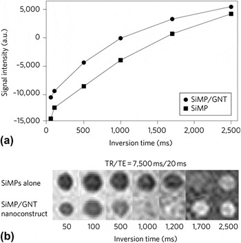
One important limitation of the in vivo fluorescence imaging technique is that it is not fully quantitative due to the signal attenuation in tissue, especially when the object is located deep in the body.Reference Ntziachristos, Bremer and Weissleder73 Thus, the in vivo optical imaging is limited to small laboratory animals and other imaging techniques need to be considered for large animals or the human body. In this case MRI, a translational imaging modality that can be used especially for tracing targeted particles, should be considered. MRI is currently one of the most powerful noninvasive diagnostic imaging techniques. The majority of clinically used MRI contrast agents are based on Gd3+ ions, which are highly toxic in a free form and, thus, have to be chelated. By confining gadolinium contrast agents inside the nanoporous structure of microfabricated PSi,Reference Tasciotti, Liu, Bhavane, Plant, Leonard, Price, Cheng, Decuzzi, Tour, Robertson and Ferrari38 the T1 contrast of these agents was improved (Fig. 5).Reference Ananta, Godin, Sethi, Moriggi, Liu, Serda, Krishnamurthy, Muthupillai, Bolskar, Helm, Ferrari, Wilson and Decuzzi74, Reference Serda, Mack, Pulikkathara, Zaske, Chiappini, Fakhoury, Webb, Godin, Conyers, Liu, Bankson and Ferrari75 Magnevist® (Bayer Schering Pharma, Berlin-Wedding, Germany), gadofullerenes, and gadonanotubes were loaded inside the pores of quasihemispherical and discoidal PSi particles, and the nanoconstructed combinations boosted the longitudinal proton relaxivity (r1) with: Magnevist®, r1 ≈ 14 mM−1s −1/Gd3+ ion (∼8.15 × 10+7 mM−1s −1/construct); gadofullerenes, r1 ≈ 200 mM−1s−1/Gd3+ ion (∼73 × 10+9 mM−1s−1/construct); gadonanotubes, r1 ≈ 150 mM−1s−1/Gd3+ ion (∼23 × 10+9 mM−1s−1/construct). These relaxivity values were about 4–50 times larger than those of clinically available gadolinium-based agents (∼4 mM−1s−1/Gd3+ ion). Such an enhancement in contrast was attributed to the geometrical confinement of the agents in the PSi nanopores, which influenced the paramagnetic behavior of the Gd3+ ions. The geometrical confinement altered the original parameters, such as the number of fast-exchanging water molecules within the inner sphere, the characteristic tumbling time of the agent, the characteristic water proton residence lifetime of the inner-sphere water molecules, and the diffusion correlation time. The change in the parameters were attributed to PSi nanostructures and to their ability to reduce the contrast agents to tumble, to decrease the mobility of the water molecules, and to favor the clustering and mutual interactions among the loaded contrast agents.
MRI characterization using a clinical scanner of (a) the signal intensity compared between PSi microparticles (SiMP) alone and PSi/gadonanotubes (SiMP/GNT) nanoconstruct, and (b) the inversion recovery phantoms for SiMP and SiMP/GNT nanoconstruct, clearly showing faster recovery for the nanoconstruct. The H-SiMP/GNT construct showed a significantly lower inversion time (T inv ≈ 1200 ms) compared with empty SiMPs (T inv ≈ 1700 ms), demonstrating that the contrast enhancement efficacy is due to the GNTs within the H-SiMP/GNT nanoconstruct. Reprinted with permission from Ref. Reference Ananta, Godin, Sethi, Moriggi, Liu, Serda, Krishnamurthy, Muthupillai, Bolskar, Helm, Ferrari, Wilson and Decuzzi74. Copyright © (2010) Macmillan Publishers Ltd.

Superparamagnetic iron oxide nanoparticles (SPIONs) are also widely used as MRI T2 contrast agents, which are applied in clinical oncology in the localization and identification of metastatic hepatic tumors.Reference Stark, Weissleder, Elizondo, Hahn, Saini, Todd, Wittenberg and Ferrucci76 SPIONs have a greater impact on transverse spin−spin relaxation (T2), leading to areas of negative contrast. An alternative approach to increase the magnetic saturation is to create clusters of smaller nanoparticles in such a way that it will induce dipole–dipole coupling. This allows for the creation of superparamagnetic cores with substantial MRI signal enhancement.Reference Canham58, Reference Jurbergs, Rogojina, Mangolini and Kortshagen59 Kinsella et al. prepared PSi nanoparticles (irregular-shaped of average size of 180–220 nm) with pore morphology (∼16 nm in pore diameter) that facilitated the proximal loading and alignment of Fe3O4 nanoparticles and have characterized them also with the MRI.Reference Kinsella, Ananda, Andrew, Grondek, Chien, Scadeng, Gianneschi, Ruoslahti and Sailor77 The anisotropic pore morphology in the PSi nanoparticles provided a host matrix that allowed control over the clustering of the Fe3O4 nanoparticle guests of 25% by mass, yielding increased magnetization and enhanced transverse relaxivity with a maximal T2 value of 556 mM Fe−1s−1 as a result of the greater magnetic strength of the interacting Fe3O4 nanoparticles in the PSi matrix.Reference Kinsella, Ananda, Andrew, Grondek, Chien, Scadeng, Gianneschi, Ruoslahti and Sailor77
D. PET/SPECT, autoradiography, and CT
PET and SPECT are noninvasive functional imaging techniques within the so-called nuclear medicine that produce a 3D-image of the functional processes in the body as a result of the gamma rays emitted indirectly by a positron-emitting radionuclide (tracer).Reference Hamoudeh, Kamleh, Diab and Fessi78 These gamma photons are used to reconstruct a 3D-image, which shows the distribution of the radiotracer in the subject’s body.Reference Debbage and Jaschke79–Reference Vallabhajosula, Solnes and Vallabhajosula81
PET is a fully translational, highly sensitive and qualitative in vivo imaging modality offering important information on physiological, biochemical, and pharmacological processes in living subjects.Reference Tu, Ma, House, Kauzlarich and Louie82 This technique is widely used in drug development, evaluation and in clinical diagnostics in the fields of oncology and neurology.Reference Atri18, Reference Choy, Choyke and Libutti57, Reference Pysz, Gambhir and Willmann83 Combining PET with high-resolution CT provides a better localization of the detected events.Reference Debbage and Jaschke79 PET requires the use of specific radiolabeled probes in picomolar concentrations with, usually, a short-lived positron-emitting radionuclide [e.g., carbon-11 (∼20 min) and fluorine-18 (∼110 min)].Reference Li and Conti80
SPECT is also based on the use of radiotracers labeled with a radionuclide. However, unlike PET, SPECT detects single photons with different energy emitted by the radiotracer. It is the most widely used nuclear imaging technique in clinical practice, but most clinically useful SPECT tracers are of limited interest for molecular and cellular imaging research applications.Reference Lecchi, Ottobrini, Martelli, Del Sole and Lucignani84
Autoradiography, a technique using x-ray film to visualize molecules or fragments of molecules that have been radioactively labeled, can also be used for imaging purposes in clinics, although it is a much less sensitive technique than PET or SPECT for 3D localization of the radiation. Nevertheless, autoradiography is often used in combination with microPET for imaging in small animals.
The ideal radiotracer should present the following characteristics: an adequate chemistry to label the biologically active compounds; appropriate energy to be detected by the imaging device; sufficient half-life to accomplish the radiolabeling process; easy production; and high target and fast blood clearance to minimize background radioactivity.Reference Fass4, Reference Massoud and Gambhir14 For use in tumor imaging, the radiotracer should be highly uptaken by tumors and be able to target to the biomarkers present in the majority of human cancer lesions.Reference Li and Conti80, Reference Vallabhajosula, Solnes and Vallabhajosula81, 85
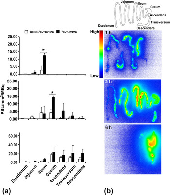
Fluorine-18 (18F) is the most commonly used and ideal radionuclide for PET, because of its ∼110 min half-life, large enough to accomplish synthesis, transportation, and imaging; and because of its low positron energy that minimizes the amount of radiation doses for the patient and ease of production. With the idea of possible application of PSi materials with PET imaging, thermally hydrocarbonized PSi nanoparticles (∼142 nm in size) were successfully labeled using 18F for assessing the biodistribution of PSi particles after enteral and parenteral administration in rats.Reference Sarparanta, Bimbo, Makila, Salonen, Laaksonen, Helariutta, Linder, Hirvonen, Laaksonen, Santos and Airaksinen41, Reference Bimbo, Sarparanta, Santos, Airaksinen, Makila, Laaksonen, Peltonen, Lehto, Hirvonen and Salonen42 The biodistribution of nanosized 18F-labeled thermally hydrocarbonized porous silicon ([18F]THCPSi) nanoparticles after oral, subcutaneous and intravenous administration in rats confirmed that the 110 min half-life of the 18F was adequate for determining the blood circulation life-time of the labeled particles and for the tracking of the particles through the gastrointestinal (GI) tract of rats (Fig. 6). The radiolabeled nanosystem exhibited excellent in vivo stability due to the fluorine’s high affinity to Si, and was even able to display a clear distinction in the biodistribution of functionalized and nonfunctionalized PSi nanoparticles, which renders the PSi-based material a flexible platform for further functionalization and development for in vivo imaging purposes.Reference Sarparanta, Bimbo, Makila, Salonen, Laaksonen, Helariutta, Linder, Hirvonen, Laaksonen, Santos and Airaksinen41
(a) Distribution of protein-coated (hydrophobin; HFBII)-18F-THCPSi and non protein-coated 18F-THCPSi labeled nanoparticles in the rat lower GI tract as a function of time quantified from macroautoradiography. The results are expressed as units of photostimulable luminescence (PSL) per mm2 normalized to the administered dose of 18F-radiolabeled particles in MBq. (b) Representative macroautoradiographs of the rat GI tract at 1, 3, and 6 h after oral administration of the HFBII-18F-THCPSi nanoparticles are also shown. A schematic diagram on top shows the arrangement of the intestines in the autoradiography cassette. Reprinted and modified with permission from Ref. Reference Sarparanta, Bimbo, Makila, Salonen, Laaksonen, Helariutta, Linder, Hirvonen, Laaksonen, Santos and Airaksinen41. Copyright © (2012) American Chemistry Society.

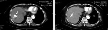
Another widely used imaging technique in clinic is CT, a special x-ray equipment that in some cases also uses a contrast material to produce multiple images or pictures of the inside of the body. This technique can be used to diagnostic, e.g., liver tumors.Reference Goh, Chung, Lo, Lau, Yu, Chng, Satchithanantham, Loong, Ng, Lim, Connor and Chow86 CT imaging also provides excellent anatomic information. In this respect, in 2007, Goh et al. reported the first-in-man study and determine the safety profile and therapeutic efficacy of a phosphorous (32P) brachytherapy device [BrachySil® (pSivida Corp., Watertown, MA) comprising PSi particles of 30 μm in size] in patients with unresectable hepatocellular carcinoma.Reference Goh, Chung, Lo, Lau, Yu, Chng, Satchithanantham, Loong, Ng, Lim, Connor and Chow86 BrachySil device was implanted in the patients directly into liver tumors under ultrasound and CT guidance, at an activity level of 4 MBq/cc of tumor. All targeted tumors have responded at 12 wk. Percutaneous implantation of 32P brachytherapy device into hepatocellular carcinoma was demonstrated to be safe and well tolerated, inducing a reduction in tumor volume (16–100%) of target tumors in all 8 patients at 12 wk postimplantation (Fig. 7). A further reduction in tumor volume was also seen in 4 of 5 patients (43–100%) after 24 wk. In addition to liver cancer treatment, PSi-based devices are also currently in clinical trial phase III for the treatment of pancreatic cancer.87
CTs showing a significant tumor response over a 12-wk (right CT) period after implantation of 32P-BioSilicon system comprising PSi microparticles. Reprinted with permission from Ref. Reference Goh, Chung, Lo, Lau, Yu, Chng, Satchithanantham, Loong, Ng, Lim, Connor and Chow86. Copyright © (2007) Elsevier B V.

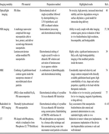
An overall summary of the key studies described above on the application of the different imaging techniques using PSi-based materials are presented in Table II.
Summary of the key studies described in this mini-review on the different imaging techniques using PSi-based materials for biomedical applications.

VI. CONCLUDING REMARKS
The emergence of nanosystems is expected to result in substantial benefits to the field of cancer therapy. PSi is an acceptable biomaterial with several advantageous features that render it an efficient drug carrier and imaging agent. The great advantages of the nanostructured PSi materials are: the good biocompatibility, biodegradability, high pore volume necessary for hosting large amounts of therapeutics, tunable pore sizes for fine-control of drug loads and release kinetics, high surface area for drug adsorption, easy surface chemistry modification for further biofunctionalization, and control of drug loading and release (Fig. 1). One can envisage that future generations of nanostructured PSi-based theranostic tools will effectively improve the transport of drugs to the targeted areas with simultaneous real-time imaging, without damaging the healthy cells. Nanostructured PSi-based materials can be strictly designed for specific applications, with a proper response and, in the future, also for the delivery of drug dosages according to the clinical needs of the patient and pathology. The surface of the nanostructured PSi materials provides a suitable platform for covalent conjugation and electrostatic attachment of a vast spectrum of dyes, fluorophores, fluorescent tags, radioactive molecules, and other imaging contrast agents. The versatility of the nanostructured PSi platform and its emerging properties will also enable the creation of personalized solutions with broad clinical implications within and beyond the realm of cancer theranostics.
In this mini-review, we have presented few examples of the potential advantages of using the PSi materials as nanostructured imaging platforms that aid the design of new generations of imaging contrast agents and their potential application in clinical imaging. These PSi tools comprising multimodal imaging agents hold considerable promise as the next generation of medicine that enables the early detection of disease, simultaneous monitoring and treatment with minimal toxicity, aiding to overcome the specific limitations of sensitivity and resolution of many imaging techniques in progressing toward the clinical applications. Tracking of the nanoparticles systemically prevalidates appropriate targeting and provides real-time information on tumor response.
The current findings have provided encouraging support for further development of even more sophisticated nanostructured PSi-based materials for imaging purposes for fulfilling the great potential and promise of PSi as a multifunctional platform—a solid part of the clinical “tool box.”
ACKNOWLEDGMENTS
H.A.S. acknowledges financial support from the Academy of Finland (Decision No. 252215 and 256394) and from the Centre for International Mobility, CIMO (decision no. TM-12-8201). L.M.B. acknowledges the Finnish Cultural Foundation for financial support.