Introduction
Epistaxis is the commonest ENT emergency.Reference Small and Maran1 Between 90 and 95 per centReference Josephson, Godley and Stierna2, Reference Viducich, Blanda and Gerson3 of nosebleeds arise from the anterior part of the septum. There remains a common area of debate and uncertainty about the location of bleeding sites in cases of posterior epistaxis. Historically, some authorsReference Padgham4 have supported the idea that the septum is the commonest site of bleeding, whilst othersReference Hallberg5, Reference Jackson and Jackson6 have favoured the lateral nasal wall, commonly referring to Woodruff's plexusReference Woodruff7 as a source.
In 1949, Woodruff, using a rigid nasopharyngoscope, noticed ‘a group or plexus of rather large blood vessels in the lateral wall of the inferior wall of the inferior meatus well posteriorly’ which he termed the ‘naso-nasopharyngeal plexus’. Reference to the original article shows that Woodruff himself merely suggested an association between this plexus and epistaxis, without offering evidence to support this view. However, from this time, numerous textbooksReference Lepore and Bailey8, Reference Watkinson and Kerr9 and journal articlesReference Pearson10–Reference Thornton, Mahesh and Lang12 have asserted that Woodruff's plexus is the commonest source of bleeding in posterior epistaxis, despite the continued lack of supporting evidence. In spite of its supposed importance, the nature of Woodruff's plexus is unclear and poorly understood.
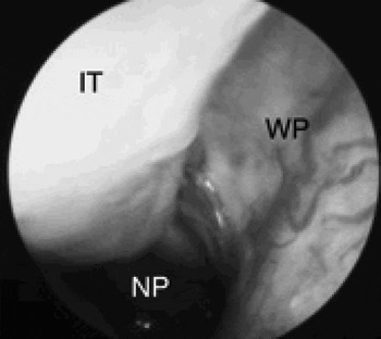
Some authorsReference Shin and Murr13, Reference Bishow14 state that the plexus is arterial; however, on endoscopy (Figure 1), it looks more likely to be venous in nature.
An endoscopic view of the posterior nasal cavity. Woodruff's plexus (WP) is easily seen in most patients. Anecdotally, it is said to be more prominent with increasing age.7 IT = inferior turbinate; NP = nasopharynx

An anatomical investigation was undertaken to test the hypothesis that Woodruff's plexus was indeed a venous plexus.
Materials and methods
A number of complementary methods were combined to establish the exact nature of Woodruff's plexus: (1) anatomical dissection and microdissection, (2) histological analysis, and (3) Spalteholz ‘clearing’ techniques.
A total of 21 cadaveric heads belonging to the anatomy department of the University of Glasgow were used in this anatomical study.
Microdissection
After normal embalming by arterial injection through the common carotid artery, seven cadavers were further injected through the same cannula with a mixture of latex and Indian ink, and another seven were left uninjected as controls. Subsequently, in all 14 specimens, the heads were divided and the area of the lateral nasal wall mucosa expected to contain Woodruff's plexus was microdissected, using a Watson Barnet binocular dissecting microscope (2.5 times magnification).
Spalteholz clearing
Three cadavers were injected with latex and Indian ink via a standard approach through the external carotid artery. A tissue block containing the nasal cavity was excised and decalcified in dilute acid solutions and then cleared through a standard multistage procedure.Reference Bancroft and Stevens16 A final, prolonged immersion in methylsalicylate (oil of wintergreen) rendered the decalcified tissues ‘transparent’. In this way, selected features of the deeper parts of the tissue block, such as the injected vessels, became clearly visible in situ.
Histology
From four cadavers, undissected mucosa from the posterior inferior meatus of one side was removed for histological analysis. From these four specimens, a total of 14 000 sections were cut at 10 µm thickness. According to standard practice, every 15th section was selected for staining (haematoxylin and eosin) and then mounted. A total of 932 sections were stained, mounted and then examined under low and high power light microscopy.
Results were recorded photographically at all stages.
Results
Microdissection
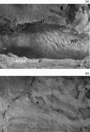
Delicate microdissection of the mucosa of the posterior inferior meatus revealed a very superficial collection of fragile and fairly large calibre (range 1.2–2.0 mm, mean 1.6 mm) vessels. These lay just beneath the surface mucosa, with little intervening connective tissue, and great care was needed to peel the surface layers away without destroying the vessels (Figure 2).
Microdissections of the inferior meatus, seen at (a) low power (×2) and (b) high power (×10). An array of superficial vessels in a configuration consistent with that of Woodruff's plexus (WP) is seen. The vessels are fairly discrete, with occasional interconnections. Smaller arterial vessels (stained black due to latex Indian ink) are seen at a deeper level. IT = cut edge of inferior turbinate; F = floor of nasal cavity (i.e. hard palate)

Spalteholz clearing
Using this technique, the vessels of a vascular plexus were demonstrated quite clearly in the posterior part of the inferior meatus. These vessels could be distinguished from the arteries, which had been injected with black latex (Figure 3).
Lateral nasal wall taken from a specimen injected with latex Indian ink then ‘cleared’ by the Spalteholz technique. Arterial vessels filled with ink are black, whilst venous vessels filled with blood appear as dark grey. Larger branches of the posterior nasal artery (0.6–1.2 mm) are diverted to the turbinates. The arteries that supply the lateral wall itself are fine branches (0.1–0.2 mm) and are few in number, lying deep to the veins. SPF = sphenopalatine foramen; WP = Woodruff's plexus; ETO = eustachian tube opening; IT = inferior turbinate

Histology
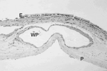
Histological analysis revealed that the epithelium overlying the posterior inferior meatus was typical respiratory epithelium, i.e. ciliated, pseudostratified, columnar epithelium with goblet cells. There were occasional secretory glands in this thin mucosa, but the most prominent features were large, discrete, thin, sinus-like blood vessels with very little muscle or fibrous tissue in their walls (Figure 4). The average vessel diameter was of the order of 1–2 mm.
Photomicrograph of section taken from inferior meatus, demonstrating the discrete, large, thin walled, superficial vessels of Woodruff's plexus. Note the thinness of the mucosa, with few structures apart from these veins. E = epithelium; WP = large, thin-walled vein of Woodruff's plexus; P = periosteum (H&E; x 50)

Discussion
One of the strengths of this study was the combination of different investigative methods in order to define a well known anatomical structure which has thus far escaped definitive characterisation. Shaheen (unpublished data) attempted to isolate Woodruff's plexus in nasoethmoidal blocks but was unsuccessful. No other articles specifically studying Woodruff's plexus exist in the English literature. Woodruff's plexus has been described as an arterial plexus formed by the anastomosis of several branches, including the pharyngeal,Reference Shin and Murr13 posterior nasal, sphenopalatineReference Bishow14 and posterior septal arteries.Reference Pope and Hobbs15 Others have suggested it to be venous, but evidence for these opinions has been absent.
Through microdissection, histological examination and clearing studies, we identified a plexus or array of large, thin walled veins in the inferior meatus, corresponding to the vessels described by Woodruff.
The association between Woodruff's plexus and posterior epistaxis was probably made simply because the plexus is so striking in appearance.Reference Yanagisawa and Citardi17 This relates to the properties of the mucosa: the mucosa is thin and relatively free of glandular or lymphoid tissue, and thus is dominated by a single layer of discrete, interconnecting veins which lie superficially enough to be visible. More anteriorly, a combination of a more complex, denser network of veins and a thicker mucosa gives a uniform pink colour. It is uncertain whether these venous vessels of Woodruff's plexus have any physiological significance beyond being a venous drainage route for the posterior part of the lateral wall. One may speculate that the thinness of the mucosa in this region represents an adaptive change to reduce obstruction at the posterior choanae.
Comment on the relationship of Woodruff's plexus with the origin of epistaxis, if any, would also be speculation. In many cases, the exact source of bleeding from the lateral wall is unclear, because the irregular contour created by the turbinates makes lateral wall epistaxis difficult to visualise and to assess regarding its source (venous or arterial). In turn, it is more difficult to treat posterior epistaxis from the lateral wall directly, and, more often than not, tamponade by nasal packing is needed. Bleeding from the wide, interconnected veins of Woodruff's plexus would be expected to result in a relatively slow but prolonged ‘ooze’ – the lack of muscle means that the injured vessels would not contract well.
• Woodruff's plexus has been frequently mentioned in the rhinological literature as a source of epistaxis, but has never been properly characterised
• This study combined a number of investigative techniques to demonstrate that Woodruff's plexus is a venous plexus
Some regard epistaxis as mainly an arterial phenomenon, although definitive proof of even this fundamental point is lacking. Epistaxis as a symptom is likely to result from a variety of causes, and will sometimes be arterial and sometimes venous. The arteries in the lateral wall are deeper and rather small in calibre; there are larger vessels supplying the nasal turbinates. It has been said that bleeding from the turbinates is less frequent because the arteries run through bony canals.Reference Burnham18 However, any tamponading or other protective effect of the bony canals is unlikely to be effective as they are generally incomplete.
Conclusions
Woodruff's plexus is a venous plexus in the posterior part of the inferior meatus. Whilst its physiological function and pathophysiological significance is uncertain, its definition as a venous plexus is important, in order to allow future discussion to be evidence-based rather than conjectural.