Introduction
Vitrification of human blastocysts is being used increasingly to cryopreserve supernumerary embryos following extended culture, especially in in vitro fertilization (IVF) cycles, in which a single embryo transfer is performed. Clinical trials have shown high survival rates and promising implantation rates following transfer of warmed blastocysts (Vanderzwalmen et al., 2002, 2003, Reference Vanderzwalmen, Ectors, Grobet, Prapas, Panagiotidis, Vanderzwalmen, Stecher, Frias, Liebermann and Zech2009; Liebermann & Tucker Reference Liebermann and Tucker2006; Mukaida et al., Reference Mukaida, Oka, Goto and Akahashi2006; Hong et al., Reference Hong, Sepilian, Chung and Kim2009; Wikland et al., Reference Wikland, Hardarson, Hillensjö, Westin, Westlander, Wood and Wennerholm2010; Coello et al., Reference Coello, Meseguer, Galán, Alegre, Remohí and Cobo2017; Youm et al., Reference Youm, Choi, Oh and Rho2017) and the existing data on the safety of vitrification in terms of obstetric and perinatal outcomes is reassuring (Mukaida et al., Reference Mukaida, Takahashi, Goto and Oka2008; Liebermann, Reference Liebermann2009; Noyes et al., Reference Noyes, Porcu and Borini2009). A recent study that looked, in addition to the survival rates, into dividing cells and the effects that vitrification may have on spindle structure and chromosome alignment suggested that vitrification at the blastocyst stage did not adversely affect overall development and the ability of most spindles to form and continue normal cell divisions (Chatzimeletiou et al., Reference Chatzimeletiou, Morrison, Panagiotidis, Vanderzwalmen, Prapas, Prapas, Tarlatzis and Handyside2012). However, in comparison with fresh embryos, vitrified embryos exhibited a significantly higher incidence of spindle abnormalities due to mechanical stress suffered by cells during exposure to high concentrations of cryoprotectants and subsequent dehydration (Chatzimeletiou et al., Reference Chatzimeletiou, Morrison, Panagiotidis, Vanderzwalmen, Prapas, Prapas, Tarlatzis and Handyside2012).
Cryopreservation of embryos with an opening in the zona following assisted hatching or biopsy for preimplantation genetic testing (PGT-A or PGT-M) represents a challenge as, in these embryos, the protective effect of the zona pellucida has been compromised. Laser zona drilling on day 3 is also often used before blastocyst biopsy to assist hatching of trophectoderm cells. Slow freezing of biopsied embryos has been previously shown to have reduced survival following thawing (Keskintepe et al., Reference Keskintepe, Sher, Machnicka, Tortoriello, Bayrak, Fisch and Agca2009). However, survival and implantation rates of vitrified blastocysts with an opening in the zona following either assisted hatching or embryo biopsy are more promising (Zech et al., Reference Zech, Lejeune, Zech and Vanderzwalmen2005; Keskintepe et al., Reference Keskintepe, Sher, Machnicka, Tortoriello, Bayrak, Fisch and Agca2009; Van Landuyt et al., Reference Van Landuyt, Verpoest, Verheyen, De Vos, Van de Velde, Liebaers, Devroey and van den Abbeel2011; Friedenthal et al., Reference Friedenthal, Maxwell, Munné, Kramer, McCulloh, McCaffrey and Grifo2018).
In this study, we investigated the effects of zona drilling and biopsy on day 3, followed by vitrification on day 5, on the cytoskeleton and development of human embryos, by analysing survival rates and spindle and chromosome configurations by fluorescence and confocal laser scanning microscopy in human biopsied and non-biopsied embryos.
Materials and methods
Source of human preimplantation embryos
In total, 98 human blastocysts were donated for research from consenting couples undergoing routine IVF/intracytoplasmic sperm injection (ICSI) treatment (n=50) and ICSI treatment with preimplantation genetic testing for aneuploidy (PGT-A), or single gene defects (PGT-M) (n=48) in the Assisted Conception Units of Biogenesis and the Iakentro Advanced Medical Centre in Greece, in which the embryos were vitrified, fixed and labelled. This work was approved by the ethical committee of the Aristotle University Medical School (licence no. A 10570).
Ovarian stimulation
Pituitary downregulation was achieved by administration of gonadotrophin-releasing hormone analogues (Suprefact, Hoechst Marrion Roussel; Arvekap, Ipsen; Cetrotide, Serono; or Orgalutran, MSD). Ovarian stimulation was performed using recombinant follicle-stimulating hormone (FSH) (Puregon, MSD or Gonal-F, Serono). Patients were monitored regularly by ultrasound and assessment of oestradiol (E2) levels. When adequate follicular development was demonstrated (≥3 follicles of 17 mm in diameter), 10,000 IU of human chorionic gonadotrophin (hCG) (Pregnyl, MSD) was administered to trigger final oocyte maturation. At 36 h after hCG administration, transvaginal ultrasound-guided egg retrieval was performed.
Oocyte retrieval and embryo culture
Oocytes were retrieved by flushing ovarian follicles with human tubal fluid (HTF) medium (Sage) incubated in 5% CO2 in air at 37°C and subsequently fertilized by conventional IVF or ICSI and cultured in SAGE or VitroLife culture medium until the day of transfer. Following transfer and vitrification of suitable embryos, spare day 5 human blastocysts were vitrified (n=50) and, following warming and culture for a further 24 h, were subjected to cytoskeletal analysis.
Embryo biopsy
Embryos from patients undergoing PGT-A (n=42) or PGT-M (n=6) were biopsied on day 3. A hole in the zona was created with the aid of a Saturn laser (RI, UK) and a single blastomere was aspirated for genetic analysis (Chatzimeletiou et al., Reference Chatzimeletiou, Morrison, Panagiotidis, Prapas, Prapas, Rutherford, Grudzinskas and Handyside2005a). All embryos were cultured up to day 5. Those embryos found to be normal following PGD/PGS were transferred on day 5 and any surplus normal embryos were vitrified for clinical purposes. All remaining spare day-5 human blastocysts that were not selected for transfer because they were diagnosed as abnormal (n=48) were vitrified and, following warming and culture for a further 24 h, were subjected to cytoskeletal analysis.
Human blastocyst vitrification
All blastocysts (50 non-biopsied and 48 biopsied) were vitrified on day 5 using either a commercial DMSO-free vitrification kit according to the manufacturer’s instructions (VitroLife) (24 non-biopsied and 23 biopsied) or increasing concentrations of DMSO/EG (5%/5–10%/10–20%/20%) (closed system VitriSafe, AstroMed-tec, Austria) (26 non-biopsied and 25 biopsied) (Vanderzwalmen et al., Reference Vanderzwalmen, Ectors, Grobet, Prapas, Panagiotidis, Vanderzwalmen, Stecher, Frias, Liebermann and Zech2009; Chatzimeletiou et al., Reference Chatzimeletiou, Morrison, Panagiotidis, Vanderzwalmen, Prapas, Prapas, Tarlatzis and Handyside2012). The blastocysts were of similar quality and were assigned randomly in the DMSO/EG or the DMSO-free group. Following warming in 1 M, 0.5 M or 0.25 M sucrose solutions, they were allowed to recover in culture for 24 h before being treated for cytoskeletal analysis. Survival was defined as full re-expansion of the blastocyst.
Cytoskeletal analysis
All blastocysts were rapidly fixed in ice-cold methanol, and immunostained with an α-tubulin antibody to visualise microtubules, acetylated tubulin (to visualise spindles, poles and mid bodies) and/or γ-tubulin (to identify spindle poles), in combination with 4′,6-diamidino-2-phenylidole (DAPI) or propidium iodide (PI) to visualise DNA (Chatzimeletiou et al., Reference Chatzimeletiou, Morrison, Panagiotidis, Vanderzwalmen, Prapas, Prapas, Tarlatzis and Handyside2012; Chatzimeletiou et al., 2005a,Reference Chatzimeletiou, Morrison, Prapas, Prapas and Handysideb).
In brief, all blastocysts were transferred into 10-μl drops of the primary antibodies under mineral oil (Sigma) and incubated at 4°C for 1 h. The blastocysts were then washed in PBS/BSA and transferred into 10-μl drops of the secondary antibodies (highly cross-adsorbed Alexa Fluor 488 or 594 conjugates (Molecular Probes), Following 1 h incubation in the secondary antibodies the blastocysts were washed in PBS/BSA and mounted on slides (BDH) in Vectashield antifade medium (Vector Laboratories, CA, USA) containing DAPI (Sigma) or PI (Sigma) under a coverslip. The coverslips were then sealed with nail varnish according to Chatzimeletiou et al. (2005b; Reference Chatzimeletiou, Morrison, Panagiotidis, Vanderzwalmen, Prapas, Prapas, Tarlatzis and Handyside2012).
All the blastocysts were analysed using a Zeiss fluorescence microscope and/or a Leica TCS-SP laser scanning confocal microscope. Standard fluorescence images were captured using the ISIS software (Metasystems) and confocal image analysis was typically accomplished by capturing a z-series stack of 1-μm thick sections encompassing the entire blastocyst (Chatzimeletiou et al., Reference Chatzimeletiou, Morrison, Panagiotidis, Vanderzwalmen, Prapas, Prapas, Tarlatzis and Handyside2012).
Classification of abnormalities in spindle/chromosome configurations
Metaphase/anaphase spindle and chromosome configurations were carefully examined and recorded for all blastocysts. The criteria for classifying spindle abnormalities were as previously described by Chatzimeletiou et al. (2005b, Reference Chatzimeletiou, Morrison, Panagiotidis, Vanderzwalmen, Prapas, Prapas, Tarlatzis and Handyside2012). For a spindle to be classified as normal, it should have had astral-shaped or fusiform poles and the chromosomes aligned at the equator. Spindles with one or two poorly defined or absent poles and misaligned chromosomes were classified in the abnormal shape category. Spindles with more than two clearly defined astral poles and the characteristic ‘Y’- or ‘X’-shaped arrangement of chromosomes were classified as multipolar. Chromosomes not aligned with the other chromosomes on the spindle (through the process of congression failure or anaphase lagging) were classified as chromosome loss (Chatzimeletiou et al., 2005b, Reference Chatzimeletiou, Morrison, Panagiotidis, Vanderzwalmen, Prapas, Prapas, Tarlatzis and Handyside2012).
Statistical analysis
Statistical analysis of the results was carried out with the SPSS 16 (SPSS Inc. Headquarters, Chicago, IL, USA) statistical package for Windows. Fisher’s exact test was used to determine whether the incidence of spindle abnormalities differed in the biopsied and non-biopsied blastocysts.
Results
Post warming survival and spindle abnormalities in non-biopsied vitrified blastocysts
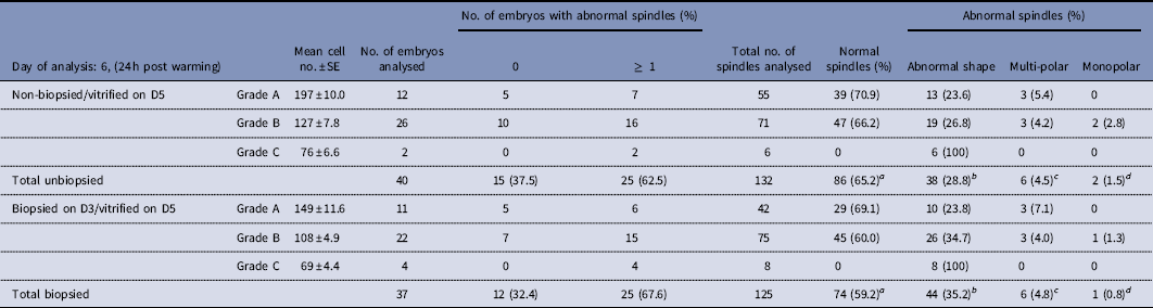
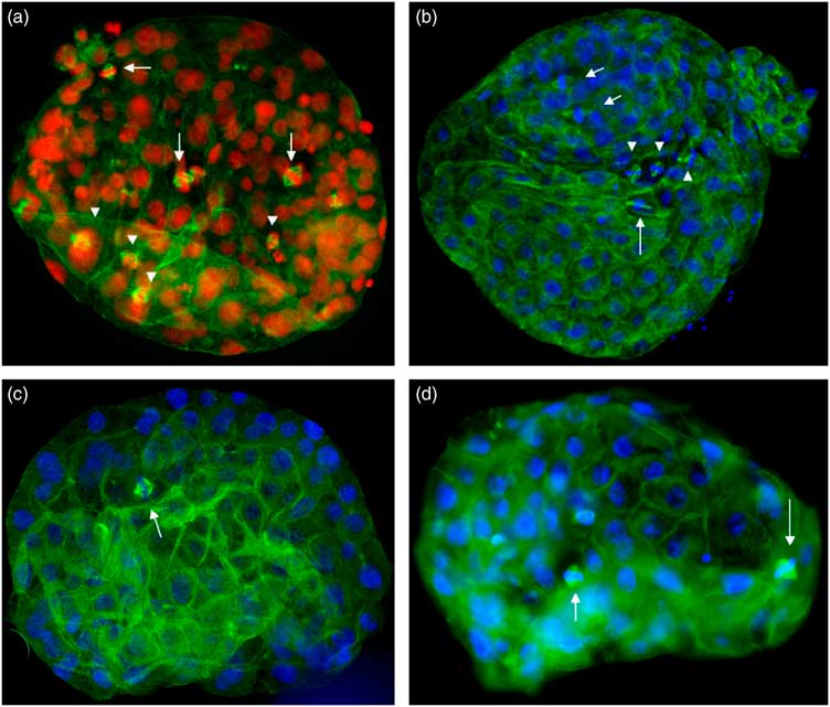
In total, 46 out of 50 (92%) human non-biopsied blastocysts that were vitrified on day 5 survived post warming. From these blastocysts 40/46 had cells with mitotic spindles and were analysed. The range of mitotic spindles was 1–8 per human vitrified blastocyst. The mean cell numbers of the grade A (n=12), B (n=26) and C (n=2) blastocysts analysed 24 h post warming, on day 6, were 197±10.0, 127±7.8 and 76±6.6 respectively (Table 1). Cytoskeletal analysis revealed that 5/12 (41.7%) of grade A blastocysts and 10/26 (38.5%) of grade B blastocysts had only normal spindles, 7/12 (58.3%) of grade A blastocysts and 12/26 (46.2%) of grade B blastocysts had at least one abnormal spindle in addition to normal spindles, while 4/26 of grade B blastocysts and all grade C blastocysts had only abnormal spindles, including abnormally shaped, monopolar and multipolar spindles, disorganised prometaphases and disorganised middle spindle fibres at telophase, chromosome lagging, congression failure and chromosome bridging. In total, 132 spindles were analysed of which 86/132 (65.2%) were normal, 38/132 (28.8%) were abnormally shaped and 6/58 (4.5%) were multipolar and 2/132 (1.5%) were monopolar (Table 1 and Figs 2a, b and 3). The distribution of spindles in the inner cell mass (ICM) and the trophectoderm (TE) was as follows: 14/86 (16.3%) normal spindles were identified in the ICM and 72/86 (83.7%) in the TE, 8/38 (21.1%) abnormally shaped spindles were in the ICM and 30/38 (79.9%) were in the TE while all the multipolar and monopolar spindles were in the TE.
Spindle abnormalities in human biopsied and non-biopsied embryos following vitrification on day 5

Blastocyst classification – grade A: top quality with well defined ICM and TE (Aa); grade B: medium quality (Bb); grade C: poor quality (Cb or Cc).
There was no significant difference in the incidence of normal and abnormal spindle chromosome configurations between the vitrified non-biopsied and the vitrified biopsied embryos a,b,c,d P>0.05).
Post warming survival and spindle abnormalities in vitrified blastocysts that were biopsied on day 3
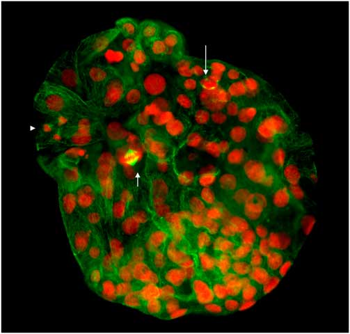
Here, 40 out of 48 (83.3%) human biopsied blastocysts that were vitrified on day 5 survived post warming and 37/40 had cells with mitotic spindles and were analysed. The range of mitotic spindles was 1–8 per human biopsied vitrified blastocyst. The mean cell numbers of the grade A (n=11), B (n=22) and C (n=4) blastocysts analysed 24 h post warming, on day 6, were 149±11.6, 108±4.9 and 69±4.4 respectively (Table 1). Cytoskeletal analysis revealed that 5/11 (45.4%) of grade A blastocysts and 7/22 (31.8%) of grade B blastocysts had only normal spindles, 6/11 (54.6%) of grade A blastocysts and 12/22 (54.6%) of grade B blastocysts had at least one abnormal spindle in addition to normal spindles, while 3/22 (13.6) of grade B blastocysts and all grade C blastocysts had only abnormal spindles. In total, 125 spindles were analysed of which 74/125 (59.2%) were normal, 44/125 (35.2%) were abnormally shaped and 6/125 (4.8%) were multipolar and 1/132 (0.8%) was monopolar (Table 1 and Figs 1 and 2c, d). The distribution of spindles in the ICM and the trophectoderm (TE) was as follows: 11/74 (14.9%) normal spindles were identified in the ICM and 63/74 (85.1%) in the TE, 6/44 (13.6%) abnormally shaped spindles were in the ICM and 38/44 (86.4%) were in the TE while all the multipolar and monopolar spindles were in the TE.
Human embryo that was biopsied on day 3, developed to the blastocyst stage on day 5 and was vitrified with the DMSO-free kit. Staining of the blastocyst 24 h post warming with α-tubulin (green) and PI (red) shows a normal astral bipolar spindle (short arrow), a tetrapolar spindle (long arrow) and a tripolar telophase (arrowhead).

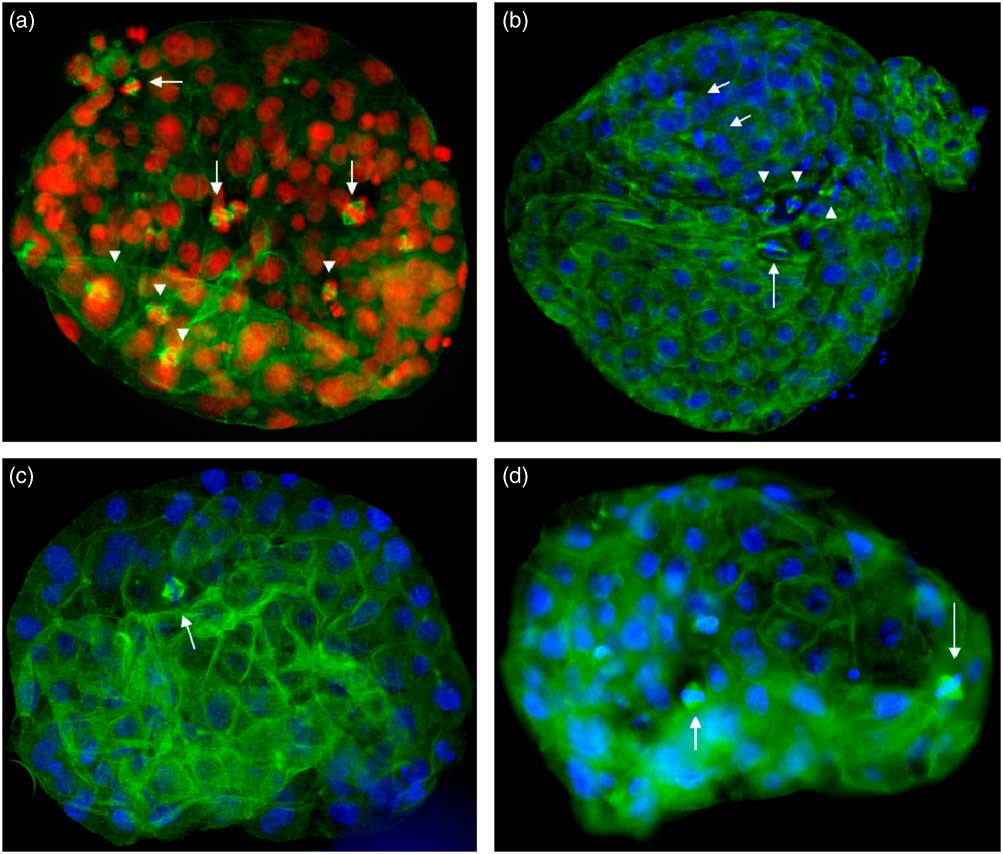
(a) Human non-biopsied blastocyst vitrified with the DMSO-free kit and stained 24 h post warming with α-tubulin (green) and PI (red). Note the normal astral bipolar spindles (arrows) and the abnormally shaped spindles with a well focussed and an unfocussed pole (arrowheads). (b) Human non-biopsied blastocyst vitrified with DMSO/EG and stained 24 h post warming with α-tubulin (green) and DAPI (blue). Note the normal astral bipolar spindles (arrows) and the abnormally shaped spindles with a well focussed and an unfocussed pole (arrowheads) Also note the elongated spindle (long arrow). (c, d) Human biopsied embryos vitrified at the blastocyst stage with DMSO/EG and stained 24 h post warming with α-tubulin (green) and DAPI (blue). Note the normal astral bipolar spindles (arrows) and the abnormally shaped spindle in (d) with a well focussed and an unfocussed pole (long arrow).
Abnormalities in biopsied vs non-biopsied vitrified blastocysts: comparison of results
The embryos assigned in the vitrified biopsied and non-biopsied group were of similar quality. Overall, the vitrified biopsied blastocysts showed lower survival post warming (83.3%) compared with the non-biopsied blastocysts (92%), but this decrease was not statistically significant (P>0.05). Cytoskeletal analysis revealed that the majority of mitotic spindles examined by confocal laser scanning microscopy in both groups had normal astral or fusiform shaped poles and were bipolar (65.2% non-biopsied vs 59.2% biopsied). The incidence of normal spindle chromosome configurations was not statistically different between the two groups (P>0.05). Specific spindle abnormalities were also observed in similar levels in the two groups (P>0.05), including abnormally shaped, monopolar and multipolar spindles, disorganised prometaphases and disorganised middle spindle fibres at telophase, chromosome lagging, congression failure and chromosome bridging. The most frequently observed type of spindle abnormality was that of a spindle with one well focussed pole and another unfocussed pole (Fig. 2a, b, d). Elongated spindles were also occasionally observed (Fig. 2c).
Post warming survival rate and incidence of spindle abnormalities in human non-biopsied embryos following vitrification with a DMSO-based and a DMSO-free kit
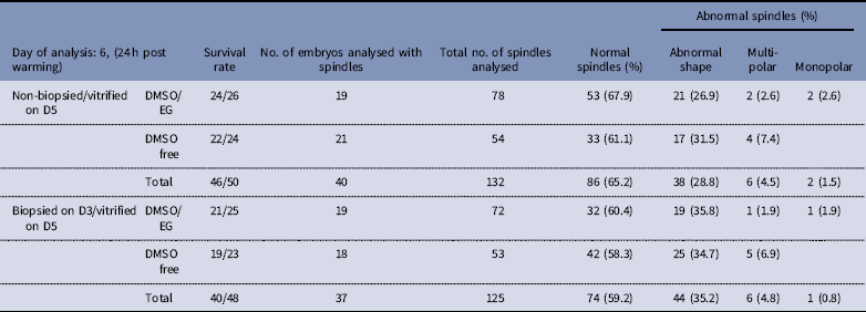
There was no significant difference in the survival rate following warming of the human non-biopsied embryos that were vitrified with DMSO/EG (24/26 92.3%) and the DMSO-free kit (22/24 91.7%). The incidence of normal and abnormal spindle chromosome configurations was also similar between the two groups (P>0.05) (Table 2); 19/24 DMSO/EG vitrified non-biopsied embryos had cells in metaphase and were analysed. In total, 78 spindles were identified of which 53 (67.9%) were normal, 21 (26.9%) were abnormally shaped 2 (2.6%) were multipolar and 2 (2.6%) were monopolar. Here, 21/22 DMSO-free vitrified non-biopsied embryos had cells in metaphase and were analysed. In total, 54 spindles were identified of which 33 (61.1%) were normal, 17 (31.5%) were abnormally shaped and 4 (7.4%) were multipolar.
Spindle abnormalities in human biopsied and non-biopsied embryos following vitrification on day 5 with the DMSO/EG or the DMSO-free kit

There was no significant difference in the survival rates and the incidence of normal and abnormal spindle chromosome configurations between the DMSO/EG and the DMSO-free vitrified embryos (P>0.05).
Survival rate and incidence of spindle abnormalities in human biopsied embryos following vitrification with a DMSO-based and a DMSO-free kit
There was no significant difference in the survival rate following warming of the human biopsied embryos that were vitrified with DMSO/EG (21/25, 84%) and the DMSO-free kit (19/23, 82.6%). The incidence of normal and abnormal spindle chromosome configurations was also similar between the two groups (P>0.05) (Table 2). 19/21 DMSO/EG vitrified biopsied embryos had cells in metaphase and were analysed. In total, 72 spindles were identified of which 32 (60.4%) were normal, 19 (35.8%) were abnormally shaped 1 (1.9%) was multipolar and 1 (1.9%) was monopolar. Here, 18/19 DMSO-free vitrified biopsied embryos had cells in metaphase and were analysed. In total, 53 spindles were identified of which 42 (58.3%) were normal, 25 (34.7%) were abnormally shaped and 5 (6.9%) were multipolar.
Distribution of γ-tubulin and acetylated tubulin
Acetylated tubulin antibodies strongly labelled the spindle poles during both metaphase and anaphase and the spindle midbodies during telophase in both biopsied and non-biopsied vitrified/warmed blastocysts. In some cases, disorganised middle spindle fibres at telophase were evident. γ-Tubulin strongly labelled the spindle poles during metaphase and anaphase and confirmed bipolarity, multipolarity or the absence of poles in certain cases.
Discussion
This study provides the first cytoskeletal analysis of human day 3 biopsied embryos that were vitrified at the blastocyst stage and compares the type and incidence of spindle abnormalities to those observed in non-biopsied vitrified blastocysts. The results suggest that there is no significant difference in the survival rate following warming between the two groups (92% non-biopsied vs 83.3% biopsied) (P>0.05). This indicates that the opening in the zona and aspiration of a single blastomere on day 3 followed by vitrification at the blastocyst stage does not adversely affect the development of human embryos. These results are supported by previous studies showing promising survival and implantation rates following transfer of warmed blastocysts (Vanderzwalmen et al., 2002, 2003, Reference Vanderzwalmen, Ectors, Grobet, Prapas, Panagiotidis, Vanderzwalmen, Stecher, Frias, Liebermann and Zech2009; Zech et al., Reference Zech, Lejeune, Zech and Vanderzwalmen2005; Liebermann & Tucker, Reference Liebermann and Tucker2006; Mukaida et al., 2006, Reference Mukaida, Takahashi, Goto and Oka2008; Hong et al., Reference Hong, Sepilian, Chung and Kim2009; Keskintepe et al., Reference Keskintepe, Sher, Machnicka, Tortoriello, Bayrak, Fisch and Agca2009; Liebermann, Reference Liebermann2009; Noyes et al., Reference Noyes, Porcu and Borini2009; Wikland et al., Reference Wikland, Hardarson, Hillensjö, Westin, Westlander, Wood and Wennerholm2010; Van et al. , 2011).
Detailed cell counts revealed that the total numbers of nuclei in the vitrified biopsied blastocysts, 24 h post warming on day 6 were slightly lower than the non-biopsied blastocysts on day 6, due to the 1/8th to 1/7th cell reduction following biopsy, but this difference was not statistically significant (P>0.05) (Table 1). These findings are in accordance with cell numbers at the blastocyst stage that have been previously reported by Hardy et al. (Reference Hardy, Handyside and Winston1989). The majority of mitotic spindles examined by confocal laser scanning microscopy in both groups had normal astral or fusiform shaped poles and were bipolar (Figs 1 and 2a–d). Specific spindle abnormalities were also observed in similar levels in the two groups with more prominent the type of spindle with one well focussed and another unfocussed pole. In general spindle abnormalities in both groups included abnormally shaped, monopolar and multipolar spindles, disorganised prometaphases and disorganised middle spindle fibres at telophase, chromosome lagging, congression failure and chromosome bridging (Figs 2 and 3). These spindle abnormalities are in agreement with previous published data (Chatzimeletiou et al., Reference Chatzimeletiou, Morrison, Panagiotidis, Vanderzwalmen, Prapas, Prapas, Tarlatzis and Handyside2012) and constitute mechanisms that may lead to postzygotic chromosomal abnormalities in early human development (Chatzimeletiou et al., 2005a,Reference Chatzimeletiou, Morrison, Prapas, Prapas and Handysideb, 2008; Ottolini et al., Reference Ottolini, Kitchen, Xanthopoulou, Gordon, Summers and Handyside2017; McCoy et al., Reference McCoy, Newnham, Ottolini, Hoffmann, Chatzimeletiou, Cornejo, Zhan, Zaninovic, Rosenwaks, Petrov, Demko, Sigurjonsson and Handyside2018). These findings also emphasise that mosaicism, which is prevalent at cleavage stages, is also evident in the trophectoderm of human blastocysts raising concern about diagnostic issues following blastocyst biopsies.
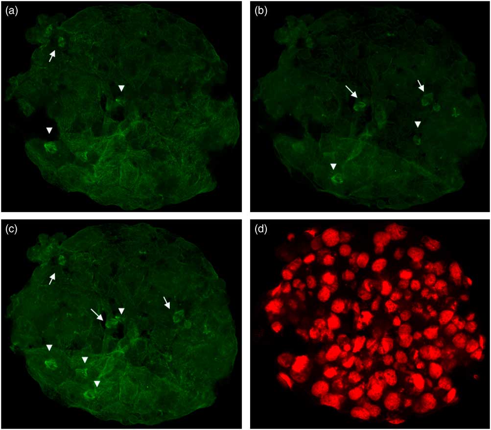
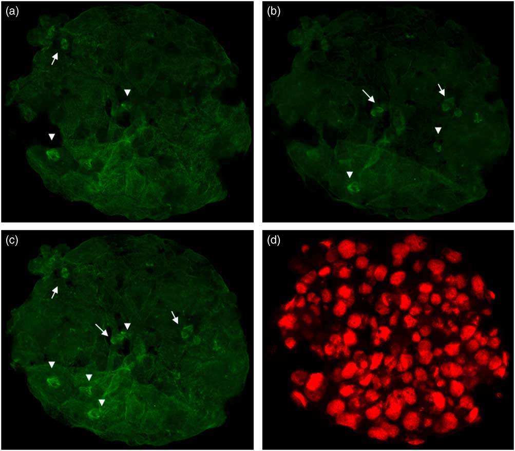
Confocal sections of the non-biopsied embryo in Fig. 2a showing a-tubulin staining only (a–c, green) and propidium iodide (PI) staining only (d, red). Note the normal astral bipolar spindles (arrows) and the abnormally shaped spindles with a well focussed and an unfocussed pole (arrowheads).
Exposure to high concentrations of cryoprotectants, which causes the cells to shrink, is expected to be the primary cause giving rise to the abnormally shaped spindles observed (Chatzimeletiou et al., Reference Chatzimeletiou, Tarlatzis, Morrison, Vanderzwalmen, Panagiotidis, Papatheodorou, Prapas, Prapas and Handyside2010). Whether it is the metaphase/anaphase stage more vulnerable to damage, than the point of centrosome duplication and movement to opposite poles, or any other mitotic stage warrants further investigation (Chatzimeletiou et al., Reference Chatzimeletiou, Morrison, Panagiotidis, Vanderzwalmen, Prapas, Prapas, Tarlatzis and Handyside2012). It is also necessary to establish if the morphological changes in spindle structure have a functional effect on further mitosis, the fate of the abnormal spindles and the potential effects these may have on the chromosomal constitution of the developing blastocysts (Chatzimeletiou et al., Reference Chatzimeletiou, Morrison, Panagiotidis, Vanderzwalmen, Prapas, Prapas, Tarlatzis and Handyside2012; Oikonomou et al., Reference Oikonomou, Chatzimeletiou, Sioga, Oikonomou, Tarlatzis and Kolibianakis2017). A superficial abnormality in morphology is not likely to affect completion of normal mitosis. However, in the case of spindles with a function abnormality, including association with chromosome lagging and bridging, further progression through mitosis may fail or, if it succeeds, may lead to daughter cell lineage with abnormal chromosomal constitutions (Chatzimeletiou et al., Reference Chatzimeletiou, Morrison, Panagiotidis, Vanderzwalmen, Prapas, Prapas, Tarlatzis and Handyside2012). As there is no checkpoint control to monitor excess spindle poles, tripolar and tetrapolar spindles are also likely to progress, giving rise to cells with chaotic chromosomal constitutions, as the segregating chromosomes would be pulled to three or four directions respectively (Sluder et al., Reference Sluder, Thompson, Miller, Hayes and Rieder1997; Musacchio & Hardwick, Reference Musacchio and Hardwick2002; Chatzimeletiou et al., 2005, Reference Chatzimeletiou, Morrison, Panagiotidis, Vanderzwalmen, Prapas, Prapas, Tarlatzis and Handyside2012; Ottolini et al., Reference Ottolini, Kitchen, Xanthopoulou, Gordon, Summers and Handyside2017).
The only case in which a detrimental effect may occur from the progression of abnormal spindles through mitosis is if the derivative abnormal cells gave rise to the fetus. Indeed, in this cohort of embryos, most spindles examined were in the trophectoderm (TE) and a minority were in the ICM. However, even in the case of abnormal spindles progressing into mitosis in the ICM, there may be no detrimental effect if the abnormal cells produced give rise to the extrafetal membranes, while a low level mosaicism localised in specific tissues of the fetus may remain phenotypically invisible in adulthood (Chatzimeletiou et al., Reference Chatzimeletiou, Morrison, Panagiotidis, Vanderzwalmen, Prapas, Prapas, Tarlatzis and Handyside2012).
Considering the new advances in preimplantation genetic testing (PGT-A) and the incorporation of 24 chromosome screening by array CGH or NGS which have recently been shown to decrease miscarriage rates and increase implantation rates, vitrification of supernumerary normal embryos following cleavage biopsy and of all embryos following blastocyst biopsy becomes more and more a necessity (Schoolcraft et al., Reference Schoolcraft, Treff, Stevens, Ferry, Katz-Jaffe and Scott2011; Forman et al., Reference Forman, Tao, Ferry, Taylor, Treff and Scott2012; Yang et al., Reference Yang, Liu, Collins, Salem, Liu, Lyle, Peck, Sills and Salem2012; Rubio et al., 2013, Reference Rubio, Bellver, Rodrigo, Castillón, Guillén, Vidal, Giles, Ferrando, Cabanillas, Remohí, Pellicer and Simón2017; Fiorentino et al., Reference Fiorentino, Bono, Biricik, Nuccitelli, Cotroneo, Cottone, Kokocinski, Michel, Minasi and Greco2014; Natesan et al., Reference Natesan, Handyside, Thornhill, Ottolini, Sage, Summers, Konstantinidis, Wells and Griffin2014; Handyside, Reference Handyside2015; Minasi et al., Reference Minasi, Fiorentino, Ruberti, Biricik, Cursio, Cotroneo, Varricchio, Surdo, Spinella and Greco2017; Friedenthal et al., Reference Friedenthal, Maxwell, Munné, Kramer, McCulloh, McCaffrey and Grifo2018). Here we document for the first time that biopsied embryos that were vitrified at the blastocyst stage showed similar survival rates following warming and no significant differences in the incidence of normal and abnormal spindle/chromosome configurations when compared with non-biopsied embryos.
Financial support
This research received no specific grant from any funding agency, commercial or not-for-profit sectors.
Ethical standards
This work was approved by the ethical committee of the Aristotle University Medical School (licence no. A 10570).
Conflicts of interest
None.