Introduction
Angiostrongylus cantonensis is a blood-feeding nematode usually inhabiting the pulmonary arteries and right ventricles of rats. The complex life cycle of A. cantonensis, also known as rat lungworm, has been described previously, including the details of migration of third-stage larvae (Alicata, Reference Alicata1965; Jindrak & Alicata, Reference Jindrak and Alicata1968). Angiostrongylus cantonensis infection in humans is gained by eating raw or improperly cooked freshwater snails or paratenic hosts; it causes eosinophilic meningitis or meningoencephalitis (Wang et al., Reference Wang, Lai, Zhu, Chen and Lun2008). In the past 10 years, hundreds of cases and several outbreaks of the disease have been reported in endemic regions, especially in China (nine outbreaks in mainland China and three in Taiwan). However, the pathogenesis of angiostrongyliasis cantonensis is complicated and its details and more effective diagnoses and treatment methods need exploration.
Eosinophilia is the main reason for the development of features when humans or mice are infected with A. cantonensis (Chuang et al., Reference Chuang, Su, Chen, Fan, Lin, Chen and Du2010). In the production of the Th2-type immune response, significantly increased immunoglobulin E (IgE), interleukin-5 (IL-5) and eotaxin concentrations in serum and cerebrospinal fluid (CSF) are important in the recruitment and activation of eosinophils to induce inflammation and damage (Yen & Chen, Reference Yen and Chen1991; Intapan et al., Reference Intapan, Kittimongkolma, Niwattayakul, Sawanyawisuth and Maleewong2008; Chuang et al., Reference Chuang, Su, Chen, Fan, Lin, Chen and Du2010). CD4+CD25+ regulatory T cells (Treg) are involved in the pathogenesis of parasitic diseases by regulating the balance of Th1/Th2 (Ismael et al., Reference Ismael, Dimier-Poisson, Lebrun, Dubremetz, Bout and Mevelec2006, Reference Ismael, Hedhli, Cerede, Lebrun, Dimier-Poisson and Mevelec2009; Fang et al., Reference Fang, Nie, Wang, Tu, Zhou, Wang, He, Zhou and Zhao2009). Our previous study (Li et al., unpublished) suggests the involvement of CD4+CD25+ Treg in A. cantonensis. However, we do not know whether treatment influences CD4+CD25+ Treg. Dexamethasone, combined with albendazole, has been widely used to treat eosinophilic meningitis caused by A. cantonensis, due to its anti-inflammatory properties (Chotmongkol et al., Reference Chotmongkol, Wongjitrat, Sawadpanit and Sawanyawisuth2004). However, dexamethasone therapy is still controversial due to immune system rebound after withdrawal of the drug and the risk of adverse drug reactions.
Glycyrrhizin (GL) is a triterpene glycoside extracted from liquorice root (Glycyrrhiza glabra) consisting of one molecule of 18-β-glycyrrhetinic acid and two molecules of glucuronic acid with the following structure: 18-β-glycyrrhetinic acid-3-O-β-d-glucuronopyranosyl-(1 → 2)-β-d-glucuronide (Matsui et al., Reference Matsui, Matsumoto, Sonoda, Ando, Aizu-Yokota, Sato and Kasahara2004). GL and other related compounds have been reported to have a variety of pharmacological activities, including anti-inflammatory, anti-allergic, anti-viral and anti-carcinoma activities, and also immune regulation activities (Sato et al., Reference Sato, Goto, Yamamura, Kurokawa, Kageyama, Takahara, Watanabe and Shiraki1996). They have been widely used for treating hepatitis due to their anti-inflammatory properties (Feng et al., Reference Feng, Wang, Yao, Zhang and Tian2007). Diammonium glycyrrhizinate (DG) is derived from the glycyrrhizin, modified in the two carboxyl groups at the 6' and 6” position to enhance stability, and has the capacity of regulating Th1/Th2 polarization (Song et al., Reference Song, Wang, Chi and Yang2007). Thus we hypothesized that DG might play a role in treating angiostrongyliasis cantonensis. We do not know whether CD4+CD25+ Treg are involved in this mechanism.
We therefore evaluated the curative effects and the mechanisms by which DG acts against A. cantonensis, and compared these with dexamethasone treatment. Signs, eosinophil percentage, Th2-type immune responses and CD4+CD25+ Treg were considered.
Materials and methods
Parasitological procedures
Female mice, aged 6 weeks, of the BALB/c strain were purchased from the Laboratory Animal Centre of Southern Medical University (Guangzhou, Guangdong, China). Mice were subjected to a 12 h light–dark cycle photoperiod for more than 1 week before experimental infection. All procedures involving animals and their care were approved by the Ethics Committee for Animal Experimentation and were conducted according to our university's guidelines for animal experimentation. The life cycle of A. cantonensis was maintained in the laboratory of the Department of Etiology, School of Public Health and Tropical Medicine, Southern Medical University, by cycling the parasite through rats and Achatina fulica snails. Infective larvae (L3) of A. cantonensis were recovered using an optimized method based on Parsons & Grieve (Reference Parsons and Grieve1990). Briefly, the snail shells were crushed, tissues were homogenized and digested in a pepsin–HCl solution (pH 1–2, 500 IU pepsin/g tissue) and incubated at 37°C for 2 h. Host cellular debris was removed from the digested product by filtration. Larvae in the filtrate were collected by serial washing in phosphate buffered saline (PBS), and were then counted under a microscope.
Experimental infection of mice and treatment
Observation group
Twenty-four female mice were randomly allocated to four groups, with the following treatment in each group: control (C), experimental 50 (E50), treatment 50 AD (T50AD) and treatment 50 AG (T50AG). Food and water were withheld for 12 h before infection. Control mice were orally dosed with distilled water; the other groups were given 50 A. cantonensis larvae intragastrically (i.g.). In addition to A. cantonensis, mice in the T50AD group were given a combination therapy of albendazole (Zentel®, GlaxoSmithKline, Research Triangle Park, North Carolina, USA), 20 mg/kg/day i.g., and dexamethasone (Sigma, St Louis, Missouri, USA), 0.5 mg/kg/day i.g., for 7 consecutive days, beginning on the fifth day post-inoculation (dpi). This was called ‘AD therapy’. The mice in the T50AG group were given a combination therapy of albendazole and DG (Lianyungang Chia Tai Tianqing Pharmaceuticals, Jiangsu, China), 50 mg/kg/day i.g., for 7 consecutive days, from 5 dpi; this was called ‘AG therapy’. Signs of all groups were observed.
Detection group
Forty-eight female mice were randomly allocated to eight groups, with the following treatment in each group: C-10, C-15, E50-10, E50-15, T50AD-10, T50AD-15, T50AG-10 and T50AG-15. Infection and treatment were the same as in the above observation groups.
According to previous results from our study (Li et al., unpublished), the survival time of infected mice is about 18 days. Mice in the groups C-10, E50-10, T50AD-10 and T50AG-10 were bled to death following heart puncture, under deep ether anaesthesia, on 10 dpi. The same procedure was performed on 15 dpi in the groups C-15, E50-15, T50AD-15 and T50AG-15. Brains were removed into a 35-mm Petri dish. The cranial cavity and cerebral ventricles (lateral, third and fourth ventricles) were rinsed with 350 μl of 0.15 m PBS, which acted as CSF, so-called ‘CSF-like fluid’ (Chung et al., Reference Chung, Chen, Wang, Chang and Yen2010), and then centrifuged at 260 g for 10 min. Serum and the supernatant fraction of CSF were stored at − 80°C until further use for detection of IgE, IL-5 and eotaxin. Peripheral blood and the precipitate of CSF were collected for eosinophil counting. Fresh anticoagulant blood was collected for CD4+CD25+ Treg analysis. Brains were taken on 15 dpi, three for worm recovery and three for histopathological examinations.
Eosinophil, immunoglobulin E, interleukin-5 and eotaxin detection
Peripheral blood and the precipitate of CSF were smeared on to a glass slide. All smears were stained with Wright–Giemsa solution. The dry slides were examined for cells under a microscope. Eosinophil percentages were made on the basis of 500 nucleated cells. The levels of IgE, IL-5 and eotaxin in the serum were determined by quantitative sandwich enzyme-linked immunosorbent assay (ELISA) using commercial kits (IgE: Biolegend, San Diego, California, USA; IL-5 and eotaxin: R&D, Minneapolis, Minnesota, USA), according to the manufacturers' instructions. The sensitivity of these kits was 0.1 ng/ml, 7 pg/ml and 3 pg/ml, respectively.
Flow cytometry
Fresh anticoagulant blood samples were immuno-stained with the following monoclonal antibodies: anti-CD4-fluorescein isothiocyanate (FITC) plus anti-CD25-phycoerythrin (PE), and washed. Anti-Foxp3-allophycocyanin (APC) was used after permeabilization with a fixation/permeabilization set. The stained cells were analysed using Cell Quest software and a FACSCaliburTM flow cytometer (Becton-Dickinson, Franklin Lakes, New Jersey, USA). All antibodies and the fixation/permeabilization set were purchased from eBioscience (San Diego, California, USA). The results were expressed as percentages.
Worm recovery and histopathology
Mouse brains were cut into pieces and homogenized separately in 15 ml PBS followed by centrifugation. Larval counts were measured under 10 × magnification using a dissecting microscope. Mouse brains were fixed separately in 10% neutral buffered formalin. Samples were embedded in paraffin wax and cut into 5 μm sections. Sections were stained with haematoxylin and eosin. At least ten fields were observed in each section under a light microscope at high power.
Data analysis
Data were analysed by SPSS (version 13.0; SPSS Inc., Chicago, Illinois, USA). Mean values are expressed as means ± standard deviations. The difference of survival time among four groups was determined by Kaplan–Meier's survival analysis. The significance of differences in body weight measurements was determined by analysis of variance (ANOVA) (repeated measures); missing data were replaced provisionally by last data. This was followed by a post hoc Bonferroni test when the ANOVA suggested a significant difference between groups. The differences of the beginning and duration of signs among the four groups were determined by an ANOVA (one-way ANOVA). This was followed by a post hoc Bonferroni test when the ANOVA suggested a significant difference between groups. The significance of differences in eosinophil percentages, concentrations of IgE, IL-5, eotaxin and CD4+CD25+ Treg between the four groups were determined by an ANOVA (factorial analysis), followed by a post hoc Bonferroni test when the ANOVA suggested significant difference. Differences were considered significant at P< 0.05.
Results
Diammonium glycyrrhizinate therapy and body weight, survival and somatic and behavioural changes
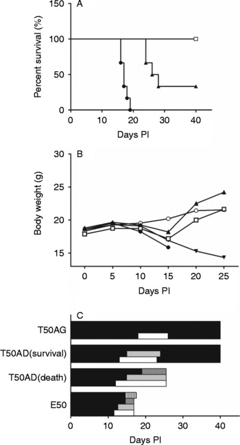
All of the mice in the E50 group were dead by about 17 dpi. In the T50AD group, two mice out of six could be cured and survived up to 40 dpi; the other four mice could not be cured and died on about 26 dpi. All of the mice in the T50AG group were cured and survived up to 40 days. AD and AG therapies were shown to significantly prolong survival time; AG therapy was more effective (P< 0.001) (fig. 1A). The difference in weight among all groups was significant (P< 0.001). The weight of E50 mice decreased significantly compared with the control group (P< 0.001). Compared with E50, AD therapy was able to recover the body weight of cured mice (P< 0.001), but unable to recover it in uncured mice (P>0.05). In AG therapy groups, the weight of all mice was recovered without difference from the control group (P>0.05) (fig. 1B). AD therapy can delay the beginning and prolong the duration of signs, such as moist hair, kyphosis, hemiplegia, and turning around, while AG can also relieve signs (P< 0.001) (fig. 1C). AG therapy was more effective than AD in decreasing weight loss, relieving signs and increasing the cure rate.
The relationship between diammonium glycyrrhizinate (DG) therapy and survival (A), body weight (B) and somatic/behavioural changes (C) in each of four groups of mice. PI, post-infection; ○, control mice (group C); ●, untreated infected (group E50); ▲, treatment with albendazole and dexamethasone (group T50AD); □, treatment with albendazole and DG (group T50AG). In (A) the line of the control overlapped the line T50AG, so was not expressed. In (B) and (C), 2 of 6 mice in T50AD died and the other 4 mice survived, their body weight and somatic/behavioural changes were different, so T50AD was divided into T50AD (death) (▾), 2 mice, and T50AD (survival) (▲), 4 mice. In (C) the symbols indicate no sign (■), moist hair (□), kyphosis ( ), mania (
 ), hemiplegia and turning around (
 ).

Diammonium glycyrrhizinate therapy and eosinophilia and Th2-type responses
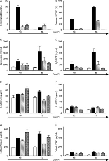
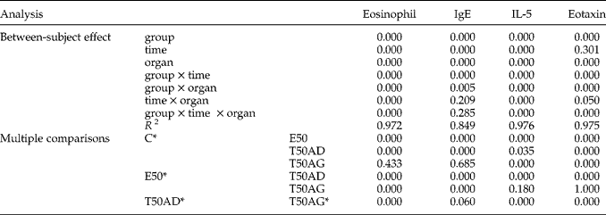
Eosinophil percentages and IgE level in peripheral blood and CSF increased in infected mice. These increases could be temporarily inhibited by AD therapy but showed a rebound effect after treatment was withdrawn. AG therapy was able to persistently inhibit eosinophil (fig. 2A and B) and IgE (fig. 2C and D) production, to a level with no difference from the control. Levels of IL-5 and eotaxin in the serum and CSF of infected mice were significantly higher than in the control mice. AD therapy was able to inhibit these increases. Interestingly, AG therapy was able to enhance levels of IL-5 (fig. 2E and F) and eotaxin (fig. 2G and H) in serum on 10 dpi, and then inhibit them, but this inhibition was persistent for CSF. Results of post hoc multiple comparisons are shown in table 1.
The relationship between diammonium glycyrrhizinate (DG) therapy and eosinophilia and Th2-type responses in each of four groups of mice in blood (A, C, E) and in CSF (B, D, F). PI, post-infection; control mice, group C (□); untreated infected, group E50 (■); treatment with albendazole and dexamethasone, group T50AD (); treatment with albendazole and DG, group T50AG (). Results are expressed as means ± standard deviations.

Significant differences in the percentages of eosinophils, IgE, IL-5 and eotaxin levels between groups.
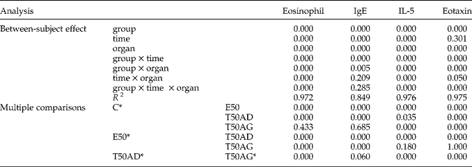
* Mice were divided into four groups: C, E50, AD50 and AG50. C, uninfected control group; E50, untreated infected group; T50AD, treatment with albendazole (A) and dexamethasone (D); T50AG, treatment with A and diammonium glycyrrhizinate (DG). Each group consisted of six mice. The mean difference is significant at the 0.05 level.
Diammonium glycyrrhizinate therapy and numbers of CD4+CD25+ regulatory T cells
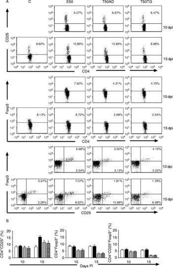
It has been demonstrated that Foxp3 expression is related to the development and function of CD4+CD25+ Treg cells in the thymus (Fontenot et al., Reference Fontenot, Gavin and Rudensky2003). We analysed the number and Foxp3 expression of CD4+CD25+ Treg. The numbers of CD4+CD25+ Treg in E50 mice were increased on 15 dpi, but the expression of Foxp3 in Treg cells was not increased. AD and AG therapies were able to inhibit the increasing numbers of CD4+CD25+ Treg significantly, and decrease the expression of Foxp3 in Treg to below the normal level. However, there was no difference in this decrease between AD therapy and AG therapy (fig. 3).
The relationship between diammonium glycyrrhizinate (DG) therapy and numbers of CD4+CD25+ Treg cells in each of four groups of mice, shown as flow cytometry plots (A) and histograms (B). PI, post-infection; control mice, group C (□); untreated infected mice, group E50 (■); treatment with albendazole and dexamethasone, group T50AD (); treatment with albendazole and DG, group T50AG (). Each group consisted of six mice.

Diammonium glycyrrhizinate therapy and worm recovery/histopathology
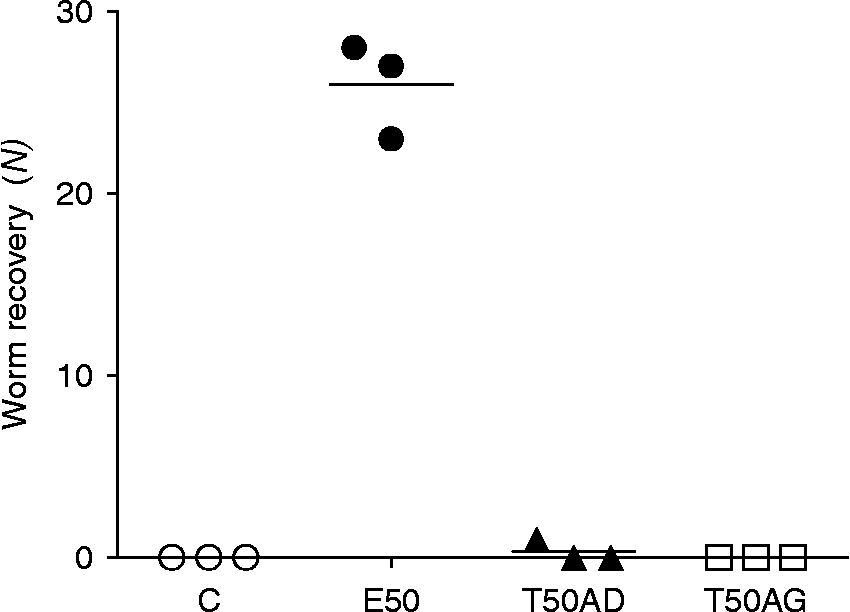
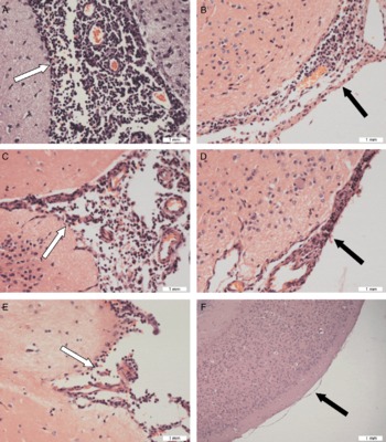
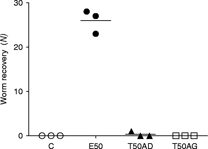
AG therapy was able to kill all of the larvae, while AD therapy killed most of the larvae, but not all (fig. 4). In mice of the E50 group, histological sections revealed meningitis with thickened meninges, infiltration of eosinophil cells and mechanical damage, including haemorrhage, traumatic lesions, subarachnoid haemorrhage and larvae in the subarachnoid space. In most of T50AD mice and all of T50AG mice, meningitis and infiltration by eosinophil cells were obviously improved; however, in one of the T50AD mice the improvement was not obvious (fig. 5). Mechanical damage caused by migrating larvae was not noted in the T50AD and T50AG groups.
The relationship between diammonium glycyrrhizinate (DG) therapy and worm recovery in each of four groups of mice. ○, control mice, group C; ●, untreated infected, group E50; ▲, treatment with albendazole and dexamethasone, group T50AD; □, treatment with albendazole and DG, group T50AG. Three mice of each group were used to detect worm recovery; the other three mice were used to examine the histopathology of the brain.
The relationship between diammonium glycyrrhizinate (DG) therapy and histopathology of the brain in untreated infected mice, group E50 (A and B); mice treated with albendazole and dexamethasone, group T50AD (C and D); and mice treated with albendazole and DG, group T50AG (E and F); shown as eosinophil infiltration into the subarachnoid space ( ) and meninges (
 ).

Discussion
We have determined the curative effects and mechanisms by which DG therapy acts in mice infected with A. cantonensis, and compared this with dexamethasone therapy. We found that DG has a better curative effect in A. cantonensis than dexamethasone, because of more capacity to relieve signs and enhance the rate of cure. Alleviating eosinophil infiltration, and inhibiting Th2-type inflammation and CD4+CD25+ Treg are involved in its therapeutic mechanism.
In mice infected with A. cantonensis, central nervous system dysfunction is the main clinical feature. Hypereosinophilic inflammation of the central nervous system is the crucial pathogenic factor. As effector cells, eosinophils are recruited from the circulation into inflamed or damaged tissues by eosinophil-selective chemokines (Rosenberg et al., Reference Rosenberg, Phipps and Foster2007). Histotoxic or helminthotoxic reactive oxygen species and granular proteins such as cytokines and chemokines are liberated to kill infectious worms; at the same time tissues are damaged (Someya et al., Reference Someya, Nishijima, Nunoi, Irie and Nagaoka1997; Ramos et al., Reference Ramos, Discipio and Ferreira2006; Hogan et al., Reference Hogan, Rosenberg, Moqbel, Phipps, Foster, Lacy, Kay and Rothenberg2008). Eosinophil numbers and degranulation of eosinophils are both increased in A. cantonensis-infected mice (Lan & Lai, Reference Lan and Lai2009). As the upstream mechanism of eosinophilia, the Th2-type immune response is important in the recruitment and activation of eosinophils (Yamaguchi et al., Reference Yamaguchi, Hayashi, Sugama, Miura, Kasahara, Kitamura, Torisu, Mita, Tominaga and Takatsu1988; Wang et al., Reference Wang, Rambaldi, Biondi, Chen, Sanderson and Mantovani1989; Sasaki et al., Reference Sasaki, Sugaya, Ishida and Yoshimura1993). Levels of IgE, IL-5 and eotaxin, as Th2-type cytokines/chemokines, are induced to elevate in angiostrongyliasis cantonensis (Yen & Chen, Reference Yen and Chen1991; Intapan et al., Reference Intapan, Niwattayakul, Sawanyawisuth, Chotmongkol and Maleewong2007; Diao et al., Reference Diao, Chen, Yin, Wang, Qi and Ji2009). However, at the late stage of infection a large amount of non-specific IgE competitively inhibits the function of eosinophils by binding to the receptor (Seminario et al., Reference Seminario, Saini, Macglashan and Bochner1999). Besides regulating the function of eosinophils by the recruitment and activation or competitive inhibition of eosinophils, the Th2-type immune response can also damage tissues by complement activation (Shaio et al., Reference Shaio, Hou, Chen, Wu, Yang and Chang1990; Padilla-Docal et al., Reference Padilla-Docal, Dorta-Contreras, Bu-Coifiu-Fanego, Rodriguez-Rey, Gutierrez-Hernandez and De Paula-Almeida2010). Th2 immune responses have to be regulated to a suitable level to prevent tissue damage. Upstream, Th2 polarization is regulated by a group of CD4+ T cells expressing CD25 on their surface, regulatory T cells (Treg). Under normal conditions, Treg are anergic, but are capable of direct proliferation in response to stimulation by Toll-like receptor (TLR) ligands, expressed on microbes and parasites. The Foxp3 gene is constitutively and specifically expressed in Treg and plays an indispensable role in their development and suppression function (Raghavan & Holmgren, Reference Raghavan and Holmgren2005). Treg suppress proliferation as well as the cytotoxic effect and the secretion of Th1 and Th2 cytokines by the production of regulatory cytokines, including transforming growth factor-β and IL-10, thus limiting the strength of the immune response of effector T cells, which makes them unable to adequately control viral and bacterial infections (Belkaid, Reference Belkaid2007).
We found that mice infected with A. cantonensis displayed weight loss, moist hair, and central nervous system dysfunction, such as kyphosis, hemiplegia, mania and turning around. The present results showed that nerve dysfunction was accompanied by eosinophilia, and that alleviation of these signs was accompanied by recovery from eosinophilia. This eosinophilia in CSF is more serious than in peripheral blood. We confirmed that increasing eosinophil numbers accompanied an increase in IL-5, eotaxin, and IgE production in infected mice. However, these increases in Th2-type immune responses in serum are more serious than in CSF. Therefore, the critical mechanism of central nervous system damage is eosinophilia. In the meantime, the number of CD4+CD25+ Treg is increased, but the examination of more cases is needed to prove an increase of Foxp3 expression. Treg might be enhanced to inhibit the Th2 immune response and to avoid strong immune damage of the host.
A combination therapy of albendazole and dexamethasone has been widely used to treat A. cantonensis. Albendazole, a benzimidazole derivative, kills parasites by binding to β-tubulin, inhibiting its polymerization and impairing glucose uptake (Kwon et al., Reference Kwon, Au, Collins, Adcock, Mak, Robbins, Chung and Barnes1994; Chotmongkol et al., Reference Chotmongkol, Wongjitrat, Sawadpanit and Sawanyawisuth2004). Dexamethasone prevents an inflammatory reaction by decreasing the production of eosinophils and the Th2-type immune response (Narala et al., Reference Narala, Ranga, Smith, Berlin, Standiford, Lukacs and Reddy2007). The combination of albendazole and dexamethasone has thus exhibited beneficial synergistic effects in killing A. cantonensis worms and reducing inflammation (Tu & Lai, Reference Tu and Lai2006). To enhance effectiveness and avoid the side-effects of dexamethasone and rebound of the immune response after its withdrawal, several kinds of traditional Chinese medicine (TCM) have been used in combination with albendazole to replace dexamethasone (Lai et al., Reference Lai, Chen, Chang and Lee2008; He et al., Reference He, Lv, Li, Zhang, Liao, Zheng, Su, Rao, Yu and Wu2011). We investigated the effects of DG on murine angiostrongyliasis. GL and related compounds are considered to have glucocorticoid-like actions (Takei et al., Reference Takei, Baba, Hisatsune, Katsuki, Miyata, Yokomizo and Isohama2008), partly due to the presence of ring-like structures similar to an adrenal cortex hormone. DG is soluble, more stable and convenient to administer. In addition to inhibition of inflammation, DG reduces oxidative stress by inhibiting cyclo-oxygenase-2 and inducible nitric oxide (NO) synthase expression (Ni et al., Reference Ni, Kuai, Lu, Yang, Fu, Wang, Tian, Yan, Zhao, Wang and Jiang2011), and down-regulating matrix metalloproteinase-9 (Abe et al., Reference Abe, Ikeda, Wake, Sato, Sato and Inoue2008). These abilities may be part of the therapeutic mechanism of DG in murine angiostrongyliasis.
We confirmed the effectiveness of AD therapy in killing worms, inhibiting eosinophilia and the Th2-type immune response. However, unfortunately, we found the curative effect of AD therapy to be more limited than in previous reports. AD therapy cured some of the mice infected (two mice out of six). However, central nervous system dysfunctions were present and sustained in almost all mice treated with AD therapy, although signs were delayed. In uncured mice, signs and weight loss were not relieved. At last, these mice become unable to tolerate a rebound eosinophilia and IgE after drug withdrawal and died by 26 dpi. Even in one of the cured mice, signs were sustained after drug withdrawal until 23 dpi. Although AD therapy kills almost all worms, it modifies histopathology changes slightly in one mouse out of six. Considering that the survival time of infected mice was about 18 days in the present study rather than 25 days in previous reports (Lin et al., Reference Lin, Chen, Lan, Lee and Lai2010), the difference in the efficiency of dexamethasone may relate to the different pathogenicity of various A. cantonensis strains (Tu & Lai, Reference Tu and Lai2006; Eamsobhana et al., Reference Eamsobhana, Lim, Zhang, Gan and Yong2010). Many reports recommend administration for 7 consecutive days, from 5 days onwards, to gain the most beneficial effect. If our strain has stronger pathogenicity, then extending the course of dexamethasone dosing to 20–23 dpi may be effective, without consideration of the adverse reactions of dexamethasone.
We also demonstrated that AG therapy is more efficient in alleviating signs and weight loss, prolonging survival time and enhancing curative rate than AD therapy. As was found in previous reports (Matsui et al., Reference Matsui, Sonoda, Sekiya, Aizu-Yokota and Kasahara2006; Ram et al., Reference Ram, Mabalirajan, Das, Bhattacharya, Dinda, Gangal and Ghosh2006), AG therapy was able to decrease eosinophil, IgE, IL-5 and eotaxin production without any rebound after withdrawal of the drug. Interestingly, DG caused increased levels of IL-5 and eotaxin in serum on 10 dpi and decreased levels by 15 dpi. We presume that DG inhibits one or some factors that act as inhibitors of IL-5 or eotaxin. CD4+CD25+ Foxp3+ Treg might be one of these factors.
The number and Foxp3 expression of CD4+CD25+ Treg in infected mice treated by AD and AG therapy were decreased to values lower than those in control animals. The rebound of the Th2-type immune response might be related to a decreasing of Treg. However, previous reports have pointed out that intraperitoneal injection of glucocorticoids increases the proportion of CD4+CD25+ T cells in lymph nodes. The mechanism is not clear, but differences in immune state, the route and dose of administration, and the combination of drugs might cause the different effects. We have made a preliminary exploration of the potential of DG on the basis of previous reports; and there is no report about DG being used to treat angiostrongyliasis cantonensis. Further studies are in progress.
In conclusion, we have demonstrated that combination therapy with albendazole and DG has more capacity to enhance survival rate, alleviate signs, inhibit eosinophilia and the Th2-type immune response than the combination therapy with albendazole and dexamethasone, and has the same capacity to decrease the number and Foxp3 expression of CD4+CD25+ Treg as the combination therapy with albendazole and dexamethasone. We suggest that the combination therapy with albendazole and dexamethasone could be replaced by the combination therapy with albendazole and DG.
Acknowledgements
We are grateful to Xin Zhang for help in the larvae preparation. Financial support was provided by the National Natural Science Foundation of China (no. 30972577) and the Natural Science Foundation of Guangdong Province (no. 9151051501000073).