INTRODUCTION
Commercial aquaculture of bivalve molluscs in Brazil was established recently (1990), with Pacific oysters Crassostrea gigas (3000 mt) and brown mussels Perna perna (11,000 mt) as the main species currently produced. Since then, the activity has expanded to include production of other native bivalve species including Crassostrea brasiliana, Crassostrea rhizophorae and Nodipecten nodosus. The State of Santa Catarina in southern Brazil is the most important Brazilian producer of bivalves. In this region, known episodes of mortalities were only those related to summer mortality of Pacific oysters C. gigas that may occur during summer months each year. However, little else is known about the occurrence and impacts of diseases on bivalves from other cultured populations.
Worldwide, bivalves have been suffering mass mortalities caused by parasitic organisms, including bacteria affecting bivalve larvae and juveniles (Elston, Reference Elston1999; Boardman et al., Reference Boardman, Maloy and Boettcher2008) and protozoans affecting juveniles and adults. Among the most important protozoans are Perkinsus spp. (Villalba et al., Reference Villalba, Reece, Ordás, Casas and Figueras2004; Choi & Park, Reference Choi, Park, Ishimatsu and Lie2010), Bonamia ostreae (Carnegie & Cochennec-Laureau, Reference Carnegie and Cochennec-Laureau2004), Haplosporidium nelsoni (Burreson & Ford, Reference Burreson and Ford2004) and Marteilia refringens (Berthe et al., Reference Berthe, Le Roux, Adlard and Figueras2004). Some of these protozoans (Perkinsus marinus and Bonamia ostreae) have demonstrated high virulences, causing sharp declines in natural populations of Crassostrea virginica and Ostreae edulis in the USA and Spain, respectively. In contrast, metazoan parasites cause mainly disruption of organ functions in bivalves, such as trematodes whose sporocysts infect bivalve gonads causing host castration (Lauckner, Reference Lauckner and Kinne1983) or Mytilicola intestinalis and Pseudomyicola spinosus copepods that cause digestive system dysfunction (Lauckner, Reference Lauckner and Kinne1983; Olivas-Valdez & Cáceres-Martínez, Reference Olivas-Valdez and Cáceres-Martínez2002).
The occurrence and impact of parasites on bivalves from natural or cultured populations from Brazil are still poorly studied, and disease mortalities may occur without notice since there is no systematic surveillance programme. In 2008, an opportunistic investigation of a Tagelus plebeius razor clam mortality event in a natural population at Santa Catarina Island registered for the first time high prevalences (up to 100%) of metacercariae larvae of Parvatrema sp. (Digenea, Gymnophallidae, da Silva et al., Reference da Silva, Cremonte, Sabry, Rosa, Cantelli and Barracco2009). Another case of bivalve mortality, although low (around 20%), was that of the brown mussel P. perna from a cultured population located in the district of Penha (north of Santa Catarina State, which seemed to be a recurrent mortality pattern during 2007–2008) in the same region and season (July–August). Suárez-Morales et al. (Reference Suárez-Morales, Scardua and da Silva2010) analysed samples during that event, and found a prevalence of 25.6% of copepod Monstrilla sp. larvae that have never been reported before infecting a mollusc host, but which was probably associated with the mortalities.
Monitoring diseases and pathological conditions among bivalve mollusc populations is one important way to recognize potential pathogens and to establish measures to prevent future epizooties in cultured areas. In order to increase our knowledge on diseases affecting four edible bivalve species from cultured and exploited wild stocks in the most important producer region (Santa Catarina State) of Brazil, this work describes for the first time results from a pathological survey, and descriptions of several new diseases for those cultured mollusc hosts.
MATERIALS AND METHODS
Animal sampling
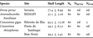
From January to March 2008 four edible bivalve species were sampled each one from one site at Santa Catarina (SC, southern Brazil), except for Crassostrea rhizophorae which was collected in two sites. A total of 80 animals were sampled from each species and site, analysed for macroscopic anomalies and processed by different techniques to survey pathologies, including haemolymph cell monolayers, tissue imprints, histological sections, Ray's fluid thioglycollate medium (RFTM) assays and polymerase chain reaction (PCR) (Table 1). Samplings were taken during the summer season, when water temperature reaches the maximum throughout the year for southern Brazil. This strategy was applied to increase the probability for detection of pathogens.
Information on bivalve's sampling in Santa Catarina State. Site, shell length (mm) and number of bivalves used for histological preparations (Nh), Ray's fluid thioglycollate medium assays (NRFTM), and polymerase chain reaction (NPCR).

Nd, not done; REMAPI, Pirajubaé's Marine Reserve.
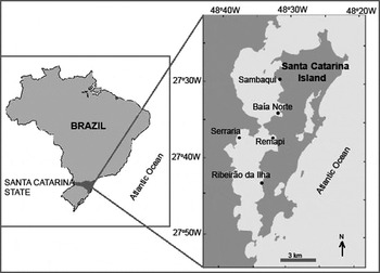
Adult mangrove oysters C. rhizophorae (Guilding 1828) were collected from the intertidal zones of two natural rocky shores, one at the Baía Norte (BN; 27°34′20″S 48°32′17″W), a bay that receives domestic effluent, and another at Sambaqui (SA; 27°29′26″S 48°32′17″W), located 9 km on north of BN and 450 m from a Crassostrea gigas culture area (Figure 1).
Map of Brazil indicating Santa Catarina State and the sampling sites, Baía Norte, Sambaqui, Ribeirão da Ilha, Serraria and Pirajubaé's Marine Reserve (REMAPI).

Adult Pacific oysters C. gigas (Thunberg 1793) were sampled from lantern nets, from a commercial culture area located in the most important producer region from Santa Catarina Island. (Ribeirão da Ilha) (RI; 27°42′56″S 48°33′47″W), on southern Santa Catarina Island (Figure 1).
The brown mussels Perna perna (Linnaeus 1758) were sampled from 1 m long ropes of one of the most intensive mussel culture area from Santa Catarina, at Serraria (SE; 27°31′58″S 48°37′39″W) (Figure 1). Mussels and Pacific oysters were sampled after 8 months of culture.
Clams Anomalocardia brasiliana (Gmelin 1791) were gathered by hand-raking from the largest natural sand bed (700 hectares) in Santa Catarina Island, a marine fishery reserve the Pirajubaé's Marine Reserve (REMAPI; 27°38′00″S 48°33′05″W), which permits controlled exploitation by fishers (Figure 1).
Haemolymph cell monolayers preparation
Haemolymph was withdrawn from adductor muscles with 21-guage needles (oysters and mussels) or 26-gauge needles (clams) attached to 1-ml syringes. Haemolymph cell monolayers (HCM) were prepared by settling 100 µl of haemolymph onto a glass slide for 15–20 minutes in a humidified chamber to permit haemocyte adhesion. Cell monolayers were then fixed with methanol and stained with Giemsa. The slides were analysed for microcell parasites, morphological cell alterations and neoplastic disorders.
Tissue imprints
Small fragments of digestive gland-gonad and gills were excised, blotted on a paper towel to remove excess moisture, and slightly impressed onto a slide. The slides were fixed with methanol and stained with Giemsa.
Histological sections
Each bivalve was shucked, and a transverse section of 5 mm (including visceral mass, gills and foot) was excised and fixed in Davidson's solution (Shaw & Battle, Reference Shaw and Battle1957). Fixed tissues were embedded in paraffin, sectioned at 5 µm thickness, and stained with Harris' haematoxylin and eosin (Howard et al., Reference Howard, Lewis, Keller and Smith2004).
Detection of Perkinsus sp. organisms by RFTM assays
The presence of protozoan Perkinsus spp. was investigated with Ray's fluid thioglycollate medium assays (RFTM; Ray, Reference Ray1966). Each animal was shucked, and the two demibranchs from one side of the body and the rectum were excised. Tissues were incubated in RFTM for 7 days in the dark at room temperature. The gills/rectum were prepared by chopping the tissues with a scalpel on a microscope slide in a pool of Lugol's iodine solution, and examinations made by light microscopy for the dark-stained spheres of Perkinsus hypnospores. Mussels P. perna and Pacific oysters C. gigas were not analysed by RFTM because they are not habitual hosts for Perkinsus spp.
Detection of Marteilioides chungmuensis by PCR
DNAs extracted from gonad tissues that were preserved in 95% ethanol, were used as samples for a nested PCR assay for detection of Marteilioides chungmuensis, using two sets of specific primers, OPF-2/OPR-2 for the first PCR and OPF-3/OPR-3 for the second PCR (Itoh et al., Reference Itoh, Oda, Yoshinaga and Ogawa2003). DNA extractions were performed with DNAzol® reagent (Invitrogen) following the manufacturer's protocol. A positive control used M. chungmuensis-infected oyster DNA provided by Dr Naoki Itoh. Negative controls used nuclease-free water instead of template DNA. PCR reactions were performed as described by Itoh et al. (Reference Itoh, Oda, Yoshinaga and Ogawa2003). The PCR was performed only for two oysters showing unknown ovarian parasites.
RESULTS
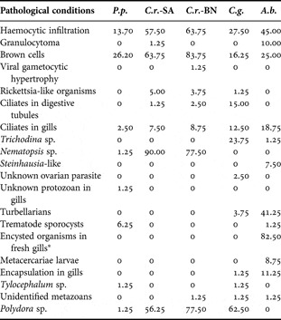
Several pathological conditions and parasites were detected in a total of 400 bivalves belonging to 4 species and examined from five sites along the coasts of Santa Catarina State (Table 2).
Prevalence (%) of pathological conditions and parasites in four edible bivalve species: Perna perna (P.p.), Crassostrea rhizophorae from Sambaqui (C.r.-SA) and Baía Norte (C.r.-BN), Crassostrea gigas (C.g.) from Ribeirão da Ilha and Anomalocardia brasiliana (A.b.) from Pirajubaé's Marine Reserve. N = 80 for each species. *Observations under microscopy on gills prepared by Ray's fluid thioglycollate medium assays.

Nonspecific histopathological symptoms of stress were observed with different prevalences among species (Table 2). One of them involved haemocytic infiltration of tissues, mainly in the connective tissue of different organs. Crassostrea rhizophorae showed the highest prevalence observed in both sites, Baía Norte (64%) and Sambaqui (58%), followed by Anomalocardia brasiliana (45%). Another symptom involved concentration of infiltrating haemocytes in foci forming so-called granulocytomas which were prevalent in clams (10%). Brown cells occurred in connective tissues of different organs in all species; mangrove oysters had the highest prevalence (84% and 64%) in Baía Norte and Sambaqui, respectively. Brown cells showed varying sizes and forms, according to bivalve species. Among mangrove oysters C. rhizophorae, brown cells were largest (around 50 µm in diameter), contained small or large granules, and were often associated with haemocytic infiltration; while C. gigas oysters had brown cells mainly with small granules inside. Perna perna mussels showed the smallest brown cells (around 15 µm) often present in clusters, and A. brasiliana showed brown cells similar to those in mussels but not in clusters (Table 2).
Parasites, including putative viruses, bacteria, protozoa and metazoa were observed in the present study and are described below.
Virus
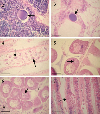
One mangrove oyster from Baía Norte showed follicles with atypical male gametes. The spermataozoa had intranuclear basophilic inclusions and condensed peripheral material (Figure 2), around 60 µm long. There was no female affected with this disease.
Pathogens found in edible bivalves from Santa Catarina State, Brazil. (2) Intranuclear basophilic inclusions and perinuclear condensed material (arrows) in a male follicle of Crassostrea rhizhophorae; (3) intracytoplasmic colonies of rickettsia-like organisms (arrow) in epithelial cells of digestive tubule; (4) oocysts of Nematopsis sp. (arrows) in gill filaments of Perna perna; (5) sporoblast of Steinhausia-like microsporidian (arrow) infecting the cytoplasm of oocyte of Anomalocardia brasiliana; (6) vacuole containing three cells (arrow) of an unidentified protozoan infecting the cytoplasm of a mature oocyte of Crassostrea gigas. Note the presence of infiltrated haemocytes in the follicle (*); (7) sporonts-like of an unidentified protozoan (arrow) in filament gills of the brown mussel P. perna. Note the numerous cells inside a parasite membrane (arrow). All scale bars: 50 µm.

Bacteria
Colonies of rickettsia-like organisms (RLOs) were found at very low prevalences in cells of the digestive diverticula of oysters (Table 2). They appeared as oval or kidney-shaped intracytoplasmic inclusions, around 70 µm long, with different degrees of basophilia and granulated texture and no haemocytic reaction was associated (Figure 3).
Protozoa
Three types of ciliates were found in bivalves in the present study. Ellipsoidal (around 40 µm-long) ciliates were observed in the lumens of digestive tubules and they were detected only in oysters at very low prevalences (< 2.5%), except for Crassostrea gigas (15%). Pear-shaped to oval (around 20 µm-long) ciliates were observed on gill filaments, gill water tubes and labial palps from all bivalve species at low prevalences (Table 2). Trichodina sp. ciliates were detected in C. gigas at moderate (24%) prevalences, and one case was detected in Anomalocardia brasiliana (Table 2).
Host phagocytes containing oocysts of a Nematopsis sp. gregarine (around 20 µm long) were found in connective tissues of labial palps, gills, mantle, digestive gland and gonads. High prevalences of oocysts were detected in mangrove oysters from both sites (90% and 78%), Sambaqui and Baía Norte, respectively (Table 2). Infection intensities were low, and there was no immune response. Remarkably, Nematopsis sp. could also be detected easily in oyster gills prepared by RFTM assays which showed an increase of 3% in the total prevalence of Nematopsis sp. when using just histology. One Perna perna mussel was infected by a Nematopsis sp. at very low intensity in gills (Figure 4).
Steinhausia-like microsporidian parasites infecting the cytoplasm of oocytes of clams A. brasiliana were found at low prevalence (7.5%; Table 2) and intensity. Sporocysts of the parasite measure around 20 µm in diameter containing spores in different development stages, and were detected without host reaction (Figure 5).
An unidentified protozoan infecting oocytes was detected in two Pacific oysters C. gigas at very low intensity. Two to six protozoan cells were observed inside a vacuole in the cytoplasm of the oocyte (Figure 6). A slight haemocytic infiltration was observed in infected follicles. The nested-PCR assays excluded Marteilioides chungmuensis as the protozoan infecting the oysters' oocytes.
An unidentified protozoan was detected in filament gills of the brown mussel P. perna. The cell appearance of this protozoan resembled a sporont containing numerous cells in development (7 µm) (Figure 7).
Metazoa
Turbellarians were observed on gill filaments of Crassostrea gigas and Anomalocardia brasiliana with low (4%) and moderate (41%) prevalences, respectively.
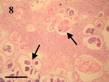
Sporocysts of bucephalid trematodes were observed in 6% of brown mussels Perna perna studied. The mantle of infected mussels always showed heavy intensities of infection, with sporocysts containing developed germ balls and mature cercariae. Few or no gonad follicles were observed among infected mussels (Figure 8). In one clam A. brasiliana sporocysts of a trematode contained germ balls in early developmental stages, without mature cercariae infecting the kidneys at moderate intensity. That animal did not show any other organ infected with sporocysts or metacercareae.
Mantle of Perna perna infected by sporocysts of trematode bucephalid (arrows). Note the absence of gonad follicles. Scale bar: 400 µm.

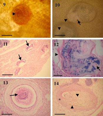
Encysted unidentified metazoans were only observed in gills of clams A. brasiliana incubated in RFTM at high prevalence (82%). The majority of organisms seemed to be turbellarians (Figure 9) since they showed a typical pair of pigmented eyes, while one resembled a metacercariae because of the head spines (Figure 10).
Pathogens found in edible bivalves from Santa Catarina State, Brazil. (9) Turbellarian encysted in gills of Anomalocardia brasiliana incubated in Ray's fluid thioglycollate medium. Note the pair of pigmented eyes (arrowhead). Scale bar: 300 µm; (10) unknown metazoan encysted in gills of A. brasiliana showing a head spine (arrowheads) and eye spot (arrow). Scale bar: 150 µm; (11) trematode metacercariae larvae (arrows) encysted in kidney of A. brasiliana. Scale bar: 300 µm; (12) magnification of a metacercaria showing the eosinophilic cyst (arrowhead) surrounding the larvae. Observe the oral sucker (arrow). Scale bar: 50 µm; (13) Metacestoda Tylocephalum sp. in Perna perna host-formed capsule. Note the apical sucker (arrow). Scale bar: 150 µm; (14) encapsulated unidentified metazoan, probably a copepod, in connective tissue nearby male follicle and mantle epithelium of Crassotrea gigas. Note the thick layer of haemocyte infiltration reacting against the invader and the brown colour of the chitinous exoskeleton of the copepod appendages (arrowheads). Scale bar: 300 µm.

Metacercariae larvae were only found in clams A. brasiliana at low prevalence (8%; Table 2). They were frequently observed in kidneys (Figures 11–12), associated with haemocytic infiltration and were sporadically concurrent with infections of mantle, gonad follicles or foot. Anomalocardia brasiliana was almost the only species showing gill-encapsulation of a partially destroyed, unidentified organism (11%) observed by histological section.
Larvae of Tylocephalum sp. (Platyhelminthes: Nematoda) were observed encapsulated in connective tissues of one brown mussel P. perna and one Pacific oyster C. gigas (Table 2; Figure 13).
Unknown metazoans, probably belonging to copepods because of the presence of chitinous exoskeleton, appendages and segmentation (Figure 14) were observed in three individuals from each of the species A. brasiliana, C. gigas and Crassostrea rhizophorae (Table 2). In the clam, the copepod was observed in mantle cavity, close to the gills' filaments; in the mangrove oyster it was found in the lumen of the intestine and in the Pacific oyster it was found encapsulated in connective tissue between gonad follicles and mantle epithelia (Figure 14).
Infestation of shells by spionid polychaete worms, probably Polydora sp., were identified by the presence of pores and detrital tubes on inner oyster shells present with variable intensities, from few and small tubes to almost covering the whole internal shell. Prevalence was high in oysters (56–77%). One mussel was infested, but no clam was infested.
Specific techniques
In the present work, the use of specific techniques such as RFTM assays to screen for Perkinsus spp. and haemolymph cell monolayers to screen for intrahaemocytic parasites or neoplasia, did not reveal the presence of any pathology in the bivalves studied. The use of imprints of tissue did not reveal any different disease or parasite from those observed by histological sections.
DISCUSSION
The present work describes the occurrence of several pathologies affecting important commercial and unexploited bivalve populations from Santa Catarina (SC) State, including mussels P. perna; clams A. brasiliana; and oysters C. gigas and C. rhizophorae. Despite the great importance of SC State as a bivalve producer in Brazil, there is little research on pathology for those productive stocks (Magalhães, Reference Magalhães1998; da Silva et al., Reference da Silva, Magalhães and Barracco2002; Boehs & Magalhães, Reference Boehs and Magalhães2004; Sabry & Magalhães, Reference Sabry and Magalhães2005; da Costa, Reference da Costa2007; Sabry et al., Reference Sabry, Rosa, Magalhães, Barracco, Gesteira and da Silva2009). Those studies described some commensals and parasites for the first time in those bivalves, including ciliates; the protozoan Nematopsis sp.; trematodes bucephalids; the polychaete Polydora websteri and the metacestode Tylocephalum sp. The more recent reports of diseases on Santa Catarina bivalves were the occurrence of Parvatrema sp. trematode and Monstrilla sp. copepod associated with mortalities among wild razor clam Tagelus plebeius (da Silva et al., Reference da Silva, Cremonte, Sabry, Rosa, Cantelli and Barracco2009) and culture mussel P. perna (Suárez-Morales et al., Reference Suárez-Morales, Scardua and da Silva2010).
Based on morphological and staining characteristics, the hypertrophied cells in male gonad follicles observed here appear similar to those described among C. gigas oysters in Korea (Choi et al., Reference Choi, Lee, Choi, Park, McGladdery and Park2004) and France (Garcia et al., Reference Garcia, Robert, Arzul, Chollet, Joly, Miossec, Comtet and Berthe2006) causing the disease viral gametocytic hypertrophy. In oysters C. gigas from France both male and female were infected (Garcia et al., Reference Garcia, Robert, Arzul, Chollet, Joly, Miossec, Comtet and Berthe2006); while in Korea, only male oysters were infected (Choi et al., Reference Choi, Lee, Choi, Park, McGladdery and Park2004), as observed in the present work.
Rickettsia-like organisms found in epithelial cells of digestive glands of oysters resembled those reported from other bivalve species (Harshbarger et al., Reference Harshbarger, Chang and Otto1977; Bower et al., Reference Bower, McGladdery and Price1994). RLO was very mildly damaging for the oysters. As observed here, Boehs et al. (Reference Boehs, Villalba, Ceuta and Luz2010) found RLOs infecting digestive gland epithelia of A. brasiliana and the mangrove mussel Mytella guayanensis from Bahia (north-east Brazil) at low prevalences (<5%).
Ciliates observed in the present study are common commensals living in paleal cavity of bivalves without causing damage.
Protozoan parasites are considered a high threat to bivalves (Lauckner, Reference Lauckner and Kinne1983), especially those listed by the World Organization for Animal Health (OIE). In the present work, we did not find any OIE-notifiable parasites. Recently, Sabry et al. (Reference Sabry, Rosa, Magalhães, Barracco, Gesteira and da Silva2009) reported Perkinsus sp. for the first time infecting the mangrove oyster C. rhizophorae from north-east Brazil, but the authors did not detect Perkinsus sp. in C. rhizophorae from Santa Catarina. In the present work we report Nematopsis sp. infecting one mussel P. perna at very low intensity. Lima et al. (Reference Lima, Abreu and Mesquita2001) reported Nematopsis sp. at high prevalences (up to 59%) and intensities in gills and digestive glands of P. perna from Itaipu Lagoon, Rio de Janeiro State. Moreover, the prevalence of Nematopsis sp. in the mangrove oysters C. rhizophorae from the present work at Sambaqui was very high (90%) even more than that (60%) observed by Sabry & Magalhães (Reference Sabry and Magalhães2005) in the same region, although the authors reported low intensities of infection. Mangrove oysters from Baía Norte, a more polluted area, had a lower prevalence of Nematopsis sp. than oysters from Sambaqui. There is no report studying the effect of pollution on prevalence of this protozoan. Surprisingly, Pacific oysters from Ribeirão da Ilha were not infected by Nematopsis sp., in contrast to oysters cultured in Sambaqui (Sabry & Magalhães, Reference Sabry and Magalhães2005).
Steinhausia-like was found for the first time infecting oocytes of the clam A. brasiliana which corresponds to the second host species reported for the Steinhausia genus in Brazil. In the first description, Matos et al. (Reference Matos, Matos and Azevedo2005) described ultrastructural aspects of the parasite and suggested Steinhausia mytilovum as the species infecting the oocytes of mangrove mussel M. guyanensis. Since few host oocytes were infected, we believe that the reproductive activity and fecundity were not affected, as observed by Carballal et al. (Reference Carballal, Iglesias, Santamarina, Ferro-Soto and Villalba2001). In contrast, Rayyan & Chintiroglou (Reference Rayyan and Chintiroglou2003) described a reduction of the condition index of mussels Mytilus galloprovincialis caused by S. mytilovum.
Another unknown ovarian protozoan was found in the Pacific oysters analysed here. Due to its localization and morphological characteristics by optical microscopy we first supposed that it might be related to the microsporidian Steinhausia sp. or to the paramyxean Marteilioides chungmuensis, since the Pacific oyster is a highly susceptible host for the former. Marteilioides chungmuensis infects oocytes of Pacific oysters from Asia (Korea and Japan) and leads to the formation of nodules on gonad; decreasing commercial value (Ngo et al., Reference Ngo, Berthe and Choi2003; Itoh et al., Reference Itoh, Komiyama, Ueki and Ogawa2004) and even causes mortality (Park et al., Reference Park, Kang, Choi and Jee2003; Tun et al., Reference Tun, Itoh, Shimizu, Yamanoi, Yoshinaga and Ogawa2008). No such nodules were observed in the two infected oysters from the current survey. Nevertheless, the specific nested-PCR assay excluded M. chungmuensis as the pathogen, thus suggesting a different parasite. To confirm the aetiological agent observation of electron micrographs is required.
The sporonts-like observed in P. perna gills resembled those from haplosporidians. There are few cases of haplosporidian-infected mussels, such as Haplosporidium tumefacientis in the mussel Mytilus californianus; Minchinia sp. in M. galloprovincialis from France (Comps & Tigé, Reference Comps and Tigé1997) and two unnamed Haplosporidium sp. in Mytilus edulis mussels from Maine, USA (Figueras et al., Reference Figueras, Jardon and Caldas1991) and Nova Scotia, Canada (Stephenson et al., Reference Stephenson, McGladdery and Stokes2002). Nevertheless, to ascertain the protozoan affiliation, more individuals showing different parasite sporogony stages should be analysed.
The turbellarians observed in the present work are probably Urastoma cyprinae since they usually occur in mantle cavities and gills of bivalves (Brun et al., Reference Brun, Boghen and Allard1999; Rayyan et al., Reference Rayyan, Photis and Chintiroglou2004; Crespo-González et al., Reference Crespo-González, Rodríguez-Domínguez, Soto-Búa, Segade, Iglesias, Arias-Fernández and García-Estévez2008). The encysted organisms found in A. brasiliana probably resulted from the incubation of gills in RFTM that induced the formation of cysts coating the turbellarian to protect it from the unusual external environmental. Crespo-González et al. (Reference Crespo-González, Álvarez, Domínguez, Búa, Iglesias, Fernández and Estévez2005) described in vitro the formation of mucus-like coating (cocoon) secreted by U. cyprinae to lay eggs inside during its life cycle. Accordingly, two main forms of U. cyprinae can be found, one elongated (free) and the other round-shaped when the turbellarian is covered by a cocoon (Noury-Sraïri et al., Reference Noury-Sraïri, Justine and Euzet1990), as observed herein by RFTM. Cáceres-Martínez et al. (Reference Cáceres-Martínez, Vásquez-Yeomans and Padilla-Lardizábal2010) also reported U. cyprinae occasionally inside of a capsule. In the present work clams A. brasiliana that live in sand–muddy bottom show higher prevalence of turbellarian than oysters C. rhizophorae that live attached on rocky or in lantern nets hanging on long line ropes. It seems that prevalence and intensity of this turbellarian are influenced by the type of host habitat (substrate and degree of exposures). Low prevalences of U. cyprinae were observed on mussels from exposed sites as well from rafts where they were permanently immersed (Robledo et al., Reference Robledo, Cáceres-Martínez, Sluys and Figueras1994; Cáceres-Martínez et al., 1998). Cáceres-Martínez et al. (Reference Cáceres-Martínez, Vásquez-Yeomans and Sluys1998) found that polluted areas had lowest prevalence.
Trematode sporocysts found in P. perna mussels probably belong to Bucephalus margaritae that infects P. perna with variable prevalence and intensities among different sites in Santa Catarina Island (da Silva et al., Reference da Silva, Magalhães and Barracco2002; da Costa, Reference da Costa2007) and whose life cycle has been recently clarified (Marchiori et al., Reference Marchiori, Magalhães and Pereira2010). All infected mussels had heavy infections with reduced gonad follicles indicating host castration. Curiously, A. brasiliana showed trematode sporocysts located in kidneys, instead of gonads, the common site (Lauckner, Reference Lauckner and Kinne1983). Boehs & Magalhães (Reference Boehs and Magalhães2004) observed high prevalences (to 33%) of trematode bucephalid sporocysts in gonads of A. brasiliana from Santa Catarina Island, but never in kidneys. Sporocysts in kidneys of A. brasiliana did not house mature cercariae, indicating a recent infection. Moreover, metacercariae larvae from an unidentified family were observed infecting kidney of A. brasiliana, which might suggest that the trematode agent could belong to a family whose bivalves behave as first and second intermediate hosts, such as Monochiirdae, although further studies should be conducted to assess this hypothesis (Lauckner, Reference Lauckner and Kinne1983).
The other type of encysted organism found in gills of A. brasiliana incubated in RFTM remains unresolved, although its morphology to some extent was similar to a metacercariae of the Echinostomatidae family; except for the eye spot (Lauckner, Reference Lauckner and Kinne1983; Cremonte, personal communication). By histology, encysted and unrecognizable organisms were observed at high prevalence. Those organisms were partially destroyed by host defences.
The occurrence of metacestode larvae of the genus Tylocephalum in the present work is not a surprise, since it is usual in bivalves (Lauckner, Reference Lauckner and Kinne1983) and has been already reported as an intermediate host of Brazilian bivalves (Nascimento et al., Reference Nascimento, Smith, Kern and Pereira1986; Lima et al., Reference Lima, Abreu and Mesquita2001; Boehs & Magalhães, Reference Boehs and Magalhães2004; Sabry & Magalhães, Reference Sabry and Magalhães2005; Sabry et al., Reference Sabry, Gesteira and Boehs2007; Boehs et al., Reference Boehs, Villalba, Ceuta and Luz2010), as observed here always without host damage.
Unidentified metazoans observed in bivalves (A. brasiliana, C. rhizophorae and C. gigas) from the present work are probably copepods. Pseudomiycola spinosus (Myicolidae) is one of the most common poecilostomatoid copepods found in marine invertebrates, occurring in the mantle cavity of more than 50 bivalve species worldwide (Ho & Kim, Reference Ho and Kim1991; Ho, Reference Ho2001; Cáceres-Martínez et al., Reference Cáceres-Martínez, Chávez-Villalba and Garduño-Méndez2005), including A. brasiliana (Narchi, Reference Narchi1965). In the Pacific oyster C. gigas, it is most likely that the copepod reached the connective tissue between follicles and mantle epithelia by accident, when it crawled to a follicle via the gonoduct and consequently evoked a haemocytic response. In contrast, the copepod observed inside the lumen of the intestine of the mangrove oyster C. rhizophorae may be related to the Mytilicola genus, although this needs to be confirmed.
Burrowing worms that belong to the Polydora genus were found infesting the bivalves studied here. In this work, oysters were the most infested bivalve especially the mangrove oysters from a polluted area (Baía Norte). Polydora websteri is frequently found infesting C. gigas with high prevalences (to 100%) throughout the year in Santa Catarina Island (Sabry & Magalhães, Reference Sabry and Magalhães2005), causing problems for marketing; while mussels P. perna cultured in Santa Catarina are more mildly infested (>22%; da Costa, Reference da Costa2007). Sabry et al. (Reference Sabry, Gesteira and Boehs2007) did not describe spionid polychaeta in C. rhizophorae from a natural bed located in Jaguaribe River (north-east Brazil), but found 100% of Polydora sp. in C. rhizophorae from Sambaqui culture in Santa Catarina Island (Sabry & Magalhães, Reference Sabry and Magalhães2005).
The results obtained in this study contribute to expand the knowledge on bivalve diseases from Brazil and will provide a database for the Brazilian government to establish future procedures for bivalve sanitary control in priority areas of bivalve culture such as Santa Catarina State.
ACKNOWLEDGEMENTS
The authors are grateful to Dr Naoki Itoh from the Graduate School of Agricultural Science, Tohoku University, Miyagi, Japan, who kindly provided preserved Marteilioides chungmuensis DNA for PCR analyses. We thank the bivalve fishers who provided bivalves for this study. We acknowledge a post-doctoral grant to Dr P.M. da Silva from CNPq (Conselho Nacional de Desenvolvimento Científico e Technológico).