Introduction
The recent edition of the International Classification of Diseases (ICD 11) introduces a new variant of posttraumatic stress disorder (PTSD), termed complex posttraumatic stress disorder (CPTSD), which is most commonly associated with severe childhood trauma (Knefel & Lueger-Schuster, Reference Knefel and Lueger-Schuster2013). This diagnosis requires the same symptoms as PTSD, with the addition of disturbances in self-organization as reflected in: (a) excessive deficits in regulating emotions, (b) negative self-concept, and (c) dysfunctions in interpersonal relations (World Health Organization, 2018). There is evidence that CPTSD is distinct from both PTSD (Karatzias et al., Reference Karatzias, Shevlin, Fyvie, Hyland, Efthymiadou, Wilson and Cloitre2017) and borderline personality disorder (BPD) (Cloitre, Garvert, Weiss, Carlson, & Bryant, Reference Cloitre, Garvert, Weiss, Carlson and Bryant2014) (for reviews, see Brewin et al., Reference Bremner, Krystal, Putnam, Southwick, Marmar, Charney and Mazure2017; Schmahl et al., Reference Schmahl, Herpertz, Bertsch, Ende, Flor, Kirsch and Bohus2014). Despite this, the recognition of CPTSD is controversial because some commentators argue that there is insufficient evidence to conclude that CPTSD is distinct from PTSD (Resick et al., Reference Resick, Bovin, Calloway, Dick, King, Mitchell and Wolf2012). This led to the decision to omit this diagnosis in CPTSD in DSM-5, which instead recognized a dissociative subtype of PTSD (Friedman, Resick, Bryant, & Brewin, Reference Friedman, Resick, Bryant and Brewin2011).
Although there is evidence of distinct neural patterns of dissociative PTSD (Lanius et al., Reference Lanius, Vermetten, Loewenstein, Brand, Schmahl, Bremner and Spiegel2010), there is no evidence regarding relative neural profiles of PTSD and CPTSD. Of relevance are findings that adult survivors of childhood abuse have abnormalities in the hippocampus, corpus callosum, anterior cingulate cortex (ACC), orbitofrontal cortex, and dorsolateral prefrontal cortex (dlPFC) (Hanson et al., Reference Hanson, Nacewicz, Sutterer, Cayo, Schaefer, Rudolph and … Davidson2015). Further, survivors of childhood abuse display functional deficits in the amygdala when processing emotional stimuli and in the striatum during reward processing (van Harmelen et al., Reference van Harmelen, van Tol, Demenescu, van der Wee, Veltman, Aleman and Elzinga2013). Also, one meta-analysis reported that BPD was distinguished from PTSD and depression by reduced ventral premotor cortex and right posterior parietal cortex activation (Schulze, Schulze, Renneberg, Schmahl, & Niedtfeld, Reference Schulze, Schulze, Renneberg, Schmahl and Niedtfeld2019). Taken together, this evidence reflects persistent neural effects of childhood abuse rather than understanding neural profiles associated with CPTSD. To advance our understanding of the validity of the CPTSD construct, there is a need to understand if the neural circuitry of CPTSD is similar or distinct to that of PTSD. Accordingly, the aim of the current study was to conduct the first investigation of neural responses and connectivity during fear processing in participants with CPTSD. On the basis of the documented disturbances in CPTSD of emotion regulation and disturbances of self-concept, we expected differences between CPTSD and PTSD in activations and connectivity related to the ACC, dlPFC, amygdala, and insula.
Method
Participants
Sixty participants of mean age 39.3 ± 12.6 years were recruited from referrals from clinical services. Inclusion criteria were satisfaction of the diagnostic and statistical manual of mental disorders-fourth edition (DSM-IV) criterion DSM, for PTSD, as diagnosed by clinical psychologists using the Clinician-Administered PTSD Scale (CAPS); (Blake et al., Reference Blake, Weathers, Nagy, Kaloupek, Gusman, Charney and Keane1995). Participants with a history of neurological disorder, psychosis, or current substance dependence were excluded. Trauma-exposed controls (TC) were also recruited from the general community. TC participants were defined as being exposed to a traumatic event but not meeting criteria for more than one cluster of PTSD symptoms. The PTSD (n = 32), CPTSD (n = 28), and TC (n = 39) participants were matched for age and gender.
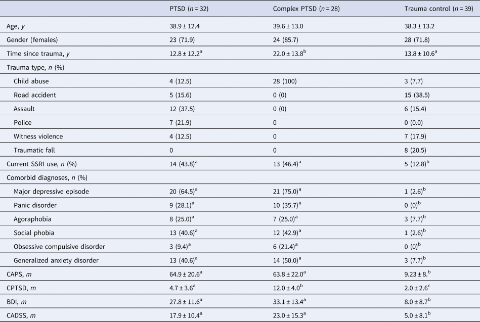
Table 1 presents the participant characteristics. Although 27 (45.0%) participants were currently using selective serotonin uptake inhibitors (SSRIs), the use of SSRIs was comparable between the PTSD and CPTSD conditions; further, participants were on a stable dosage for at least 2 months prior to the scan. Whereas all CPTSD participants developed PTSD as a result of childhood sexual or physical abuse, only 5 (15.6%) of PTSD participants reported childhood abuse as their primary trauma assault. The comorbid major depressive episode was evident in most PTSD (13; 61.9%) and CPTSD (18; 75%) participants.
Participant characteristics

CAPS, Clinician Administered PTSD Scale; CPTSD, complex PTSD total score; SSRI, selective serotonin reuptake inhibitor; BDI, Beck Depression Inventory; CADSS, Clinician-Administered Dissociative States Scale.
Different superscript letter indicates significant difference between groups.
Procedure
The study was approved by the Western Sydney Area Health Service Human Ethics Committee and participants provided written informed consent. After assessment for PTSD, participants were assessed for comorbid Axis I disorders using the Mini International Neuropsychiatric Interview [MINI version 5.5 (Sheehan et al., Reference Sheehan, Lecrubier, Harnett-Sheehan, Amorim, Janavs, Weiller and Dunbar1998)] that was used to assess for current major depressive episode (MDE), panic disorder, agoraphobia, social phobia, obsessive-compulsive disorder, and generalized anxiety disorder. Participants were assessed for CPTSD by the administration of an initial version of the International Trauma Questionnaire [ITQ; (Cloitre et al., Reference Cloitre, Shevlin, Brewin, Bisson, Roberts, Maercker and Hyland2018)], which indexes ICD-11 CPTSD symptoms. Various versions of the ITQ have validated the CPTSD construct in multinational studies. The version used in this study represents an initial version that was available at the time of this study and comprised two questions that addressed each of the three core clusters of emotion dysregulation (e.g. when I am upset, I find it hard to calm myself down afterwards), negative self-concept (e.g. I feel empty or hollow inside) and disturbed relationships (e.g. my relationships have extreme ups and downs). Each item was scored on a 4-point scale (1 = not at all, 4 = extremely), and a threshold for CPTSD scored at least 3 (moderately) on at least one item of each of the 3 CPTSD clusters. The internal consistency of this measure in the current sample was strong (α = 0.89). Participants also completed the Beck Depression Inventory-2 (Beck, Steer, & Brown, Reference Beck, Steer and Brown1996) to assess the severity of depressive symptoms and the Clinician-Administered Dissociative States Scale [CADSS; (Bremner et al., Reference Bremner, Krystal, Putnam, Southwick, Marmar, Charney and Mazure1998)] to measure dissociative symptoms.
Facial emotional paradigm
The facial emotional paradigm comprised subliminal and supraliminal emotion processing conditions presented as two separate runs. Both tasks used facial expressions depicting different emotions selected from standardized series of facial expressions of fear, anger, disgust, sadness, happiness, and neutral (Korgaonkar, Grieve, Etkin, Koslow, & Williams, Reference Korgaonkar, Grieve, Etkin, Koslow and Williams2013). To address the hypotheses, we focus here on the processing of threat faces (i.e. fear, anger, and disgust). As both tasks used the same stimuli, the subliminal task was always presented first to control the potential priming effects of prior exposure to consciously appraised stimuli on the subsequent subliminal presentation of the same stimuli. In the subliminal condition, emotional faces were presented for 16.7 ms, followed immediately by a neutral face mask for 150 ms, with an interstimulus interval of 1233.3 ms. To control conscious detection on the basis of perceptual features, the neutral masked faces were offset by 1 degree in random directions. Prior testing using behavioral psychophysiological testing indicates that the presentation of facial stimuli at ⩽20 ms meets signal detection criteria for discrimination of emotional expression (Korgaonkar et al., Reference Korgaonkar, Grieve, Etkin, Koslow and Williams2013). In the supraliminal condition, each face was presented for 500 ms separated by a 750 ms interstimulus interval, based on evidence that conscious discrimination of emotions is reliably achieved at durations ⩾330 ms (Williams et al., Reference Williams, Liddell, Rathjen, Brown, Gray, Phillips and Gordon2004). In both subliminal and supraliminal conditions, each emotion was presented in a 10s block comprising eight different faces depicting the same emotion. Each emotion was repeated over five blocks in a pseudorandom sequence. A total of 240 facial stimuli were presented in each of the supraliminal and subliminal conditions. In both tasks, participants were instructed to pay attention to each emotion face in order to respond to post-test questions. Both these tasks have been well established in both healthy individuals and clinical samples (Bryant et al., Reference Bryant, Kemp, Felmingham, Liddell, Olivieri, Peduto and Williams2008).
Imaging acquisition
Imaging was performed on a 3T GE Signa HDx scanner (GE Healthcare, Milwaukee, Wisconsin) using an eight-channel head coil. Functional images were acquired using an echo-planar imaging MR sequence with parameters: TR = 2500 ms, TE = 27.5 ms, Flip angle = 90°; FOV = 24 cm, matrix size = 64 × 64. A total of 120 functional T2*-weighted volumes (one per two faces) were acquired, comprising 40 contiguous slices parallel to the intercommissural (AC-PC) line, with 3.5 mm thickness resulting in a voxel resolution of 3.75 mm × 3.75 mm × 3.5 mm. Three dummy scans were obtained prior to the start of each acquisition. High resolution T1-weighted anatomical structural images were also acquired in the sagittal plane using a 3D spoiled gradient echo (SPGR) sequence: TR = 8.3 ms; TE = 3.2 ms; flip angle = 11°; TI = 500 ms; NEX = 1; ASSET = 1.5; frequency direction: S/I; matrix size = 256 × 256. A total of 180 contiguous 1 mm slices were acquired covering the whole brain resulting in 1 mm3 isotropic voxel resolution. This sequence was collected for use in the normalization of the functional magnetic resonance imaging (fMRI) data to standard space.
fMRI data analysis
fMRI data pre-processing and analyses were done using SPM8 (http://www.fil.oin.ucl.ac.uk/spm) and has been described previously (Korgaonkar et al., Reference Korgaonkar, Grieve, Etkin, Koslow and Williams2013) and in detail in the Supplementary Materials. Briefly, data were first realigned and unwarped, screened for motion artifacts using the Artifact Detection Tools (http://www.nitrc.org/projects/artifact_detect/), normalization parameters to transfer to standard space were estimated, the global signal from CSF/white matter was removed, followed by smoothing and high pass filtering.
In the first-level analysis, a hemodynamic response convolved boxcar function was used to model the Blood Oxygenation Level-Dependent (BOLD) response for each of the emotion blocks in a general linear model framework. Separate contrast images were calculated for fear, anger, and disgust v. neutral images to determine response to threat stimuli. These contrast images were normalized to standard space (voxel size 2 mm3 isotropic) using normalization warps estimated in the preprocessing steps outlined above. Standard space contrast maps for the three threat stimuli were then entered for a second-level random-effects analyses using a repeated measures analysis of variance (ANOVA) design for each task (emotion as within-subject variable and group as between-subject variable) to evaluate neural differences between CPTSD and PTSD groups. We chose neutral as a baseline for comparison because the previous published work has largely compared emotional face processing relative to neutral. However, considering the possibility that ‘Neutral’ stimuli may not be an appropriate baseline when evaluating emotion processing, we checked whether group differences were not driven due to a difference in neutral processing. To achieve this, we additionally evaluated group differences for each emotion relative to the implicit baseline.
We also evaluated neural abnormalities that were common to both CPTSD and PTSD groups relative to TC using conjunction analyses. To evaluate our hypothesized regions of interest of the negative affect circuitry, we used the AAL atlas to define the bilateral amygdala, rostral ACC, and insula regions whereas the DLPFC was defined using a 10 mm sphere centered around coordinates (L: −36, 20, 26; R: 46, 30, 18) identified from a meta-analysis of 105 functional imaging studies of facial emotion processing (Fusar-Poli et al., Reference Fusar-Poli, Placentino, Carletti, Landi, Allen, Surguladze and Politi2009). To evaluate any neural differences beyond our hypothesized ROIs, we also performed an exploratory whole-brain voxel-wise analysis. We controlled for type 1 errors by using a voxel-wise family-wise error correction (p FWE = 0.05) for the ROI and whole-brain analyses. Post hoc tests were conducted on significant clusters to evaluate group effects for the individual threat emotions.
Functional connectivity analyses [generalized psychophysiological interactions: gPPI (McLaren, Ries, Xu, & Johnson, Reference McLaren, Ries, Xu and Johnson2012)] were performed using seed regions selected based on the group-by-emotion activation findings i.e. neural regions distinct between the CPTSD and PTSD groups. The details for the gPPI model and analysis are described in the Supplementary Materials. As done for the activation analyses, connectivity maps were compared voxel-wise in a second level repeated measures ANOVA analysis between the groups. We first tested group connectivity differences using ROI analyses for our predefined ROIs but also performed exploratory whole-brain level analyses. As done for the activation analyses, a voxel-wise family-wise error correction (p FWE = 0.05) was used for statistical inferences.
For significant activations/connectivity clusters distinct between the CPTSD and PTSD groups, we extracted mean beta values and secondary analyses were conducted to index the correlations between neural and symptom measures (CAPS & CPTSD scores), time since trauma, and also with total scores on the CADDS to determine the extent to which the observed effects are associated with dissociative responses. Correlations were conducted across the entire sample (both groups combined). As these analyses were exploratory, we used an uncorrected p < 0.05 to assess statistical significance.
Results
Participant characteristics
Table 1 presents participant characteristics. There was no significant difference between groups on age [F (2, 96 = 0.03, p = 0.97)]. There were differences between groups in terms of years since initial trauma [F (2, 96 = 5.16, p = 0.007)], CAPS total score [F (2, 96 = 120.12, p < 0.001)], and the CPTSD measure [F (2, 96 = 73.06, p < 0.001)]. Specifically, PTSD and CPTSD did not differ on CAPS total score, dissociation (CADSS), depression (BDI score), diagnostic comorbidity, or SSRI use. Both PTSD and CPTSD groups had more severe PTSD symptoms, dissociation, depression, Axis I disorders, and SSRI use than controls. CPTSD participants reported (a) longer time since trauma than PTSD and controls, and (b) greater Complex PTSD scores than PTSD and controls.
Neural activations
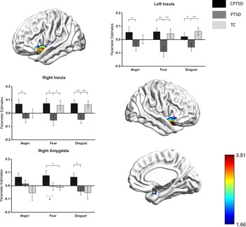
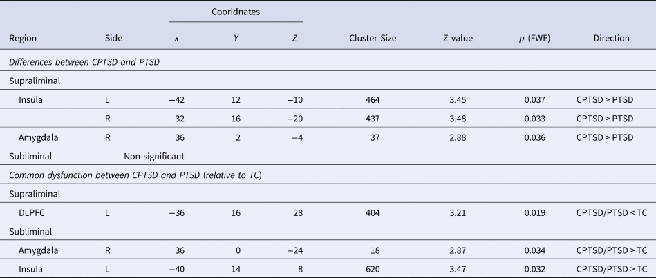
The significant main effect of group was found in the insula (bilaterally and predominantly in the anterior portion) and amygdala (right only) for the supraliminal condition (p FWE<0.05) (Table 2). Post hoc tests revealed that CPTSD had significantly greater activation relative to PTSD for each of the individual threat emotions for all of these regions with the exception of the right amygdala for anger processing (Fig. 1). No significant group differences were observed for the DLPFC or rostral ACC, or at the whole-brain level. There were no significant group differences in either of the ROIs or at the whole-brain level for subliminal processing of threat emotions. The significant group effects were not found to be driven due to differences in neutral processing as a comparative baseline. There were no group differences for the neutral contrast relative to the implicit baseline in both the right insula and right amygdala regions. Although there was a significant difference in the left insula, CPTSD had greater activation relative to PTSD.
Regions with significant differences in threat processing in CPTSD v. PTSD participants. (a) Increased activation of left insula, right insula, and right amygdala in CPTSD participants relative to PTSD participants during supraliminal presentation of threat stimuli. Voxel wise family-wise error correction set at 0.05. (b) Post hoc comparisons using mean beta estimates from these clusters indicated that CPTSD participants demonstrated increased activation in the bilateral insula during the presentation of each of the threat emotions and in the right amygdala during fear and disgust threat emotions (p < 0.05). No significant group differences were observed for subliminal threat processing.

Neural activation differences between the CPTSD and PTSD groups for supraliminal and subliminal threat processing

Using the conjunction analyses, both CPTSD and PTSD groups had significant differences in neural activation relative to TC in the left DLPFC during threat supraliminal processing (both groups <TC) and for the right amygdala and left insula during subliminal processing (both groups >TC) (Table 2). No effects were significant at the whole-brain level from this analysis for either task.
Functional connectivity
As CPTSD-PTSD activation differences were observed only for the supraliminal condition, functional connectivity analyses were performed only for this condition. Connectivity using the left and right insula and the right amygdala as seeds were evaluated relative to the hypothesized ROIs and at the whole-brain level. No differences in connectivity were significant between the two groups.
Associations with symptom measures
As we did not have a particular hypothesis with regard to the specific emotion type, we averaged activation values over the three threat emotions to test association between the CPTSD v. PTSD significant supraliminal activation clusters and symptom measures. For the entire sample, CAPS was positively correlated with the insula bilaterally (left: r = 0.269, p = 0.038; right r = 0.377, p = 0.003); while CPTSD was also positively correlated with all three regions with significant group differences (left insula: r = 0.340, p = 0.010; right insula: r = 0.388, p = 0.003; right amygdala: r = 0.318, p = 0.017).
Association with dissociative responses
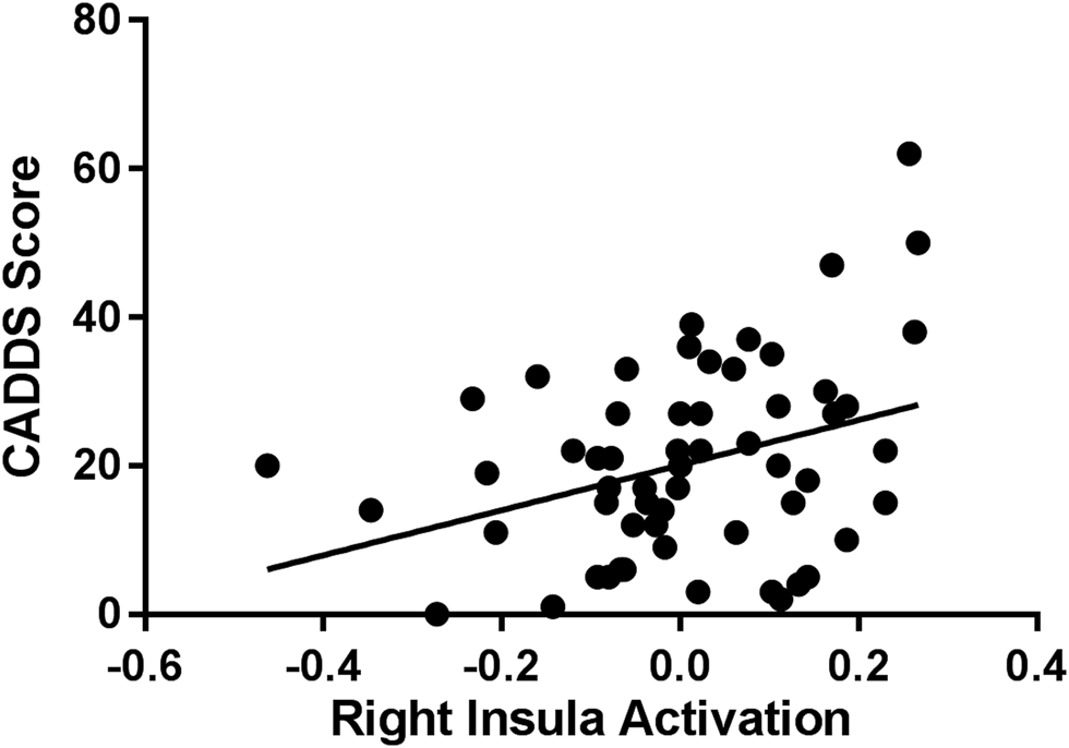
As done for the symptoms measures, we examined correlations between the bilateral insula and right amygdala activations for supraliminal threat processing with total CADDS scores in the entire sample (both groups pooled). Figure 2 demonstrates that there was a significant positive correlation between CADDS scores and right insula (r = 0.347, p < 0.007). This suggests that across the entire sample more severe dissociative responses were associated with greater activation of the right insula during threat processing.
Relationship between Clinician-Administered Dissociative States Scale and right insula activation for supraliminal threat processing. (r = 0.347, p < 0.01).

Discussion
Major findings
The key finding to emerge from this study was that participants with CPTSD displayed greater bilateral insula and right amygdala activation relative to participants with PTSD during supraliminal processing of fear, anger, and disgust faces (i.e. threat processing). This result needs to be understood in the context that the two groups did not differ in terms of severity of PTSD symptoms, and so this difference cannot be attributed to the degree of PTSD symptomatology. Numerous studies have demonstrated a distinct symptom profile of CPTSD relative to PTSD (Brewin et al., Reference Brewin, Cloitre, Hyland, Shevlin, Maercker, Bryant and Reed2017), and this investigation now provides the first evidence of neurally distinct profiles of these two disorders during threat processing. The finding that this pattern was observed in supraliminal, but not subliminal, processing accords with much research that these different stages of processing activate different neural networks (Dehaene, Changeux, Naccache, Sackur, & Sergent, Reference Dehaene, Changeux, Naccache, Sackur and Sergent2006). Some models suggest that subliminal processing may not activate comparable networks as supraliminal processing because the activation may dissipate rapidly, may be highly dependent on the salience and strength of the visual stimulus, and it may limit the depth of semantic processing (Dehaene & Changeux, Reference Dehaene and Changeux2011). It is premature to definitively infer the reasons why differences were not observed between PTSD and CPTSD during subliminal processing, and further research is needed to address this issue.
The insula is frequently activated during self-referential thinking and imagery of oneself, including interoceptive representation of bodily sensations (Craig, Reference Craig2009). It is for this reason that the insula is regarded as a region strongly underpinning self-awareness. It is also involved in the processing of negative emotions (Damasio et al., Reference Damasio, Grabowski, Bechara, Damasio, Ponto, Parvizi and Hichwa2000), which is to be expected because of its many interconnections with regions associated with the experience of emotion. The functional significance of insula activation in CPTSD may be understood in terms of the disruptions in CPTSD in self-organization, including an impoverished sense of self, low self-esteem, and poor monitoring of one's bodily state. The role of the insula in emotion processing potentially reflects the emotion dysregulation symptoms of CPTSD.
The observation of greater right amygdala activation accords with much evidence of amygdala involvement in fear expression (Phelps, Delgado, Nearing, & LeDoux, Reference Phelps, Delgado, Nearing and LeDoux2004). Neural models of PTSD that explain sensitivity to the threat have noted network involving the amygdala, insula, and dorsal anterior cingulate cortex (Shalev, Liberzon, & Marmar, Reference Shalev, Liberzon and Marmar2017), which is supported by evidence of over-reactivity in each of these networks (Lanius et al., Reference Lanius, Frewen, Girotti, Neufeld, Stevens and Densmore2007). The greater right amygdala activation in CPTSD than PTSD participants may reflect the more extreme emotion dysregulation that occurs in CPTSD, resulting in stronger fear expression and threat detection. Interestingly, CPTSD had greater activation than PTSD for each threat emotion except for anger in the right amygdala. This may be understood in the context of a meta-analysis that found that whereas fear activates the amygdala, anger does not (Fusar-Poli et al., Reference Fusar-Poli, Placentino, Carletti, Landi, Allen, Surguladze and Politi2009).
The current findings are worth considering in the context of some evidence of neural processing of negative facial expression in BPD (Schulze et al., Reference Schulze, Schmahl and Niedtfeld2016). In contrast to the current observation, several studies have found that BPD participants display diminished insula activation when processing negative emotions relative to healthy controls (Mier et al., Reference Mier, Lis, Esslinger, Sauer, Hagenhoff, Ulferts and Kirsch2013). This reduced activation in BPD participants has been interpreted in terms of deactivation of neural networks implicated in social cognition (Schulze, Schmahl, & Niedtfeld, Reference Schulze, Schmahl and Niedtfeld2016). Whereas there is some symptom overlap between CPTSD and BPD, they are strongly distinguished by the requirement that CPTSD meets PTSD criteria, and BPD is characterized by a persistent sense of abandonment. This distinction in symptom profile may in part account for the discrepancy between increased activation in CPTSD in this study and evidence of diminished activation in BPD. We also note there is contrary evidence of increased insula activation in BPD (Schulze et al., Reference Schulze, Schmahl and Niedtfeld2016), potentially reflecting the enhanced emotional reactivity commonly observed in BPD, however, these studies have used negative stimuli not involving facial expressions (Schulze et al., Reference Schulze, Domes, Kruger, Berger, Fleischer, Prehn and Herpertz2011). Moreover, a recent meta-analysis of neuroimaging studies of BPD did not identify the insula as a region that was distinctively activated in BPD during affective processing, suggesting that our finding may not be inconsistent with the prevailing evidence (Schulze et al., Reference Schulze, Schulze, Renneberg, Schmahl and Niedtfeld2019). We note that we did not formally assess for BPD in the current study and so we are unable to consider the results in relation to possible BPD diagnosis. Individuals with CPTSD and BPD can be distinguished on the basis of their symptom profiles (Brewin et al., Reference Brewin, Cloitre, Hyland, Shevlin, Maercker, Bryant and Reed2017), and so future studies could investigate differential neural patterns in individuals with BPD v. CPTSD.
The finding that activation of the bilateral insula was associated with severity of dissociative responses accords with prior evidence of a positive association between dissociation levels and insula activation during fear processing (Felmingham et al., Reference Felmingham, Kemp, Williams, Falconer, Olivieri, Peduto and Bryant2008). It also accords with evidence that for a subset of participants in a study with comorbid BPD and PTSD, dissociation levels were associated with greater right insula activity during a dissociation-inducing task (Ludascher et al., Reference Ludascher, Valerius, Stiglmayr, Mauchnik, Lanius, Bohus and Schmahl2010). The insula is implicated with monitoring of bodily and internal states (Critchley, Wiens, Rotshtein, Ohman, & Dolan, Reference Critchley, Wiens, Rotshtein, Ohman and Dolan2004), and so it is possible that more dissociative reactions are associated with a greater focus on interoceptive awareness. We note there is evidence of reduced insula activation in dissociative PTSD when processing trauma reminders, which has been interpreted as a dissociative subtype of PTSD that over-regulates distress associated with trauma memories (Lanius et al., Reference Lanius, Vermetten, Loewenstein, Brand, Schmahl, Bremner and Spiegel2010). It is possible that dissociative inhibition becomes most active in response to trauma-related material, which were not presented in the current study.
There were no significant differences in functional connectivity between CPTSD and PTSD participants. Altered connectivity between the insula and amygdala has been repeatedly observed in PTSD in response to threat-related emotional faces and during resting state (Fonzo et al., Reference Fonzo, Simmons, Thorp, Norman, Paulus and Stein2010). The lack of difference between CPTSD and PTSD in functional connectivity may be attributed to hyper-connectivity being associated with PTSD generally rather than to distinctive factors associated with CPTSD or childhood trauma. This interpretation accords with evidence that amygdala-insula connectivity has been observed in PTSD rather than childhood trauma (Jovanovic & Ressler, Reference Jovanovic and Ressler2010).
Notably, both the CPTSD and PTSD groups displayed subliminal amygdala and insula activation relative to controls. This pattern accords with the proposal that PTSD is fundamentally underpinned by disturbances in fear circuitry (Jovanovic & Ressler, Reference Jovanovic and Ressler2010). Considerable evidence points to over-reactivity of amygdala (Shin et al., Reference Shin, Wright, Cannistraro, Wedig, McMullin, Martis and Rauch2005) and insula (Morey et al., Reference Morey, Dunsmoor, Haswell, Brown, Vora, Weiner and LaBar2015) in PTSD, consistent with the role of an exaggerated response to a threat. Unexpectedly, there was evidence of greater DLPFC recruitment in PTSD and CPTSD relative to control participants, which contrasts with some models of PTSD that characterize the disorder as having hypoactivation of the DLPFC (Shalev et al., Reference Shalev, Liberzon and Marmar2017). Diminished recruitment of the DLPFC in PTSD has been reported more in the context of inhibitory or reappraisal paradigms (Rabinak et al., Reference Rabinak, MacNamara, Kennedy, Angstadt, Stein, Liberzon and Phan2014), and there are reports of increased DLPFC when PTSD participants are presented with emotional stimuli (White, Costanzo, Blair, & Roy, Reference White, Costanzo, Blair and Roy2015). The DLPFC can be understood as serving an executive control function that initiates a range of regulatory functions, including affective regulation (Fitzgerald, DiGangi, & Phan, Reference Fitzgerald, DiGangi and Phan2018), and so it is possible that the greater DLPFC observed in CPTSD and PTSD participants reflects a greater compensatory response in response to threat stimuli.
The major constraint of the current study design is that all of the current samples of CPTSD participants had experienced marked childhood maltreatment. Accordingly, we cannot disentangle the effects of CPTSD and childhood maltreatment. This potential confound is common in studies of CPTSD because it can be difficult to recruit PTSD participants with comparable levels of childhood trauma as those with CPTSD. In this context, it is noteworthy that there is evidence of distinctive insula and amygdala activity in survivors of childhood trauma (Teicher, Anderson, Ohashi, & Polcari, Reference Teicher, Anderson, Ohashi and Polcari2014). One study has found increased insula and amygdala activation in survivors of childhood abuse who were psychologically healthy (McCrory et al., Reference McCrory, De Brito, Sebastian, Mechelli, Bird, Kelly and Viding2011). This has resulted in the proposal that childhood maltreatment may lead to a specific subgroup of individuals with distinctive neural profiles (Teicher et al., Reference Teicher, Anderson, Ohashi and Polcari2014). Future studies need to study neural responding in CPTSD samples with and without a history of childhood maltreatment to determine if distinctive neural signals are associated with development trauma or CPTSD symptom presentation. In this context, it is also worth noting that we observed differences between CPTSD and PTSD in insula activation during threat processing, although CPTSD was not different from controls. This unexpected finding may be attributed, in part, to the finding that our TC were not matched to CPTSD in regard to their trauma histories.
We recognize several other limitations. First, this study relied on facial expressions as the threat stimuli. Given the diversity of insula findings in BPD in studies that have used face and non-face stimuli (Fonzo et al., Reference Fonzo, Simmons, Thorp, Norman, Paulus and Stein2010), it would be useful to use a range of threat stimuli in future studies. Second, we did not conduct a detailed history of childhood abuse; previous studies have noted that severity and timing of childhood maltreatment influences neural dysfunction (Choi, Jeong, Polcari, Rohan, & Teicher, Reference Choi, Jeong, Polcari, Rohan and Teicher2012), and measurement of these factors may have shed light on the role of childhood trauma on the distinctive profile of CPTSD participants. Third, we note that we assessed PTSD with DSM-IV criteria rather than DSM-5 or ICD-11 definitions, which raises the possibility that the current findings may not map onto replication studies using these revised definitions of PTSD. Relatedly, we did not conduct a structured clinical interview to differentiate CPTSD from BPD. Fourth, we recognize that using a symptom provocation task (such as a script-driven imagery paradigm) may have yielded different results, however, we used the face paradigm because it provides a standardized, biologically salient stimulus that can be presented both subliminally and supraliminally.
Despite these limitations, this study provides the first evidence regarding distinctive neural profiles of individuals with CPTSD and PTSD. The greater insula activation during threat processing accords with the core clinical profile of CPTSD involving emotion dysregulation and disturbances of self-concept. In the context of the debate about the merits of the CPTSD diagnosis, understanding neural substrates that differentiate this syndrome from PTSD is important in validating the new diagnosis. Further neurobiological studies are needed that examine the neural profiles of PTSD, CPTSD, and BPD to add clarity to this ongoing debate.
Supplementary material
The supplementary material for this article can be found at https://doi.org/10.1017/S0033291719003921
Acknowledgements
The study was funded by the National Health and Medical Research Council (1073041). The funder had no role in study design, data collection or analysis, or report preparation.
Conflicts of interest
No authors are declaring a conflict of interest.