Significant outcomes
-
∙ Neuropeptidases are proteolytic enzymes involved in the functional control of neuropeptides.
-
∙ Neuropeptidases interact significantly between various cortico-limbic regions in basal conditions and stress significantly change the pattern of the interactions.
-
∙ Plasma neuropeptidases correlate significantly with cortico-limbic regions in basal conditions and the pattern of these correlations was altered under stress.
Limitations
-
∙ The small sample size may limit the precision of the statistical analysis.
-
∙ Although the existence of significant correlations may be indicative of a functional association, they do not conclusively demonstrate a direct relationship between two locations.
-
∙ In general, enzymatic activities exhibit broad substrates specificity. Results may therefore be subject to various interpretations.
Introduction
Under stress conditions, the whole organism responds coordinately to maintain homeostasis in order to increase the survival of the subject (Reference Goldstein and Kopin1). The term allostasis goes beyond this concept involving mechanisms, which achieve homeostasis in response to stress conditions through anticipatory adjustments mainly of hypothalamus–pituitary–adrenal axis and autonomic nervous system (ANS) (Reference Sterling2). However, the stress response at central and peripheral level is far from being fully understood. This global response could be also framed within the neurovisceral integrative model that implies the existence of reciprocal regulatory mechanisms involving not only brain and heart (Reference Thayer and Lane3), but also virtually the whole organism that is, from brain to peripheral tissues (Reference Prieto, Villarejo and Segarra4).
The neuropeptides enkephalins and oxytocin are anxiolytic agents involved in the brain response to stress (Reference Hernández, Segarra and Ramírez5,Reference Hernández, Prieto and Segarra6). They are also released in the blood acting as neurohormones. Their role in circulation is not yet fully understood. These peptides in both brain and blood are metabolized by neuropeptidases such as enkephalin- (alanyl- and leucyl-aminopeptidase, AlaAP, LeuAP) and oxytocin- (placental-leucyl aminopeptidase, P-LeuAP) degrading activities which may be assayed fluorometrically in their soluble (Sol) and membrane-bound (MB) forms using arylamide derivatives (aminoacyl-β-naphthylamides, aaNNap) as substrates (Reference Hernández, Segarra and Ramírez5,Reference Ramírez, Prieto and Banegas7). Proteolytic enzymes, including aminopeptidases, are in general enzymes with broad substrate specificity. To analyze enkephalin- and oxytocin-degrading activities, we used AlaNNap or LeuNNap as substrates. With AlaNNap as a substrate, we can determinate MB AlaAP (EC 3.4.11.2) which hydrolyzes enkephalins and endorphins but also Ang III and bradykinins. Sol AlaAP (EC 3.4.11.14) appears more selective as it metabolizes only enkephalins. Using LeuNNap, we measure Sol and MB LeuAP (EC 3.4.11.1) that hydrolyzes enkephalins and dynorphins but also Ang II and substance P. In the presence of 20 mM of l-methionine, LeuNNap allows the determination of placental leucine-aminopeptidase (P-LeuAP; EC 3.4.11.3), also identified as the insulin-regulated aminopeptidase (IRAP) or the AT4 receptor for Ang IV that hydrolyzes not only oxytocin and vasopressin but also enkephalins, dynorphins and Ang III (reviewed in Reference Hernández, Segarra and Ramírez5,Reference Hernández, Prieto and Segarra6,Reference Segarra, Prieto and Banegas8). In the present work, we focused our attention on the role of these aminopeptidases as enkephalin- and oxytocin-degrading activities.
We have previously described the effect of acute restraint stress on these neuropeptidase activities in some cortico-limbic regions of adult male rats such as prefrontal cortex (PFC), hippocampus (HC) and amygdala (AM) (Reference Hernández, Segarra and Ramírez5,Reference Hernández, Prieto and Segarra6). Significant correlations between these brain areas were observed in control animals which were markedly changed under stress conditions (Reference Hernández, Prieto and Segarra6). However, the behavior of these neuropeptidase activities to an acute restraint stress may also be modified at the periphery, as a response to changes in plasma levels of their endogenous substrates such as enkephalin and oxytocin, acting as neurohormones that are metabolized by the same aminopeptidases. Indeed, it was previously reported that neuropeptidase activities interact significantly between brain and plasma as well as with several peripheral tissues in various experimental conditions (Reference Prieto, Villarejo and Segarra4,Reference Segarra, Prieto and Banegas8,Reference Segarra, Prieto and Banegas9).
Aims of the study
Since stress involves a global coordinated response of the body, there is presumably an interaction between brain and plasma aminopeptidase activities. In order to evaluate the neuroendocrine response to acute restraint stress, we studied its influence on the plasma AlaAP, LeuAP and P-LeuAP activities, as well as the possible interactions between these plasma activities and those of the cortico-limbic regions.
Materials and methods
Ten adult male Wistar rats, kept under standard conditions, were used in this experiment. To induce restraint stress, five animals were randomly selected and placed individually inside plastic cylinders (21 cm in length and 6 cm in diameter) for one hour. Both ends of the cylinders were closed with ventilated sliding doors. The other five rats were used as controls (Reference Hernández, Segarra and Ramírez5). Immediately after the end of the restraint period, the animals were anesthesized with equithensin and blood samples were obtained from the left cardiac ventricle. Subsequently, plasma was isolated by centrifugation for 10 min at 2000×g and stored at −20°C. AlaAP and LeuAP and P-LeuAP were measured fluorometrically using AlaNNap and LeuNNap as substrates as previously described (Reference Hernández, Segarra and Ramírez5,Reference Ramírez, Prieto and Banegas7). These enzymatic activities were compared with those (the same ones) assayed in the Sol and MB fractions of the PFC, HC and AM of the same animals and previously reported (Reference Hernández, Prieto and Segarra6). Sol and MB activities were assayed in their corresponding subcellular fractions obtained as previously described (Reference Hernández, Prieto and Segarra6,Reference Ramírez, Prieto and Banegas7). Briefly, after brain tissue samples homogenization in 10 volumes of 10 mM Tris-HCl buffer (pH 7.4), the homogenates were ultracentrifuged at 100 000×g for 30 min (4°C) to obtain the supernatant as the Sol fraction. The pellets (obtained after ultracentrifugation) were homogenized again in Tris-HCl buffer (pH 7.4) containing 1% of Triton X-100 to solubilize the membrane proteins and again ultracentrifuged (100 000×g, 30 min, 4°C) to obtain the supernatant corresponding to the MB fraction. To guarantee a complete recovery of enzyme activities, the detergent was removed from the supernatant by adding adsorbent polymeric Biobeads SM-2 (100 mg/ml) and shaking for 2 h at 4°C. Proteins were determined in triplicate by the Bradford method (Reference Bradford10) using BSA as a standard. The specific AP activities were expressed as pmol of AlaNNap or LeuNNap hydrolyzed per min per mg of protein. Fluorogenic assays were linear with respect to the time of hydrolysis and protein content. The experimental procedures for animal use and care were in accordance with the European Communities Council Directive 86/609/EEC. The Student’s t-test was used to determine statistical differences between groups. p-Values below 0.05 were considered significant. Pearson’s coefficient of correlation was computedto study the association between APs of the plasma versus that of PFC, HC and AM in their Sol and MB forms. Computations were performed using SPSS 13.0 and STATA 9.0. p-Values below 0.05 were considered significant.
Results
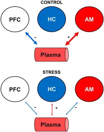
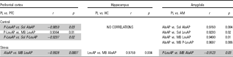
The results of enzymatic plasma activities respectively for control versus stressed animals were (mean±SEM) for AlaAP: 167.3±11.6 versus 162.5±9.7; for LeuAP: 144.1±6.4 versus 177.7±16.5; for P-LeuAP: 129.2±6.4 versus 127.4±14.4. Levels of activities in plasma and brain areas in control and under stress are provided in the figure 1 of supplementary material. Comparisons within brain areas and between control and stressed animals were previously reported (Reference Hernández, Segarra and Ramírez5,Reference Hernández, Prieto and Segarra6). No significant differences were observed between plasma of control and stressed animals. In contrast, highly significant correlations between plasma and cortico-limbic regions were demonstrated. In control rats, plasma neuropeptidase activities correlated significantly with PFC and AM but not with HC (Table 1, Fig. 1). For AM, only plasma AlaAP activity interacts positively with the other neuropeptidase activities (AlaAP, LeuAP and P-LeuAP): the higher enkephalinase activity (AlaAP) in plasma, the higher enkephalinase (AlaAP, LeuAP) and oxytocinase (P-LeuAP) activities in AM. However, for the PFC, only P-LeuAP (with oxytocinase activity) correlated (mainly negatively) significantly with the other neuropeptidase activities: the higher oxytocinase activity in plasma, the lower enkephalinase and oxytocinase activities in the PFC. In controls, no correlations were observed for plasma LeuAP with any brain area. The pattern of these correlations between plasma and brain changed radically after acute restraint stress. Only selective but highly significant correlations were observed between plasma and the three cortico-limbic areas studied: For PFC, plasma AlaAP correlated negatively with MB LeuAP (p<0.0007) (both of them having enkephalinase activity); For HC, the plasma LeuAP correlated positively with MB AlaAP (p<0.004) (both having enkephalinase activity); For AM, plasma P-LeuAP (with oxytocinase activity) correlated negatively with MB AlaAP (with enkephalinase activity) (p<0.03).
Predominance of positive (+) (red lines) or negative (−) (blue lines) correlations between plasma and the prefrontal cortex (PFC), hippocampus (HC) and amygdala (AM) in control and stressed animals. The line thickness is proportional to the number of correlations.

Plasma vs. brain

Correlations between plasma (PL) vs. soluble (Sol) or membrane-bound (MB) aminopeptidase (AP) activities in the prefrontal cortex (PFC), hippocampus (HC) and amygdala (AM), in the control and stressed animals. Pearson’s correlation coefficients (r) and p-values are indicated and specify the significance of the differences between these correlations. Negative correlations are highlighted in italics on a gray background.
Discussion
Although the enzymatic activities determined in the present study focused on enkephalinase and oxytocinase activities, it should be kept in mind that these aminopeptidases may also hydrolyze other substrates such as angiotensins, bradikinins or substance P.
Neuropeptides may be hydrolyzed by Sol or MB enzymes and, in the present and previous (Reference Hernández, Prieto and Segarra6) studies, selective significant correlations between Sol or MB activities were observed. Both pools exhibit quite different behavior and regulation mechanisms. Their abundance differs depending on the brain area and the enzyme considered, being, in general, 1–10 times higher in the Sol fraction. The existence of different functions for both Sol and MB activities was postulated (Reference Prieto, Villarejo and Segarra11). The MB activities were more tissue-specific than the Sol ones. The regulation of MB enzymes could be under nuclear control and probably functionally associated with their endogenous substrate receptors. Such regulation would be more directly linked to certain regional-selective functions and less related to more unspecific changes of their surrounding biochemical environment such as variations in pH or concentration of anions, cations, steroids, etc. The Sol enzymes may be more subject to such environmental influence (Reference Prieto, Villarejo and Segarra11).
The present results clearly support a connection between plasma and brain most likely throughout the ANS. In previous study, the correlation between the cortico-limbic structures revealed that in controls there was a positive interaction between the PFC and AM. However, there was no correlation at all between these two areas and HC. In contrast, this pattern changed markedly after restraint stress, showing mainly positive correlations between the three cortico-limbic areas: PFC, HC and AM (Reference Hernández, Prieto and Segarra6).
Stress induces an increase of plasma enkephalin levels derived from several sources: mainly from sympathetic nerves but also from the chromaffin cells of the adrenal medulla and other sites such as macrophages or the gastrointestinal tract (Reference Pierzchala and Van Loon12–Reference Barron, Pierzchala and Loon14). However, the behavior of oxytocin under restraint stress conditions is not clear at all. Sánchez et al. (Reference Sánchez, Aguado and Sánchez-Toscano15) did not find any changes of oxytocin in the paraventricular nucleus of the hypothalamus or in the median eminence of restrained rats whereas Babygirija et al. (Reference Babygirija, Bülbül and Yoshimoto16) reported an increase of oxytocin in the paraventricular nucleus together with no significant changes in plasma under restraint stress. Finally, Laguna-Abreu et al., (Reference Laguna-Abreu, Koenigkam-Santos and Colleta17) reported increased levels of plasma oxytocin under immobilization stress.
Plasma neuropeptidase activities of controls exhibited significant correlations with PFC and AM but no correlations were detected with HC. After stress there were also significant correlations between plasma and, in this case, with the three cortico-limbic regions. This suggests a certain parallelism with the correlations observed between various cortico-limbic regions themselves. We could speculate that a similar response occurs in brain and periphery mediated by the ANS. However, the functional meaning for this fact remains to be elucidated. Considering the association high neuropeptidase activity/low levels of the susceptible neuropeptides and vice-versa, the present results showing a significant inverse correlation between PFC versus plasma for, respectively, MB LeuAP and AlaAP and between plasma and AM for, respectively, P-LeuAP versus MB AlaAP under stress conditions, might suggest low levels of the corresponding neuropeptides in brain together with high levels of them in plasma. Considering that these aminopeptidase activities reflect also enkephalinase and oxytocinase activities, these results fit with the proposed role of plasma enkephalins and oxytocin as anxiolytic agents under stress conditions (Reference Lang, Heil and Ganten18,Reference Carrasco and Van De Kar19). Therefore, inhibitors of these activities could be considered as potential targets for therapeutic agents to reduce anxiety ‘as they would protect the anxiolytic agents enkephalin and oxytocin from degradation’ (Reference Loyens, De Bundel and Demaegdt20).
We have previously discussed the possibility that the brain and peripheral tissues are reciprocally connected by afferent and efferent mechanisms involving the ANS (Reference Prieto, Villarejo and Segarra4,Reference Segarra, Prieto and Banegas8,Reference Segarra, Prieto and Banegas9), thus mutually regulating the functional status of neuropeptidase activities at both locations and, as a consequence, their susceptible neuropeptidergic substrates and the functions exerted by them under certain conditions such as stress.
Therefore, in our opinion, there are enough data to hypothesize that there is a bidirectional interaction between the brain and the entire organism, carried out through the autonomic and neuroendocrine systems as communication pathways. The involved mechanisms may imply anterograde and retrograde axonal transport between nerve terminals (such as in endothelial cells of vascular walls) and neuronal bodies of motor and sensitive autonomic neurons, modulated by their functional status under stress conditions (Reference Prieto, Villarejo and Segarra4). In addition, the possible secretion of APs into the bloodstream (Reference Banegas, Prieto and Vives21), which parallel with their endothelial concentration, may be regulated by the functional status of the ANS. Neuropeptidases take a direct part in this interaction and responds in many physio-pathologic processes including the stress response. These results support a general coordinate response under stress conditions involving, in this case, a significant interaction between plasma and cortico-limbic neuropeptidase activities.
Acknowledgements
Ana Belén Segarra, Joaquín Hernández, Isabel Prieto, Marc de Gasparo and Manuel Ramírez-Sánchez contributed to the conception and design of the research, interpretation of data, critical revision of the article,and final approval of the version to be published.
Financial Support
This work was supported by the Junta de Andalucía through project no. P10-CVI6476.
Conflict of Interest
The authors report no conflict of interest.
Ethical Standards
The authors assert that all procedures used in this work comply with the ethical standards of the relevant national and institutional guides on the care and use of laboratory animals.
Supplementary material
To view supplementary material for this article, please visit http://dx.doi.org/10.1017/neu.2016.2