Introduction
More than a century of research has sought to identify the predictors of childhood cognitive function, with a growing focus on the in utero environment, thus paralleling research on predictors of childhood and adult health. Numerous precedents exist; within the framework of research on fetal origins of chronic disease, there is now considerable evidence that should direct attention upstream to those factors at play at the time a child is born, namely to the intrauterine environment. The placenta has been suggested to be a window into the intrauterine environment through which the accrued consequences of exposures, genetic susceptibilities and their interactions can be inferred from the earliest stages of pregnancy.Reference Hemachandra, Klebanoff, Duggan, Hardy and Furth 1 – Reference Thompson and Regnault 6 The data relating the intrauterine period to the origins to at least some types of altered brain function is also robust (e.g.Reference Rees and Inder 7 , Reference Coe and Lubach 8 ). Recent studies have focused on the possible role of paternal age on offspring characteristics including childhood intelligence quotient (IQ).Reference Liu, Zhi and Li 9 However, while placental growth (as represented by placental weight and its derivatives including its shape) has been linked to child and adult health,Reference Campbell, Hall and Barker 10 – Reference Misra, CM, Chadha, Charles and Miller 14 no prior published studies have reported associations with IQ in childhood or adulthood. We sought to extend our work on the link between placental growth as reflected in placental shape and childhood and adult healthReference Misra, CM, Chadha, Charles and Miller 14 – Reference Salafia, Zhang and Miller 19 to the outcome of childhood IQ. We and others assert that the delivered placenta is a representation, or a summary, of the exposures and experiences of the in utero environment of the child. This would suggest that there may be an association between measures of placenta and childhood IQ as other evidence indicates the potential importance of prenatal factors.Reference Camp, Broman, Nichols and Leff 20 – Reference Broman 27 To date, prior studies of the relationship between the placenta and child and adult outcomes have rarely utilized measures other than simply placental weight and the placental weight–birth weight ratio (or its inverse, the fetoplacental weight ratio). Placental weight is but a summary of many dimensions of placental villous growth, including the placental chorionic surface area, and the thickness of the placental chorionic disk. The placental chorionic surface area reflects the laterally expanding growth of the placenta (measured by disk shape, the distance from the cord insertion site to the nearest disk margin and by chorionic disk diameters). This aspect of placental growth serves two functions. First, the umbilical–chorionic vessels run in the chorionic plate. These large caliber vessels bear the burden of transfer of large volumes of fetal blood to and from the villus capillary bed. Thus, disk shape, distance from cord insertion to margin and large/small chorionic disk diameters are each measures that capture dimensions of this high capacitance/low resistance flow system. Second, the chorionic disk area measures the area of the uterine lining covered by the placentaReference Craven, Zhao and Ward 28 and in effect, how many maternal spiral arteries are potential suppliers of the placenta.
Placental chorionic disk thickness reflects the arborization of the fetal stem villi with their arterioles, which terminate in terminal villi, the locus of the villous and vascular nutrient exchange surface.Reference Benirschke, Kaufmann and Baergen 29 However, the fetal stem arterioles are principal sites of placental vascular resistance,Reference Sagol, Sagol and Ozdemir 30 , Reference Mitra, Seshan and Riachi 31 thus they contribute to total fetal peripheral resistance and fetal heart work.Reference Sebire and Talbert 32 In contrast to the more global measures of placental weight and the ratio to birth weight, analyses of these more nuanced measures could shed light on the mechanisms linking placental growth to later cognitive outcomes, as they have for somatic outcomes.Reference Misra, CM, Chadha, Charles and Miller 14
Using data from the U.S. Collaborative Perinatal Project (CPP), we sought to determine how placental growth measures relate to child IQ at age 7 years. We considered a number of standard placental measures of growth and development as potential predictors of childhood IQ. On the basis of recent studies implicating parental age and sex differential effects therein, we also considered the role of mother's and father's age and whether the placental measures’ effects differed for boys and girls.
Methods
Subjects were a subset of the National CPP. Details of the study have been described elsewhere.Reference Liu, Zhi and Li 9 , Reference Campbell, Hall and Barker 10 Briefly, from 1959 to 1965, women who attended prenatal care at 12 hospitals were invited to participate in the observational, prospective study. At entry, detailed demographic, socioeconomic and behavioral information was collected by in-person interview. A medical history, physical examination and blood sample were also obtained. In subsequent prenatal visits, women were repeatedly interviewed and physical findings were recorded. During labor and delivery, placental gross morphology was examined and samples were collected for histologic examination. The children were followed up to 7 years of age.
In the full CPP cohort, there were 55,740 singleton pregnancies. The full CPP data set includes more than one pregnancy to the same mother over the 7 years of recruitment, necessitating selection of a subset or use of methods to account for the lack of independence of the observations. The sample was therefore restricted to only or first singleton live births within a family. Among 41,341 eligible singleton births, 36,017 contributed placenta data (∼87%). The analytic sample was restricted to those with complete data on the necessary placental gross measures (see next paragraph), placental weight, birth weight and known gestational age ⩾34 weeks and <43 completed weeks (n = 24,152, 67%). The lowest 0.5% of placental variables was also excluded, as biologically implausible, leaving a sample of 24,061 subjects with placental and pregnancy data.Reference Salafia, Zhang and Charles 18 , Reference Salafia, Zhang and Miller 19 We then excluded observations with missing data on key variables for analyses focused on in this manuscript: maternal and/or paternal age; race categorized as African-American or white; socioeconomic status (SES); age 7-year IQ test results; exact age at the 7-year IQ testing. This sample composition (n = 12,883) is consistent with prior analyses of age 7-year IQ in this data set by other investigators,Reference Saha, Barnett and Foldi 33 albeit smaller due to the need for complete placental data (20,827 v. 12,883) and potentially a result of sampling differences (choosing a random birth rather than the first or only birth). Comparing births in the CPP with and without placental data suggests that there is no major difference in the samples (seeReference Salafia, Zhang and Charles 18 , Reference Salafia, Zhang and Miller 19 , Reference Baptiste-Roberts, Salafia and Nicholson 34 ).
Placental variables
Placental gross measures were collected according to a standard protocol;Reference Benirschke 35 the original codings and recodings used for this analysis follow:
(1) Placental weight. Placental weight was measured in decagrams to the nearest 10 g; the unit of analysis was grams.
(2, 3) Placental (chorionic plate) surface diameters (large, small). Diameters (large and small) of the chorionic plate surface were recorded in centimeters, and analyzed as coded.
(4) Placenta (chorionic plate) surface area (cm 2 ). This is a mathematical product of the larger and smaller chorionic plate surface diameters.
(5) Placental (chorionic disk) thickness. Placental chorionic disk thickness at the center of the chorionic plate was measured by piercing the disk with a knitting needle on which millimeter marks were inscribed, and analyzed in units of 0.1 cm.
(6) Placental (chorionic plate) shape. Chorionic plate shape coding was based on the gross examination of the delivered placenta. Surface shapes included round-to-oval, and a variety of atypical shapes (e.g. bipartite, tripartite, succenturiate, membranous, crescent or ‘irregular’). Only 926 (3.8%) were labeled as one of the six categories of shape other than round-to-oval. For this analysis, shape was recoded as a binary variable with ‘round-to-oval’ as ‘0’ and ‘other than round-to-oval’ as ‘1’.
(7) Umbilical cord length. Umbilical cord length was measured in the Labor and Delivery Room, and recorded in centimeters.
(8) Umbilical cord eccentricity. Distance from the umbilical cord insertion to the closest edge of the placental chorionic plate was recorded to the nearest centimeters. Umbilical cord insertion was also coded as membranous (velamentous), marginal or normal (inserted onto the chorionic plate surface). We combined these two variables into a single umbilical cord insertion distance measure, by recoding velamentous cord insertions as a negative value, cords inserted at the placental margin as ‘0’ and progressively more central cords as ‘1’ to ‘9’ (overall scale range: 13–13; further details available from the authors).
Parental characteristics
Parental characteristics were recorded at enrollment. Paternal and maternal ages were measured (at enrollment) in years and months with transformation to centered variables to minimize problems with collinearity.Reference Wood 36 Parity counted all delivered offspring and did not include miscarriages/early pregnancy losses; parity was recoded into a binary variable (0 = nulliparous, 1 = parous). SES index was a combined score for education, occupation and family income as scaled by the US Bureau of the Census.Reference Myrianthopoulos and French 37 Mother's race was coded as a binary variable denoting African-American as ‘1’ and all others as ‘0’; original data coded race as Caucasian, African-American, and ‘other’, most of whom were Puerto Ricans (9.2%).
IQ at age 7 years was assessed using the Wechsler Intelligence Scale for Children (WISC), widely regarded as a valid and reliable measure of intelligence in children. 38
Preliminary analyses indicated gender-modified effects of key variables consisted with prior work by our team.Reference Misra, Salafia, Miller and Charles 15 Therefore, analyses were stratified on gender with covariates of SES, African-American race, parity, gestational age and exact age at WISC (IQ) testing.
Placental weight (g), chorionic plate surface diameters (cm) and chorionic plate surface area (cm2), chorionic disk thickness (mm), shape, umbilical cord length (cm) and umbilical cord eccentricity were independent variables in linear regression analyses (SPSS-PC) of age 7-year IQ adjusted for the exact age at testing in 12,883 singleton term liveborns with complete placental data. Prior work further suggested the potential for nonlinearities in placental variable effectsReference Misra, Salafia, Miller and Charles 15 so we considered linear regression with categorical predictors. Because of instability of models, we explored these effects only for variables that had similar effects for boys and girls so that data could be combined.
Results
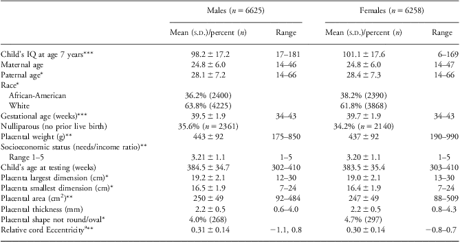
In Table 1 we described the means and distribution of a number of birth and placental characteristics measured in this study. The 12,883 singleton births were approximately evenly distributed by sex with 49% female. The racial distribution was less evenly distributed with slightly more than one-third (37.2%) reported as African-American.
Descriptive characteristics of the study sample

IQ, intelligence quotient.
aCord location relative to disk edge normalized to radius of disk.
*P < 0.05; **P < 0.01; ***P < 0.001.
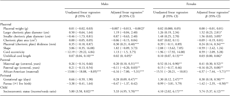
All models were adjusted for exact age at the 7-year-old IQ testing. We first examined the effects of each placental measure alone. All of the covariates we considered for inclusion have been reported by us or others to be related to both IQ and placental factors. Furthermore, there is biologic plausibility for some of these associations (e.g. nulliparity, gestational age). In addition, we conducted analyses to test the association between each of the covariates and each placental factor. Then we considered effects of placental measures in a single model together after taking account of parental, perinatal and child factors as these factors were likely to confound effects of placental factors. We compared the estimate of the regression coefficient and the model fit with the covariate both included and excluded from our models of placental variables and IQ. Table 2 provides the results for the simple models of the independent variables (placental, parental, perinatal and child) and IQ, as well as the results for the full models (see next paragraph for description of adjusted full models). Race and SES were associated with IQ in both genders. In the models considering each placental measure alone, we found that, in girls only, the greater the larger and smaller placental chorionic plate surface diameters, and the chorionic plate surface area (essentially an interaction term for the larger and smaller diameters), the greater the IQ. Maternal age and gestational age were also each positively associated with increased IQ in girls but not boys. Umbilical cord length was also positively associated with IQ in both genders.
Placental predictors of WISC Full Scale IQ (points) at age 7 years

WISC, Wechsler Intelligence Scale for Children; IQ, intelligence quotient.
All models control for child's exact chronological age at age 7-year testing.
aAdjusted models include all covariates listed in this table.
*P ⩽ 0.05; **P ⩽ 0.01; ***P ⩽ 0.001.
After adjustment for parental, perinatal and child covariates (see Table 2), several of the placental measurements independently associated with IQ at age 7 years and again, results varied by gender. The tests for interactions between gender and placental factors in these cases (placental chorionic plate surface diameters, umbilical cord length) were statistically significant (P < 0.05).
Placental weight
There was a positive association between placental weight and IQ but only in boys. However, the size of coefficient was very small.
Placenta (chorionic plate) surface diameters
Both the smaller and the larger chorionic plate surface diameters were positively associated with IQ only in girls.
Placental (chorionic disk) thickness
There was a positive association between placental chorionic disk thickness and IQ for both boys and girls with a similar magnitude of effect.
Umbilical cord length
There was a positive association with IQ for girls only, with approximately one-third the magnitude of effect of the unadjusted analysis.
The R 2 (approximate variance explained) by the covariates as a group was ∼0.218 for males and 0.257 for females. The addition of the placental variables all together incrementally increased the R 2 to 0.223 in males and 0.263 in females.
Prior work suggested the potential for nonlinearities in placental effects so additional analyses were performed to examine a range of cutpoints of the predictor as described in the section ‘Methods’. However, due to instability in these models, we have not presented these results.
Discussion
Our analysis finds that placental measures including measures of the chorionic disk shape, are related to cognitive outcome at age 7 years. These findings build on our previous work in this field. First, it should not be unexpected that placental shape measures that are reflections of the cumulative growth of the placenta across gestation affects neurodevelopment given the evidence that the intrauterine environment is significantly correlated with such outcome.Reference Rees and Inder 7 , Reference Coe and Lubach 8 , Reference Clark, Mele and Wapner 39 The placenta is the fetus’ sole source of oxygen and nutrients and as such would be reasonably expected to influence newborn outcome directly. We have recently shown an allometric scaling relation between the placental weight and the birth weight in the CPP cohort with an exponent approximately equal to 0.75, as predicted by Kleiber's Law. This implies that placental weight is a justifiable proxy for fetal metabolic rate when other measures of fetal metabolic rate are not available.Reference Salafia, Misra, Yampolsky, Charles and Miller 40 However, placental growth may also mark exposures that alter fetal neurodevelopment, and thus be more indirect measures of the intrauterine forces and factors that modulate fetal neurodevelopment. In addition, placental shape measures may provide insights into timing of gestational stressors sufficient to deform placental growth and potentially affect the fetus.Reference Salafia, Yampolsky, Shlakhter, Mandel and Schwartz 41 Within the CPP cohort, our group has produced a number of important findings of independent effects of placental growth as reflected by measures of the chorionic disk surface and chorionic disk thickness on neonatal and childhood outcomes, as well as gender specificity of such effects. These findings include:
-
1. Placental shape measures including those of the chorionic plate surface and chorionic disk thickness, have independent significant effects on fetal growth;Reference Salafia, Zhang and Charles 18 that is, placental weight does not capture all the subtlety of the information about gestational ‘adequacy’ that is encoded in placental shape.
-
2. Different placental disk proportions yield different mean birth weights, with ranges of fetoplacental weight ratios from 8.46 to 6.33. Taking the mean weight of a well-proportioned placenta (mid quartiles for chorionic plate area and thickness, 435.9 g), these different fetoplacental weight ratios would yield expected birth weights of from 3688 g (actual observed mean birth weight 2823 g) to 2759 g (observed mean birth weight 3620.5 g).Reference Salafia, Zhang and Miller 19 The impact of changing placental disk proportions, independent of any other factor, is large.
-
3. Female and male fetuses do not have the same relationships of their birth weights to placental shape measures.Reference Misra, Salafia, Miller and Charles 15 Female fetuses appear to modulate their growth more closely in step with changing placental disk proportions; male fetuses appear to grow without responding to changing placental shape measures, where these changes may reflect shifts in the intrauterine environment that require adaptation.
-
4. How large or small a newborn is relative to his/her placental shape measures (as calculated in the observed to expected birth weight ratio) is associated with significant and independent effects on body mass index and diastolic blood pressure.Reference Misra, CM, Chadha, Charles and Miller 14
In the current study, we extend these observations to IQ, a measure that has been superseded by other measures of cognitive function but which does capture aspects of function that have proved informative in the understanding of cognition in the past.Reference Naglieri and Goldstein 42 We continue to demonstrate both gender-specific and nonlinear effects of placental measures on this measure of neurodevelopmental outcome. To our knowledge, there are no published reports assessing the influence of placental factors on IQ or other measures of cognitive function.
We find the association of IQ measures of placental growth and especially placental thickness to be intriguing because the placenta is an ideal site to search for developmental alterations in angiogenesis that may mirror altered neuronal structures that depend on similar regulatory systems.Reference Zacchigna, Ruiz de Almodovar and Carmeliet 43 – Reference Autiero, De Smet, Claes and Carmeliet 45 Our current working hypothesis is that structures that develop by branching have conserved morphogenetic mechanisms.Reference Davies 46 , Reference Patel, Sharpe and Miletich 47
Placental disk thickness is a direct measure of placental villous and vascular branching growth, which involves branching angiogenesis until the early third trimester.Reference Benirschke, Kaufmann and Baergen 29 Since villous growth is driven by vascular growth,Reference Mayhew 48 , Reference Benirshcke, Kaufmann and Baergen 49 disk thickness is an indirect measure of placental angiogenesis.Reference Kingdom, Huppertz, Seaward and Kaufmann 50 Placental angiogenesis depends on the same factors that regulate angiogenesis elsewhere in the body.Reference Storkebaum, Lambrechts and Carmeliet 51 These factors have recently been shown to be deeply involved in neuronal development and maintenance. Even vascular endothelial growth factor, once thought to be exclusively involved in angiogenesis have recently been identified as a neurotrophic factor.Reference Storkebaum, Lambrechts and Carmeliet 51 Conversely, factors identified initially for their neurotrophic properties are now recognized as important angiogenic mediators.Reference Hu, Wang and Guo 52 Cross-talk between angiogenic and neurotrophic factors emphasizes the extent to which vascular and neuronal developmental programs are integrated.Reference Li, Ford, Lavik and Madri 53 – Reference Sondell, Sundler and Kanje 64 The placenta, the anatomical barrier between mother and fetus, is a complex 3D vascular tree covered by a thin veneer of supporting tissue. Forming the boundary between maternal and fetal beings, the placenta is the obvious ‘first responder’ to factors delivered to the conceptus via maternal perfusion,Reference Reynolds, Borowicz and Caton 65 including environmental toxicants (e.g. cadmiumReference Helmestam, Stavreus-Evers and Olovsson 66 ), decidually produced trophic factors such as interleukin-10 (e.g.Reference Thaxton and Sharma 67 ), as well as factors that impact on cell cycle regulation.Reference Giudice 68 Our findings that there is a direct and independent relationship between placental chorionic disk thickness, a simple albeit crude (see next paragraph) measure of placental branching vascular growth and IQ, a measure of aspects of neuronal/cognitive function, are yet another piece of evidence that may provide a link between control of vascular and neuronal development.
We cannot exclude, as an alternative hypothesis, that the correlation of reduced chorionic disk thickness and age 7-year IQ reflects instead effects of reductions in transplacental transport of nutrients and factors key to neurodevelopment. However, the reductions in birth weight we have observed are small, on the level of tens of grams.Reference Salafia, Zhang and Charles 18 We think it unlikely that reduced chorionic disk thickness is correlated with age 7-year IQ on the basis of altered placental nutrient transfer function alone.
Limitations of this study are that the standard measures of placental shape are imprecise and more poorly measure the most complicated placental shapes, which our data suggest indicate placentas that have been subject to gestational stress. Irregularly shaped placentas with variable disk thickness may have ‘struggled’ more to maintain fetal well-being; a single pair of diameters and a single disk thickness may not capture the complexity of their shape. More precise placental shape measures, as delineated in our most recent workReference Yampolsky, CM and Shlakhter 69 exist. As such, we expect that future studies that utilize more precise placental measures will not only confirm the above observations, but illuminate subtleties in the placental shape–IQ relationship that cannot be assessed using the current standard, but crude, measures. There is also an increased potential for false positive findings given the number of placental variables examined.
Conclusion
IQ at age 7 years was associated with placental factors with apparent differences by gender. Several placental measures, including the more nuanced measure of placental thickness, are also correlated with age 7-year IQ independent of parental, child and perinatal factors, but the findings vary by gender. Further exploration of the possible interaction of these factors on the arborization, reflected by placental chorionic disk thickness, that correlates with age 7-year IQ is indicated.