Introduction
Scimitar syndrome is an uncommon congenital malformation of pulmonary venous drainage to the junction of the right atrium and inferior caval vein and is often associated with lung hypoplasia and anomalous systemic arterial blood supply to the anomalously draining lung segment. Reference Canter, Martin and Spray1,Reference Alsoufi, Cai and Van Arsdell2 Rarely, there may be dual pulmonary venous drainage to both the inferior caval vein and the left atrium, often referred to as the “scimitar variant,” Reference Schramel, Westermann and Knaepen3 which we will refer to as the scimitar variant with dual pulmonary venous drainage.
Surgical correction of the anomalous pulmonary venous drainage to reduce the left-to-right shunt is usually indicated. However, when dual drainage of the pulmonary veins is identified, it may be possible to correct this anomaly with a transcatheter approach. Reference Luciano, Laux and Boudjemline4 The venous anatomy, however, is variable, and although several devices have been used for vascular closure, their safety and efficacy have not been fully established. We present a series of four patients with the scimitar variant with dual pulmonary venous drainage treated with transcatheter procedures in three institutions (Evelina London Children’s Hospital, UK; Jagiellonian University Medical College, Poland; Centre Hospitalier Universitaire Sainte-Justine, Canada).
Methods
We retrospectively collected data from four patients with the scimitar variant with dual pulmonary venous drainage, who were treated with a transcatheter procedure. The anatomical findings were delineated with either CT or MRI. All the patients had right ventricular volume overload by echocardiography and/or MRI.
Results (Table 1)
Case 1
A 19-year-old man was incidentally found to have a scimitar variant with dual pulmonary venous drainage during investigation of a shoulder injury. CT imaging demonstrated dual pulmonary venous drainage to the systemic veins and the left atrium (Figure 1a). At cardiac catheterisation, selective right lower pulmonary arteriography confirmed that the right lower pulmonary vein drained to the inferior caval vein/right atrium junction and onwards to the left atrium via a cranially draining vein. The pulmonary vein measured 11 mm in diameter at its connection with the inferior caval vein. Balloon test occlusion of the scimitar vein with a 12 mm x 3 cm Tyshak II balloon (NuMED Inc., Hopkinton, NY, USA) with concurrent selective right lower pulmonary arteriography confirmed unobstructed pulmonary venous drainage to the left atrium (Figure 1b). There was no rise of the right pulmonary capillary wedge pressure during balloon occlusion. An 18 mm Amplatzer Muscular Ventricular Septal Defect Occluder (Abbott, Abbott Park, Illinois, USA) was deployed to occlude the anomalous pulmonary venous drainage to the systemic venous circulation without complication (Figure 1c).
( a ) Three-dimensional constructed CT images demonstrating dual pulmonary venous drainage to the systemic veins and the LA. ( b ) Dual drainage was confirmed by balloon occlusion of the scimitar vein and subsequent pulmonary artery injection with levophase imaging of the pulmonary veins. ( c ) implantation of an 18 mm Amplatzer Muscular Ventricular Septal Defect Occluder in scimitar vein without obstruction of additional drainage. CT, LA: left atrium.

Characteristics of the patients and interventions. Pt. no: patient number, CHD, AMVSD: amplatzer muscular ventricular septal defect occluder, AVP: amplatzer vascular plug, AALAA: amplatzer amulet left atrial appendage occluder, APC: aortopulmonary collaterals

Case 2
A four-year-old boy, weighing 15.3 kg, presented with recurrent chest infections. Transthoracic echocardiography demonstrated that the right lower pulmonary vein drained to the inferior caval vein. Cardiac MRI demonstrated a scimitar variant with dual pulmonary venous drainage, suggesting that there was an additional communication of the anomalously draining right lower pulmonary vein with the right middle pulmonary veins, allowing alternative drainage to the left atrium if the right pulmonary venous connection to the inferior caval vein were to be occluded. At cardiac catheterisation, contrast injection in the anomalous right lower pulmonary vein showed retrograde contrast flow to the right middle pulmonary vein and into the left atrium, confirming dual drainage of the right lower lobe. A 10 mm Amplatzer Vascular Plug II (Abbott, Abbott Park, Illinois, USA) was deployed in the scimitar vein without complication. Selective pulmonary angiography demonstrated unobstructed flow from the right lower pulmonary vein to the right middle pulmonary vein and trivial residual flow to the inferior caval vein.
Case 3
An 11-year-old boy with VACTERL association (vertebral defects, anal atresia, cardiac defects, trachea-oesophageal fistula, renal anomalies, and limb abnormalities) had been initially diagnosed with PAPVD in another institution. He presented with mild dyspnoea on exertion. CT scanning demonstrated a scimitar variant with dual pulmonary venous drainage. A trans-septal puncture was performed to access the right lower pulmonary vein using an 8.5 Fr steerable sheath (Abbott, Abbott Park, Illinois, USA). Direct pulmonary venous angiography demonstrated the right lower pulmonary vein draining to both the inferior caval vein and left atrium. The proximal vessel size was 13 mm, and the distal vessel connecting with the inferior caval vein was 10 mm (Figure 2a). An 18 mm Amplatzer Amulet Left Atrial Appendage occluder (Abbott, Abbott Park, Illinois, USA) was deployed successfully in the scimitar vein from an inferior caval vein approach with the “body” of the device placed in the vessel and the disc at its connection with the inferior caval vein (Figure 2b, 2c).
( a ) (AP projection, lateral projection): right lower pulmonary vein draining to left atrium. Accessory right lower pulmonary vein (scimitar vein) draining to the IVC (arrow in lateral projection). ( b ) (AP projection, lateral projection): contrast injection from right pulmonary vein showing the occlusion of the scimitar vein with 18 mm Amplatzer Amulet Left Atrial Appendage Occluder. ( c ) (AP projection, lateral projection): contrast injection from IVC showing complete occlusion of the scimitar vein. ( d ): CT scanning one week after device implantation showing scimitar vein occlusion and pulmonary vein drainage to LA, without obstructing of the IVC. IVC: inferior caval vein, CT, LA: left atrium.

Case 4
A three-year-old girl, weighing 17.4 kg, presented with recurrent chest infections. Her CT scan demonstrated a scimitar variant with dual pulmonary venous drainage. There were also aortopulmonary collaterals originating from the abdominal aorta connecting to the right lower pulmonary artery. At cardiac catheterisation, three aortopulmonary collaterals were occluded with Amplatzer Vascular Plug IV devices (Abbott, Abbott Park, Illinois, USA). Balloon test occlusion of the scimitar vein with a 10 mm x 2 cm Tyshak II balloon (NuMED Inc., Hopkinton, NY, USA) with concurrent pulmonary angiography demonstrated unobstructed right lower pulmonary vein drainage to the left atrium without elevation of right pulmonary capillary wedge pressure. The scimitar vein was occluded with a 10 mm Amplatzer Vascular Plug II (Abbott, Abbott Park, Illinois, USA).
Follow-up
All patients were discharged without complications. Antithrombic therapy after the procedure was as follows: Case 1 received aspirin and clopidogrel for 6 months; Case 2 received aspirin for 6 months; Case 3 received aspirin and clopidogrel for 3 months followed by aspirin for a further 3 months; and Case 4 received aspirin for 6 months. These regimens reflected institutional practice, as each centre adhered to its own protocol. There was no residual shunt in two patients (50%) and minimal residual shunt in the other two (50%) patients at discharge. No residual shunt or pulmonary vein obstruction was present on follow-up echocardiography at 3 months. All patients remain asymptomatic at follow-up (29.5 (8–79) months).
Discussion
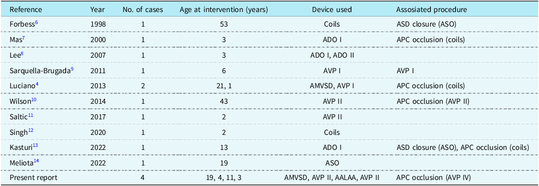
Scimitar syndrome constitutes approximately 6% of patients referred for surgical repair of PAPVD. Reference Alsoufi, Cai and Van Arsdell2 Dual drainage of the anomalous pulmonary veins with connection to both the systemic venous return and the left atrium is rare. These “dual drainage” anatomies can be further subdivided into those in whom the right middle and lower pulmonary veins drain into the inferior caval vein and also into the left atrium via connecting veins, and those in whom the left upper veins of the left lung drain to the innominate vein via a large vertical vein and to the left atrium via another developed left pulmonary vein branch. Reference Luciano, Laux and Boudjemline4 Several reports of transcatheter occlusion of the latter type (the “vertical vein variant”) have been published. Reference Luciano, Laux and Boudjemline4–Reference Forbess, O’Laughlin and Harrison6 However, only a limited number of case reports of transcatheter correction of the “scimitar variant” have been published. A review of the literature identified 11 previously reported cases of transcatheter correction of the scimitar variant with dual pulmonary venous drainage (Table 2). Reference Luciano, Laux and Boudjemline4,Reference Forbess, O’Laughlin and Harrison6–Reference Meliota, Zaza and Vairo14 The number of reported cases per study ranged from single-patient case reports to small series of two patients. Various occlusion devices have been employed, including coils, Amplatzer Vascular Plugs, Amplatzer Duct Occluders, Amplatzer Septal Occluders, and Amplatzer Muscular Ventricular Septal Defect Occluders. Associated procedures were frequently performed, such as atrial septal defect closure and aortopulmonary collateral embolisation. This heterogeneity reflects the variable venous anatomy and associated haemodynamic lesions encountered in this patient population. Our present report adds four further cases in which the Amplatzer Muscular Ventricular Septal Defect Occluder, Amplatzer Vascular Plug II, and Amplatzer Amulet Left Atrial Appendage Occluder were used. To our knowledge, this is the first report describing the use of Left Atrial Appendage occluder for occlusion of the scimitar vein in the scimitar variant with dual pulmonary venous drainage. The rationale for selecting the Left Atrial Appendage occluder was related to both size and stability. Given the vessel dimensions, a device large enough to seal the anomalous connection and equipped with an anchoring mechanism to ensure stability was deemed necessary. The Amplatzer Amulet Left Atrial Appendage occluder has hooks that deploy distally, allowing secure anchoring to the vessel wall. These cases demonstrate the feasibility of using different device platforms and combined interventions to achieve complete redirection of pulmonary venous return to the left atrium in anatomically suitable patients. Cross-sectional imaging with CT or MRI was crucial in all cases to define the venous anatomy and confirm the presence of dual drainage, although careful procedural assessment—including balloon occlusion testing with concurrent pressure measurement and selective angiography—remains essential to ensure unobstructed pulmonary venous drainage following device deployment. The expanding cumulative experience, now including our four new cases, underscores the importance of an individualised, anatomy-driven approach performed in high-volume congenital/structural catheter laboratories with a full complement of occlusion devices available.
Reported cases of transcatheter correction for scimitar variant with dual pulmonary venous drainage. No.: number, ASO: amplatzer septal occluder, ADO: amplatzer duct occluder, APC: aortopulmonary collaterals, AVP: amplatzer vascular plug, AMVSD: amplatzer muscular ventricular septal defect occluder, AALAA: amplatzer amulet left atrial appendage occluder

Conclusion
In the setting of anomalous pulmonary venous connections, a detailed assessment of the pulmonary venous drainage by CT or MRI scanning is important to define the presence of dual drainage. The identification of such alternative venous pathways to the left atrium may allow for a transcatheter approach in some patients with scimitar variant with dual pulmonary venous drainage.
Financial support
No funding was received to assist with the preparation of this manuscript.
Competing interests
Shakeel A Qureshi is a consultant for NuMED Inc. and Venus Medtech and does proctoring or lectures for NuMED Inc., Venus Medtech, Occlutech, Medtronic Inc., and Abbott. Matthew I Jones is a consultant for Abbott Medical and Venus Medtech. Mounir Riahi is a consultant for Abbott Medical and Venus Medtech. All other authors declare no conflicts of interest.