Introduction
Ultrasonography is the primary modality used for imaging thyroid nodules, as it is readily accessible, non-invasive and cost-effective. According to the American Thyroid Association guidelines for the assessment of thyroid nodules, thyroid sonography with survey of the cervical lymph nodes should be performed on all patients with known or suspected thyroid nodules.Reference Haugen, Alexander, Bible, Doherty, Mandel and Nikiforov1 An anteroposterior-to-transverse ratio of greater than 1.0, or so-called ‘taller-than-wide’ sign, is a significant sonographic feature to predict malignancy of thyroid nodules; it is typically assessed on a transverse image, with height measurements taken parallel to the sound beam and width measurements taken perpendicular to sound beam.Reference Tessler, Middleton, Grant, Hoang, Berland and Teefey2 In 2002, Kim et al.Reference Kim, Park, Chung, Oh, Kim and Lee3 introduced the concept of the ‘taller-than-wide’ shape of thyroid nodules for the first time, and presented new sonographic criteria for fine-needle aspiration biopsy of non-palpable solid thyroid nodules. The American College of Radiology launched the Thyroid Imaging, Reporting and Data System (‘TI-RADS’) in 2017,Reference Tessler, Middleton, Grant, Hoang, Berland and Teefey2 and considered the ‘taller-than-wide’ sign as an important factor to predict the malignancy of thyroid nodules.
With the development of artificial intelligence for predicting thyroid malignancy, quantitative analysis of ultrasound images is being given increasing attention. However, to date, only a few scholars have investigated the optimal cut-off value of an anteroposterior-to-transverse ratio to distinguish malignant nodules from benign ones, and few have attempted to determine whether nodule size affects the efficiency of the anteroposterior-to-transverse ratio. Hence, the present study aimed to address these problems.
Materials and methods
Study population
This study was approved by the Ethics Committee of Beijing Tongren Hospital, Capital Medical University, and informed consent for ultrasound and surgery was obtained from all patients prior to each procedure.
For this study, we collected the pre-operative ultrasound and post-operative pathological data of patients with thyroid nodules who were admitted to multiple medical centres in China (Beijing Tongren Hospital, China-Japan Union Hospital of Jilin University, and First Hospital of Qinhuangdao), from 2012 to 2019.
The inclusion criteria were as follows: (1) complete information regarding patient's age, gender and pathological diagnosis; (2) high-resolution pre-operative ultrasound images; and (3) post-operative pathological diagnosis of nodules. The exclusion criteria were as follows: (1) patients with incomplete data (e.g. age, gender and/or diagnosis); (2) absence of high-resolution pre-operative ultrasound images; and (3) lack of post-operative pathological diagnosis of nodules.
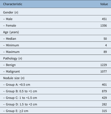
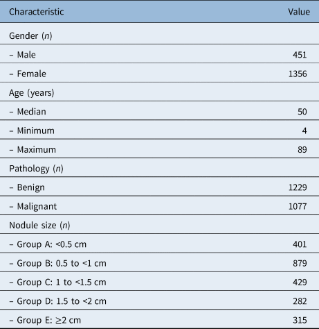
The initial population included 6395 patients with thyroid nodules who underwent surgical procedures. The sample was refined by excluding 4352 patients without high-resolution pre-operative ultrasound images and 17 patients with an undetermined pathological diagnosis of nodules. Finally, 1807 patients were enrolled in the study, including 451 male and 1356 female patients. The patients’ maximum age was 89 years old, and their minimum age was 4 years old, with a median age of 50 years old. A total of 2306 nodules (1229 benign nodules and 1077 malignant nodules) were involved. The nodules were divided into five groups according to the maximum diameter of the nodules, as shown in Table 1.
Characteristics of thyroid nodules in the study
Ratio measurement
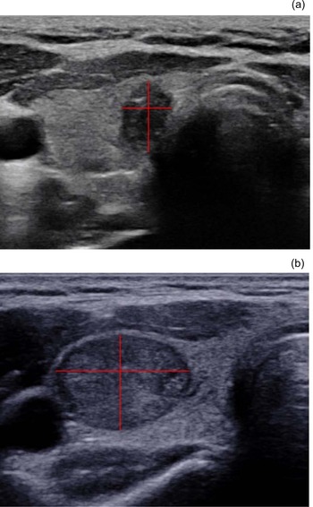
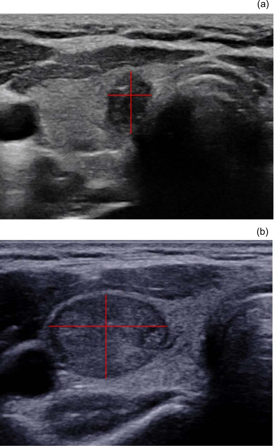
UltraLabeler, a graphics processing tool developed by the artificial intelligence team of Zhejiang University (Hangzhou, China) for this study, was used to measure anteroposterior-to-transverse ratio. This tool could be used to draw lines on the ultrasound image, and calculate the length of line segments. Hence, on the transverse ultrasound images of thyroid nodules, the anteroposterior diameter means the vertical height of the nodule, and the transverse diameter means the horizontal width of the nodule. These two line segments were drawn on the transverse image via UltraLabeler (Figure 1), and the anteroposterior-to-transverse ratio was calculated as follows: anteroposterior-to-transverse ratio = (vertical height of nodule) / (horizontal width of nodule). In fact, the anteroposterior-to-transverse ratio value was given by the UltraLabeler tool directly after the vertical and horizontal lines were drawn.
Measurement of the anteroposterior-to-transverse ratio. The ratio was calculated on the transverse ultrasound image of the thyroid nodules, as follows: anteroposterior-to-transverse ratio = (vertical height of nodule) / (horizontal width of nodule). (a) The nodule was papillary carcinoma, and its anteroposterior-to-transverse ratio was 1.42; (b) The nodule was adenoma, and its anteroposterior-to-transverse ratio was 0.71. The red lines reflect the vertical height and horizontal width of the nodules.
Statistical analysis
The receiver operating characteristic curve was drawn, and the area under the curve was calculated to assess the effectiveness of an anteroposterior-to-transverse ratio in predicting the malignancy of thyroid nodules. The sensitivity, specificity, accuracy and Youden index (Youden index = sensitivity + specificity − 1) were analysed. A p-value of less than 0.05 was considered statistically significant. All statistical analyses were carried out using SPSS software, version 28.0.1.0(142) (IBM, Armonk, New York, USA).
Results
Anteroposterior-to-transverse ratio
The anteroposterior-to-transverse ratio of thyroid nodules did not fit a normal distribution, as indicated by the histogram, normal quantile–quantile (Q–Q) plot and boxplot (Figure 2), and the Kolmogorov–Smirnov test of normality (p < 0.001). As shown in Table 2, the maximum anteroposterior-to-transverse ratio was 2.06 and the minimum was 0.32, with a median value of 0.83 and an interquartile range of 0.28. The median anteroposterior-to-transverse ratio was 0.93 for malignant nodules and 0.77 for benign nodules, which represented a statistically significant difference (p < 0.001, by Mann–Whitney U test).
Statistical characteristics of the anteroposterior-to-transverse (A/T) ratio of thyroid nodules. (a) Histogram, (b) normal quantile–quantile (Q–Q) plot and (c) boxplot of anteroposterior-to-transverse ratio, respectively. The p-value on a Kolmogorov–Smirnov test of normality was less than 0.001, indicating that the anteroposterior-to-transverse ratio was not normally distributed in this study. The median anteroposterior-to-transverse ratio was 0.83, with an interquartile range of 0.28.

Anteroposterior-to-transverse ratio values of thyroid nodules

Diagnostic value of ratio
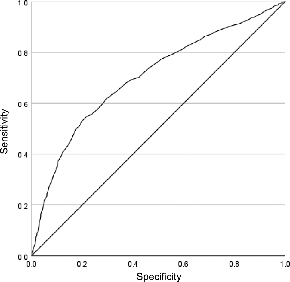
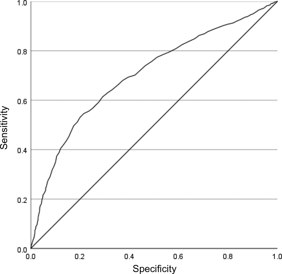
Figure 3 illustrates the receiver operating characteristic curve for the anteroposterior-to-transverse ratio predicting malignancy of thyroid nodules. The value of the area under the operating characteristic curve was 0.709 (p < 0.001), indicating that the anteroposterior-to-transverse ratio was a meaningful predictor of malignant nodules. When the cut-off value of the anteroposterior-to-transverse ratio was 0.9, that was to say, when we chose an anteroposterior-to-transverse ratio of greater than 0.9 as the sign of malignant nodules, sensitivity was 0.546, specificity was 0.785 and accuracy was 0.673, and the Youden index reached a maximum value of 0.331.
Receiver operating characteristic curve for anteroposterior-to-transverse ratio predicting thyroid nodule malignancy. The area under the receiver operating characteristic curve was 0.709 (p < 0.001). When the cut-off value of the anteroposterior-to-transverse ratio was 0.9, the sensitivity, specificity and accuracy values were 0.546, 0.785 and 0.673 respectively, and the Youden index reached a maximum value of 0.331.
Thyroid nodule size
The thyroid nodules were divided into five groups according to size: group A = less than 0.5 cm, group B = 0.5 cm to less than 1 cm, group C = 1 cm to less than 1.5 cm, group D = 1.5 cm to less than 2 cm, and group E = 2.0 cm or greater. The anteroposterior-to-transverse ratios for these five groups were statistically significantly different (p < 0.001, by Kruskal–Wallis test). Hence, we believed we should choose a different cut-off value for the anteroposterior-to-transverse ratio to predict thyroid cancer according to a specific range of nodule size.
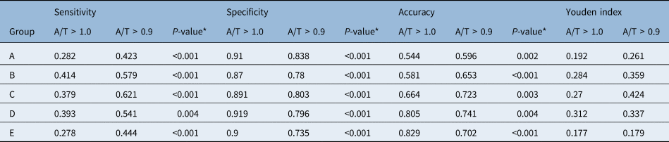
As shown in Table 3, the predictive effectiveness of anteroposterior-to-transverse ratios of greater than 0.9 and greater than 1.0 were compared according to nodule size. When the diameter of a thyroid nodule was less than 1.5 cm, an anteroposterior-to-transverse ratio of greater than 0.9 was associated with higher sensitivity, accuracy and Youden index, compared with an anteroposterior-to-transverse ratio of greater than 1.0, but specificity was lower. When the diameter of a thyroid nodule was larger than or equal to 1.5 cm, an anteroposterior-to-transverse ratio of greater than 0.9 was associated with higher sensitivity and Youden index, compared with an anteroposterior-to-transverse ratio of greater than 1.0, but specificity and accuracy were lower.
Predictive effectiveness of A/T ratios greater than 1.0 and 0.9 according to thyroid nodule diameter
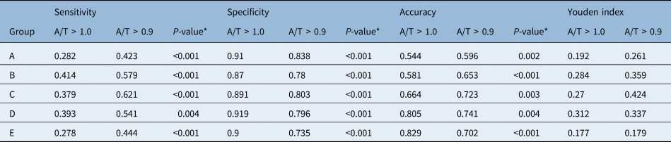
The thyroid nodules were divided into five groups according to size: group A = less than 0.5 cm, group B = 0.5 cm to less than 1 cm, group C = 1 cm to less than 1.5 cm, group D = 1.5 cm to less than 2 cm, and group E = greater than or equal to 2.0 cm. *Statistical significance was assessed using NcNemar's test or Fisher's exact test. A/T = anteroposterior-to-transverse ratio
Discussion
Ultrasonography is currently the preferred method for the clinical evaluation of thyroid nodules. According to the American College of Radiology Thyroid Imaging, Reporting and Data System (2017 edition),Reference Tessler, Middleton, Grant, Hoang, Berland and Teefey2 different signs were assigned different integral values, and the risk of malignancy in nodules was determined by the total score. The higher the total score, the greater the risk of malignancy in the thyroid nodule. A score of 3 points was given to a thyroid nodule with an anteroposterior-to-transverse ratio of greater than 1.0, and a score of 0 was given to a thyroid nodule with an anteroposterior-to-transverse ratio of 1 or lower. A nodule given a total score of 3 points could have a mildly suspicious risk of malignancy, and 4 points indicates a moderately suspicious risk. That was to say, a nodule with an anteroposterior-to-transverse ratio of greater than 1.0 would likely be malignant if it had any other ultrasonic sign of malignancy.
According to the ultrasound (‘U’) grading system, based on the 2014 British Thyroid Association guidelines, a taller-than-wide shape was one of the five key factors indicating malignancy (a grade of U5).Reference Perros, Boelaert, Colley, Evans, Evans and Gerrard Ba4 In the present study, the value of the area under the receiver operating characteristic curve for the anteroposterior-to-transverse ratio in predicting thyroid cancer was 0.709, which was statistically significant. This confirmed that the anteroposterior-to-transverse ratio could be used as an indicator to differentiate malignant thyroid nodules from benign nodules.
The mechanism of the intrinsic relationship between thyroid nodule malignancy and the anteroposterior-to-transverse ratio remains elusive. Kim et al.Reference Kim, Park, Chung, Oh, Kim and Lee3 demonstrated that benign thyroid nodules could only grow in the normal glandular tissue plane, while the malignant thyroid nodules could break through different glandular tissue planes. Yoon et al.Reference Yoon, Yoon, Chang, Seo, Yun and Choi5 pointed out that the mechanism of the ‘taller-than-wide’ sign was no or minimal compressibility of a thyroid nodule by the ultrasound probe, which occurred more frequently in malignant nodules than in benign nodules. Although it was difficult to compress and deform thyroid cancer nodules during examination, benign nodules were soft and easy to deform. The nodules were elliptic after compression; thus, benign nodules were likely associated with an anteroposterior-to-transverse ratio of 1.0 or lower.
There are two main methods for measuring the anteroposterior-to-transverse ratio of thyroid nodules. One method involves measurement on the transverse section of ultrasound images of the thyroid nodules,Reference Richman, Benson, Doubilet, Peters, Huang and Asch6 as in our study. The other method involves measurement on the longitudinal section,Reference Moon, Jung, Lee, Na, Baek and Lee7,Reference Moon, Kwak, Kim and Kim8 in which the width of the nodule is changed into the diameter from the cranial side to the caudal of the nodule. In the American College of Radiology Thyroid Imaging, Reporting and Data System,Reference Tessler, Middleton, Grant, Hoang, Berland and Teefey2 the anteroposterior-to-transverse ratio is measured on the transverse section. In some other studies, two methods of anteroposterior-to-transverse ratio measurement were adopted,Reference Moon, Jung, Lee, Na, Baek and Lee7,Reference Seo, Na, Kim, Kim and Yoon9 or the measurement method was not specified.Reference Haugen, Alexander, Bible, Doherty, Mandel and Nikiforov1,Reference Xiang, Chu, Chen, Liu, Ding and Zeng10,Reference Na, Baek, Sung, Kim, Kim and Choi11 It has been suggested that these two measurement methods are of equal significance in the prediction of thyroid cancer.Reference Chen, Hu and Chen12
The present study aimed to calculate the optimal cut-off value for the diagnosis of thyroid cancer. It was found that when the diameter of a thyroid nodule was less than 1.5 cm, an anteroposterior-to-transverse ratio of greater than 0.9 would achieve a higher accuracy and sensitivity, compared with an anteroposterior-to-transverse ratio of greater than 1.0, while the specificity was relatively lower. When the diameter of a thyroid nodule was larger than 1.5 cm, an anteroposterior-to-transverse ratio of greater than 0.9 was more sensitive, compared with an anteroposterior-to-transverse ratio of greater than 1.0, but both specificity and accuracy were lower. Therefore, the corresponding anteroposterior-to-transverse ratio can be selected as the diagnostic criterion of thyroid carcinoma according to the clinical application scenario. For instance, for a thyroid nodule with a diameter less than 1.5 cm, a greater sensitivity and accuracy can be obtained with consideration of an anteroposterior-to-transverse ratio of greater than 0.9 to predict thyroid cancer, which is more significant for clinical screening to detect suspected cancer nodules. Higher specificity can be achieved in the diagnosis of thyroid cancer using an anteroposterior-to-transverse ratio of greater than 1.0, reducing the need for fine-needle aspiration biopsy. Further research to identify the most optimal cut-off value of the anteroposterior-to-transverse ratio may facilitate development of automatic diagnosis of thyroid cancer. It will not be difficult to choose the proper cut-off value of the anteroposterior-to-transverse ratio according to nodule size to predict thyroid cancer, via artificial intelligence, in the future.
• An anteroposterior-to-transverse ratio of greater than 1.0 is equal to a ‘taller-than-wide’ sign, and is a meaningful indicator for thyroid cancer
• In this study, predictive effectiveness of the anteroposterior-to-transverse ratio was influenced by nodule size
• When a thyroid nodule diameter was less than 1.5 cm, an anteroposterior-to-transverse ratio of more than 0.9 was associated with higher sensitivity and accuracy
There are several limitations to our study. It is a retrospective study, and it is difficult to prevent selection bias completely. We enrolled only 12 patients aged under 18 years old, and more paediatric patients are needed for further study. In addition, this study mainly focused on papillary thyroid carcinoma and nodule size; the inclusion of other types of thyroid cancer and additional factors are needed in future studies. Thus, a further prospective clinical control study comprising a larger sample size will be carried out to explore the mechanism of the anteroposterior-to-transverse ratio in predicting thyroid malignancy in the future.
Conclusion
The anteroposterior-to-transverse ratio was a meaningful indicator of thyroid cancer, and its predictive effectiveness was influenced by the size of thyroid nodules. When thyroid nodules were less than 1.5 cm, an anteroposterior-to-transverse ratio of greater than 0.9 achieved a higher accuracy and sensitivity in predicting malignancy, while an anteroposterior-to-transverse ratio of greater than 1.0 had better specificity and accuracy for nodules larger than 1.5 cm.
Acknowledgements
Thanks to the artificial intelligence team (Qiang Zhu, Ming Kong, Mengze Li) from the College of Computer Science and Technology, Zhejiang University (led by Professor Qiang Zhu) for the graphics processing tool UltraLabeler. Thanks also to Professor Changdong Yang from the First Hospital of Qinhuangdao for his contribution to this study.
Competing interests
None declared