Introduction
An enlarged submandibular gland is a common presentation to the ENT surgeon, with a variety of possible diagnoses. Recently, a new entity has been described: immunoglobulin G4 (IgG4) related chronic sclerosing sialadenitis. As ENT surgeons may be the first specialists to see patients with salivary gland disease, it is important to highlight this newly described disease.
The association between increased levels of IgG4 and sclerosing inflammatory disease was first described as occurring in the pancreas in 2001.Reference Hamano, Kawa, Horiuchi, Unno, Furuya and Akamatsu1 Over the last 10 years, it has become apparent that IgG4-related inflammatory fibrosis can manifest in many organs as part of a systemic IgG4-related disease, including the pancreas, bile ducts, liver, retroperitoneum, lymph nodes, kidneys, lungs, orbital tissues, pituitary gland, aorta, parotid and salivary glands.Reference Kamisawa, Funata, Hayashi, Eishi, Koike and Tsuruta2 There is a wide spectrum of disease, and clinical presentation usually depends on the organ involved. The disease can be confined to a single organ or involve multiple organs at any one time, although not all are clinically apparent. It has many mimics including malignancy, and inflammatory, infective and other immune-mediated diseases.Reference Stone, Zen and Deshpande3
In 2010, the relationship between chronic sclerosing sialadenitis and IgG4 was described in the Western population.Reference Geyer, Ferry, Harris, Stone, Zukerberg and Lauwers4 Geyer et al. reported elevated tissue IgG4-positive plasma cells in patients with Sjögren's syndrome, chronic sclerosing inflammatory fibrosis and sialadenitis; the authors found a lymphoplasmacytic infiltrate with increased numbers of IgG4-positive plasma cells in the chronic sclerosing inflammatory fibrosis group.Reference Geyer, Ferry, Harris, Stone, Zukerberg and Lauwers4 A link was proposed between chronic sclerosing sialadenitis and IgG4, and this new disease entity was distinguished as separate from Sjögren's syndrome, sialadenitis or lymphoproliferative disease.
We present a review of the current understanding regarding the presentation, diagnosis and management of IgG4-related chronic sclerosing sialadenitis, with the aim of highlighting this new disease to ENT surgeons. We propose a diagnostic pathway to aid management of these cases.
Materials and methods
A computer literature search of Medline, Embase and Cochrane Library databases was performed (by E Crewe) to identify all studies on the subject of interest. For this purpose, the search terms used were: ‘IgG4’, ‘hyperIgG4 syndrome’ and ‘IgG4 related chronic sclerosing sialadenitis’. The abstracts of the identified manuscripts were inspected and irrelevant studies were excluded. The full text of relevant studies was then examined.
Disease presentation
Patients usually present with IgG4-related chronic sclerosing sialadenitis in the sixth decade of life and there is a male preponderance (similar to other organ manifestations of IgG4-related disease), although one study by Takano and colleagues found a slight preponderance in females.Reference Takano, Yamamoto, Takahashi, Hinomura, Imai and Himi5 The disease commonly affects the submandibular glands, but involvement of the parotid and minor salivary glands has been described.Reference Geyer and Deshpande6–Reference Blanco, Mesko, Cura and Cabello-Inchausti9
Patients with IgG4-related chronic sclerosing sialadenitis can present to the ENT surgeon with: dry eyes and mouth (sicca syndrome); diffuse enlargement of the submandibular, parotid or lacrimal glands (Mikulicz's disease); or a solitary submandibular gland mass (Küttner's tumour).Reference Yamamoto, Takahashi, Ohara, Suzuki, Naishiro and Yamamoto8, Reference Blanco, Mesko, Cura and Cabello-Inchausti9 In the absence of clinical suspicion and awareness of the condition, IgG4-related chronic sclerosing sialadenitis is under-diagnosed and mistaken for other diseases.Reference Chan10
Immunoglobulin G4 related chronic sclerosing sialadenitis can be detected incidentally after patients present to a different specialty with other organ involvement. Systemic involvement is more common in patients presenting with diffuse enlargement of submandibular glands and/or lacrimal glands, and/or sicca symptoms, compared to those who have solitary submandibular gland enlargement alone. In Japan, the salivary gland is the third most common location for extra-pancreatic lesions associated with autoimmune pancreatitis, with the incidence of IgG4-related sclerosing cholangitis and lymphadenopathy being higher than that of salivary gland enlargement.Reference Hamano, Arakura, Muraki, Ozaki, Kiyosawa and Kawa11
Disease investigation
In a patient presenting to the ENT surgeon with enlargement of the submandibular gland or diffuse enlargement of glands, or sicca syndrome, non-invasive serological evaluation for total immunoglobulin G (IgG) and IgG4 subclass can readily be performed. An elevated serum IgG4 level (greater than 1.4 g/l) alone is not sufficient for the diagnosis of IgG4-related chronic sclerosing sialadenitis, but it can be supportive in the correct clinical context and aid the decision for referral. The sensitivity of elevated serum IgG4 for IgG4-related chronic sclerosing sialadenitis is estimated at 70–80 per cent, which is similar to that described for other organ manifestations.Reference Ghazale, Chari, Smyrk, Levy, Topazian and Takahashi12 However, serum IgG4 may be elevated (usually 2–3 times the upper limit of normal levels) in a range of inflammatory and malignant diseases, including lymphoma, and so is not disease-specific.Reference Ghazale, Chari, Smyrk, Levy, Topazian and Takahashi12 The serum IgG4 level is reportedly higher in those with multi-organ rather than single-organ disease. The literature suggests that serum IgG4 levels can be normal in 20 per cent of patients with autoimmune pancreatitis, but there are limited data regarding normal IgG4 values in those with IgG4-related chronic sclerosing sialadenitis.
Ultrasound of the head and neck usually reveals enlargement of the gland(s) and the presence of local lymphadenopathy. It is useful in guiding a site for biopsy (discussed below), but is otherwise non-specific in diagnosis. A magnetic resonance imaging scan of the salivary glands can be useful to look for distribution and involvement of the disease, which may not be clinically apparent, and to differentiate it from mimics such as Sjögren's syndrome. In IgG4-related disease, the parotid, submandibular and labial glands can all be diffusely enlarged, with ill-delineated infiltration, T1 and T2 hypointensity, homogeneous marked contrast-enhancement and restricted diffusion.Reference De Cocker, D'Arco, De Beule, Tousseyn, Blockmans and Hermans13 Local lymph nodes may also be enlarged, with restricted diffusion, but this is a non-specific finding. The absence of necrosis and lack of extracapsular spread point to a benign diagnosis. However, sarcoidosis and other granulomatous diseases (e.g. Wegener's granulomatosis) may also present with the combination of generalised salivary involvement and reactive lymph node enlargement, with the former appearing on imaging as a diffuse non-nodular enlargement with homogeneous enhancement of the glands.
The primary role of fine needle aspiration (FNA) is to exclude a neoplastic process. In the presence of significant fibrosis, FNA is non-diagnostic. Furthermore, when cellular material is obtained, it comprises a mixed population of chronic inflammatory cells. This is a non-specific feature on its own. Fine needle aspiration is not suited to demonstrating an increased proportion of IgG4 plasma cells, which is one of the features in the pathological diagnosis of IgG4-related disease.
Ultrasound-guided biopsy of the enlarged salivary gland for histological diagnosis of IgG4-related chronic sclerosing sialadenitis is the preferred method for diagnosis. When representative material is obtained on a biopsy, histopathological features in conjunction with IgG4 immunohistochemistry are sufficient for a collaborative diagnosis of IgG4 disease. In our experience, sublabial biopsy has not been helpful, as the presence of salivary gland tissue is variable. Sometimes, the enlarged submandibular gland is excised to exclude malignancy and diagnosis made retrospectively. Open excision biopsy is associated with potentially significant morbidity, such as facial weakness occurs due to injury of the mandibular branch of the facial nerve, or hypoglossal or lingual nerve (a risk of approximately 1–2 per cent). Open excision biopsy would be contemplated only in the differential diagnosis of malignancy when the ultrasound-guided biopsy was suggestive of malignancy, or if it was inconclusive and there were worrying clinical signs or symptoms.
There are characteristic morphological changes in the tissue that distinguish IgG4-related chronic sclerosing sialadenitis from other forms of sialadenitis or sialectasis-related inflammation.Reference Smyrk14 The three key morphological findings in organs affected by IgG4-related disease are: marked lymphoplasmacytic infiltrate, storiform fibrosis and obliterative phlebitis (Figure 1).Reference Culver and Bateman15 In the salivary gland, there is preservation of lobular architecture, large irregular germinal centres, acinar atrophy, an absence of lymphoepithelial lesions, and, characteristically, the presence of prominent interlobular fibrosis, composed of activated fibroblasts, lymphocytes and plasma cells (both T and B cells). The plasma cells are predominantly IgG4-positive (counts of more than 10 per mean of three high power fields in biopsy sections and more than 50 per mean of three high power fields in resection specimens), with an IgG4-positive to IgG-positive plasma cell ratio of over 40 per cent (Figure 2). The use of these cut-offs has been recommended in the Boston consensus criteria to support a histopathological diagnosis of IgG4-related disease, and discriminate it from other disease mimics.Reference Deshpande, Zen, Chan, Yi, Sato and Yoshino16 Both the morphological changes and the presence of IgG4-positive cells are needed for a definite or probable histological diagnosis. It must be remembered that IgG4-positive plasma cells can be seen around an area infiltrated with malignancy, so morphological examination is crucial.Reference Strehl, Hartmann and Agaimy17
Photomicrograph of the submandibular gland showing loss of parenchyma, storiform fibrosis and chronic inflammation. (H&E; ×40)

Immunostaining for immunoglobulin G4 (IgG4) plasma cells, showing dark brown IgG4+ plasma cells. (IgG4; ×400)
Histological assessment is important in IgG4-related disease investigation in order to exclude malignancy and to carry out supportive immunohistochemical analysis. The submandibular and parotid glands are often the most accessible sites to obtain tissue, whilst the pancreas, renal and lung tissue, for example, are less accessible.
Immunoglobulin G4-related chronic sclerosing sialadenitis can mimic many other diseases. For example, a patient with Sjögren's syndrome may present to the ENT surgeon with sicca symptoms and diffuse enlargement of the salivary glands, which often includes the parotid gland. Typically, there is immune-mediated destruction of the exocrine glands, with prominent lymphoepithelial lesions and cystic dilatation of peripheral ducts.Reference Vitali, Bombardieri, Jonsson, Moutsopoulos, Alexander and Carsons18 Whilst plasma cells and lymphoid follicles can be observed, fibrosis is unusual. Sialolithiasis may present with intermittent swelling and pain affecting the submandibular gland, and may show periductal fibrosis and inflammation, and patchy, lobular atrophy and inflammation. Granulomatous diseases can present with generalised salivary involvement and reactive lymph node enlargement, with other systemic features. Sarcoidosis can be suggested by non-caseating epithelioid cell granulomas; these resolve or convert into hyaline connective tissue, and lack necrosis or an intact reticular pattern. In Wegner's granulomatosis, the hallmarks are necrotising granulomatous inflammation, and a pauci-immune vasculitis in small- and medium-sized vessels.
Importantly, IgG4-related chronic sclerosing sialadenitis should be differentiated from lymphoproliferative disease. The association of lymphoma with longstanding Sjögren's syndrome or hepatitis C infection is well known.Reference Ambrosetti, Zanotti, Pattaro, Lenzi, Chilosi and Caramaschi19 An increased risk of malignancy has been reported in patients with IgG4-related disease, but a specific association with IgG4-related chronic sclerosing sialadenitis has not been shown.Reference Ghazale and Chari20 Malignant transformation has been reported in patients with IgG4-related orbital disease.Reference Kim, Grobmyer, Dixon, Allan and Hochwald21, Reference Cheuk, Yuen, Chan, Shih, Kuo and Ma22 Lymphomas of the salivary glands such as non-Hodgkin's lymphoma and mucosa-associated lymphoid tissue should be excluded.
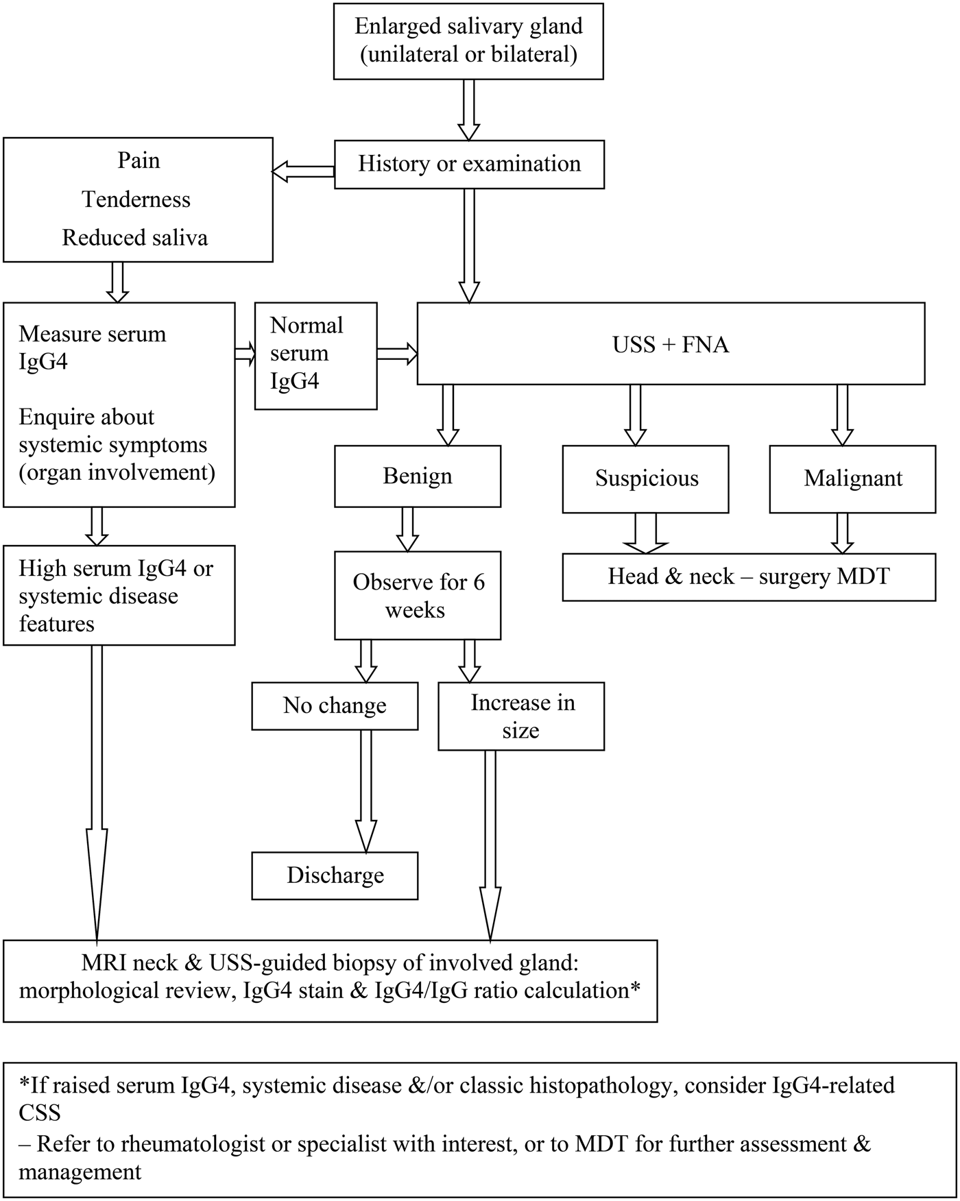
A pathway for investigation of IgG4-related chronic sclerosing sialadenitis by the ENT surgeon is proposed in Figure 3.
Pathway in the investigation of immunoglobulin G4 related chronic sclerosing sialadenitis. IgG4 = immunoglobulin G4; USS = ultrasound; FNA = fine needle aspiration; MDT = multidisciplinary team; MRI = magnetic resonance imaging; IgG = immunoglobulin G; CSS = chronic sclerosing sialadenitis
Disease management
The roles of the ENT surgeon are to exclude malignancy and to aid in the diagnosis of IgG4-related disease in cases where the glandular tissue is the most accessible to biopsy. Patients with submandibular gland enlargement and a history of sicca symptoms, in whom malignancy has been excluded, should be referred to a rheumatologist (or alternative clinician with an interest in the disease) for consideration of a systemic disease process. Evaluation of serum IgG4 levels and histopathological assessment of biopsies with IgG4 immunohistochemistry form a part of the systemic disease investigation (Figure 3).
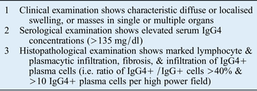
Diagnostic criteria for IgG4-related disease have been established. The original criteria were developed to diagnose autoimmune pancreatitis in Asia and the USA,Reference Chari, Smyrk, Levy, Topazian, Takahashi and Zhang23, Reference Otsuki, Chung, Okazaki, Kim, Kamisawa and Kawa24 but have since been adapted to reflect the multi-systemic nature of the disease.Reference Umehara, Okazaki, Masaki, Kawano, Yamamoto and Saeki25 These incorporate the classical histological characteristics, imaging findings, serological elevations of IgG4, organ manifestations and responses to corticosteroids (Table I). More recently, the Boston histological consensus criteria have been agreed.Reference Deshpande, Zen, Chan, Yi, Sato and Yoshino16 These reflect changes in our understanding of the importance of characteristic morphology, thresholds of IgG4 cells, and the IgG4 to IgG ratio used to distinguish between disease mimics. The presence of sialomegaly and elevated serum IgG4 in isolation do not meet the diagnostic criteria for IgG4-related chronic sclerosing sialadenitis as part of IgG4-related disease.
Japanese comprehensive diagnostic criteria*
A definite diagnosis of IgG4-related disease is made when all criteria (1, 2 and 3) are fulfilled. A probable diagnosis is made with fulfilment of criteria 1 and 3. A possible diagnosis is made with fulfilment of criteria 1 and 2. It is important to differentiate IgG4-related disease from malignant tumours of each organ (e.g. cancer and lymphoma) and similar diseases (e.g. Sjögren's syndrome, primary sclerosing cholangitis, Castleman's disease, secondary retroperitoneal fibrosis, Wegener's granulomatosis, sarcoidosis and Churg–Strauss syndrome) by additional histopathological examination. Even when patients cannot be diagnosed using the comprehensive diagnostic criteria, they may be diagnosed using organ-specific diagnostic criteria for IgG4-related disease.
* For the diagnosis of IgG4-related disease, adapted from Umehara et al.Reference Umehara, Okazaki, Masaki, Kawano, Yamamoto and Saeki25 IgG4 = immunoglobulin G4; IgG = immunoglobulin G
Ultimately, patients with IgG4-related chronic sclerosing sialadenitis will benefit from multidisciplinary discussion and management involving a rheumatologist, gastroenterologist, histopathologist and radiologist with an interest in the disease. Further biochemical and serological tests, imaging and the need for biopsies will be guided by clinical presentation and organ involvement.
The necessity and urgency of treatment depends on the involvement of vital organs, and risk of organ dysfunction or failure. Urgent treatment is needed for presentations with potentially irreversible consequences. In the case of the salivary gland, this is chronic salivary and lachrymal gland dysfunction. In the case of other organs, this can include visual impairment from compression of the optic nerve, aortic dissection from an inflammatory aortic lesion, and irreversible fibrosis within the mesentery and retroperitoneum. The first-line treatment is corticosteroids. Most patients respond rapidly to an initial high dose of prednisolone (a starting dose of 30–40 mg daily) that is tapered over three to six months.Reference Kamisawa, Shimosegawa, Okazaki, Nishino, Watanabe and Kanno26 However, relapse is common, occurring in over 50 per cent of patients in whom steroids have been discontinued.Reference Hart, Kamisawa, Brugge, Chung, Culver and Czajo27 In those with recurrent or refractory disease, second-line immunosuppressant agents have included azathioprine and mycophenolate.Reference Hart, Kamisawa, Brugge, Chung, Culver and Czajo27 More recently, B-cell depletion with rituximab, the monoclonal antibody against cluster of differentiation 20, has been used with success in those who are intolerant of steroids or those with refractory disease.Reference Khosroshahi, Carruthers, Deshpande, Unizony, Bloch and Stone28 Such cases should also be managed in a multidisciplinary setting with appropriate assessment of response to therapy using objective measures.
Conclusion
Patients with IgG4-related chronic sclerosing sialadenitis can present to the ENT surgeon with a solitary lesion, or with diffuse enlargement of the salivary glands and sicca symptoms. In those with diffuse enlargement particularly, there should be a high index of clinical suspicion for systemic disease. In those with solitary enlargement, IgG4-related chronic sclerosing sialadenitis should be considered after malignancy is excluded. It is important that the ENT community is aware of IgG4-related chronic sclerosing sialadenitis in order to facilitate early diagnosis, prevent irreversible organ dysfunction and aid detection of other systemic manifestations of disease. Diagnosis is based on identifiable criteria and is usually confirmed in the salivary gland by histological findings. If IgG4-related chronic sclerosing sialadenitis is confirmed, referral to a rheumatologist and multidisciplinary team management should be considered.
Acknowledgement
EL Culver was supported by a Wellcome Trust Research Fellowship (095160/Z/10/Z) awarded to investigate the natural history and pathogenesis of IgG4-related disease.