Introduction
Over the last decade, average litter size has been increased gradually by genetic selection in pigs. This has been associated with a reduction in the mean piglet birth weight and concomitantly with an increased within-litter variation in birth weight leading to a rise in the proportion of small piglets (<1.0 kg birth weight) in large litters (Le Dividich, Reference Le Dividich1999; Milligan et al., Reference Milligan, Fraser and Kramer2002; Quiniou et al., Reference Quiniou, Dagorn and Gaudré2002). According to the data of Quiniou et al. (Reference Quiniou, Dagorn and Gaudré2002), small piglets weighing less than 1.0 kg at birth represent on average 13% of total born piglets, ranging from 7% in litters with 11 piglets or less to 23% in litters with 16 piglets or more. Two-thirds of runt piglets (i.e. very low birth weight (VLBW) piglets weighing <0.8 kg) die during suckling. Mortality is 34% for low birth weight (LBW) piglets (0.81 to 1.0 kg body weight (BW)) born alive and less than 10% for piglets above 1.6 kg BW. As a result, 8% of weaned piglets are LBW (6%) and VLBW (2%) piglets.
Within-litter variation in birth weight results from difference in intrauterine growth. In the pig, intrauterine growth retardation, leading to LBW, occurs naturally. The aetiology and underlying mechanisms of intrauterine growth retardation in livestock as well as in humans and rodents have been recently reviewed (McMillen and Robinson, Reference McMillen and Robinson2005; Foxcroft et al., Reference Foxcroft, Dixon, Novak, Putman, Town and Vinsky2006; Murphy et al., Reference Murphy, Smith, Giles and Clifton2006; Wu et al., Reference Wu, Bazer, Wallace and Spencer2006). It results from alteration in foetal substrate supply. In agreement with the ‘thrifty phenotype’ hypothesis developed by Hales and Barker (Reference Hales and Barker1992), it has been shown in several species that there is a relationship between birth weight and subsequent pattern of growth and development of tissues and organs (Dauncey, Reference Dauncey1997; McMillen and Robinson, Reference McMillen and Robinson2005). As described in a recent review (Rehfeldt and Kuhn, Reference Rehfeldt and Kuhn2006), and in other recent studies (Bee, Reference Bee2004; Poore and Fowden, Reference Poore and Fowden2004a; Gondret et al., Reference Gondret, Lefaucheur, Juin, Louveau and Lebret2006), pigs of LBW (0.95 to 1.3 kg) exhibited consistently lower postnatal growth rates and lower lean percentage than heavy birth weight (HBW) pigs at slaughter.
The aim of the present paper is to review the influence of LBW on the growth and development of the neonatal pig during the suckling period. In the current review, LBW piglets referred to piglets weighing 0.8 to 1 kg, median birth weight (MBW) piglets referred to piglets weighing ∼1.4 kg and HBW piglets referred to piglets weighing more than 1.6 kg at birth, unless indicated in the text. The review focuses primarily on the development of adipose tissue and the digestive tract. The importance of hormones and growth factors in the regulation of these developmental processes is also considered.
Growth in low birth weight piglets
Growth and development processes of an organism involve weight gain and changes in shape, chemical composition and physiological functions. In the pig as well as in other species, these processes have been reviewed widely (Reeds et al., Reference Reeds, Burrin, Davis, Fiorotto, Mersmann and Pond1993). Mature body sizes and weights, relationships between birth weights and mature weights and the postnatal growth rates differ markedly between species. In the pig, weight at birth represents a very small proportion of mature weight compared with the majority of mammals. The mature weight/birth weight ratio is about 300, whereas it varies between 20 and 40 for many other mammals including humans and rats. The pig has a very high growth rate. During the 2- to 4-week suckling period, piglets from modern genotypes grow at the rate of about 250 to 270 g/day (King et al., Reference King, Le Dividich and Dunshea1999). The precise rate is very variable and depends mainly on the availability of milk. Weaning weight is positively correlated to birth weight with LBW piglets still exhibiting a lower BW at weaning than their counterparts with a MBW (equivalent to the mean litter BW) or a HBW (more than 1.9 kg). Compared with MBW or HBW piglets, LBW piglets exhibit a lower growth rate (15% to 30%) in the 1st month of postnatal life (Campbell and Dunkin, Reference Campbell and Dunkin1982; Wolter et al., Reference Wolter, Ellis, Corrigan and DeDecker2002; Poore and Fowden, Reference Poore and Fowden2004a; Gondret et al., Reference Gondret, Lefaucheur, Louveau, Lebret, Pichodo and Le Cozler2005 and Reference Gondret, Lefaucheur, Juin, Louveau and Lebret2006). During the first 3 weeks of suckling, it has been shown that milk intake per unit BW is similar in the two groups even though LBW piglets consume less milk per suckle than MBW piglets (Campbell and Dunkin, Reference Campbell and Dunkin1982). The higher birth weight piglets tend to select the anterior teats (Hartsock et al., Reference Hartsock, Graves and Baumgardt1977) that are easier for milk extraction. Interestingly, this difference in growth rate persists until the market weight. This is illustrated by the observation that 10 to 15 more days are needed for LBW pigs to reach the market weight (100 to 110 kg; Powell and Aberle, Reference Powell and Aberle1980; Gondret et al., Reference Gondret, Lefaucheur, Louveau, Lebret, Pichodo and Le Cozler2005 and Reference Gondret, Lefaucheur, Juin, Louveau and Lebret2006). It has been reported that the effect of birth weight on postnatal growth pattern is sex-specific with females being more dependent on originating birth weight than males (Poore and Fowden, Reference Poore and Fowden2004a). Despite these differences in growth rates, the relative growth of small piglets has been reported in some studies to be higher than that of heavier piglets. Between birth and weaning, VLBW and LBW piglets exhibit a seven-fold and six-fold increase, respectively, in BW, whereas MBW and HBW piglets exhibit a five- and fourfold increase, respectively, in BW (Quiniou et al., Reference Quiniou, Dagorn and Gaudré2002; Gondret et al., Reference Gondret, Lefaucheur, Juin, Louveau and Lebret2006).
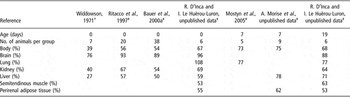
With regard to morphology, intrauterine growth retardation is associated with lower abdominal circumference and crown-rump length at birth (Poore and Fowden, Reference Poore and Fowden2004a; Mostyn et al., Reference Mostyn, Litten, Perkins, Euden, Corson, Symonds and Clarke2005). Significant differences have been also reported in the body composition of piglets during early postnatal growth. By chemical analysis of the whole body at birth, it has been reported that LBW piglets have less fat and protein and more water than their littermates (Rehfeldt and Kuhn, Reference Rehfeldt and Kuhn2006). Even though linear correlations between BWs and various organ weights have been reported, the extent of weight variation differs between the organs examined (Table 1; Widdowson, Reference Widdowson1971; Ritacco et al., Reference Ritacco, Radecki and Schoknecht1997; Bauer et al., Reference Bauer, Walter, Hoppe, Gaser, Lampe, Kauf and Zwiener1998 and Reference Bauer, Walter, Ihring, Kluge, Lampe and Zwiener2000a; Mostyn et al., Reference Mostyn, Litten, Perkins, Euden, Corson, Symonds and Clarke2005; R. D’Inca and I. Le Huërou-Luron, unpublished data). At birth, brain weight is poorly affected by birth weight. Conversely, the significant reduction in the weight of kidney, liver and skeletal muscle is proportional to that of BW (39% to 75%).
Relative body and organ weights of intrauterine growth retarded compared with median birth weight piglets (expressed as a percentage of median birth weight piglets)

For intrauterine growth-retarded piglets, piglets had a very low birth weight† (0.63 ± 0.11 kg) or a low birth weight‡ (between 0.80 and 1.0 kg). For median birth weight piglets, birth weight was 1.35 to 1.60 kg in all studies.
Adipose tissue development and low birth weight piglets
The rapid increase in BW is associated with marked changes in organ growth and body composition in the neonatal period. Pigs, like sheep and rats, are characterised by a small amount of total body fat at birth (<2%) compared with guinea pig and human neonates (∼10%). They have a remarkable capacity to deposit large amounts of fat soon after birth. Body fat is derived primarily from dietary fat. During the first day of life, carcass fat content increases linearly with colostrum fat content (Le Dividich et al., Reference Le Dividich, Tivey, Blum, Strullu and Louat1997). It continues to increase and reaches 11% by 3 weeks. During the 3-week suckling period, 54% of milk fat intake is retained in the body (Noblet and Etienne, Reference Noblet and Etienne1987) with fat accretion occurring at a mean rate of 30 to 35 g/day, depending mainly on the amount of ingested milk (Marion and Le Dividich, Reference Marion and Le Dividich1999) and on the milk fat content (Jones et al., 1999).
At the cellular level, growth of adipose tissue results from proliferation and differentiation of adipocyte precursor cells, and subsequent enlargement of the mature fat cells. It is under the control of hormones and growth factors (Grégoire et al., Reference Grégoire, Smas and Sul1998; Louveau and Gondret, Reference Louveau and Gondret2004). Marked changes occur in morphology, cell size and chemical composition of adipose tissue during the neonatal period. In the foetus, fat cell cluster differentiation begins between 45 and 60 days of gestation in subcutaneous tissue (Hausman and Kauffman, Reference Hausman and Kauffman1986). At birth, the percentage of multilocular adipocytes is very high, but by day 3 post partum, many unilocular adipocytes (one major central lipid droplet) are observed (Mersmann et al., Reference Mersmann, Goodman and Brown1975; Hauser et al., Reference Hauser, Mourot, De Clercq, Genart and Remacle1997). In subcutaneous adipose tissue, a marked increase in adipocyte size is observed with diameters increasing from 19 to 24 μm at 3 days of age to 36 μm at 23 days of age (Mersmann et al., Reference Mersmann, Underwood, Brown and Houk1973 and Reference Mersmann, Goodman and Brown1975). A similar increase has been reported in perirenal fat (Hauser et al., Reference Hauser, Mourot, De Clercq, Genart and Remacle1997). The total lipid content increases in both subcutaneous and perirenal fat between 7 and 30 days of age (Hauser et al., Reference Hauser, Mourot, De Clercq, Genart and Remacle1997). Postnatally, adipose tissue appears as a number of individual depots, some in the abdominal cavity (e.g. perirenal), some under the skin (subcutaneous depots, the more abundant in pigs) and some within the musculature (inter- and intra-muscular depots). Adipocytes from different depots, while having many features in common, are not identical, varying in size and in some of their secretory properties (Gardan et al., Reference Gardan, Gondret and Louveau2006). Unlike other tissues, adipose tissue mass has considerable capacity to expand.
During the past two decades, various clinical and experimental observations have indicated that birth weight influences the subsequent development of adipose tissue (Symonds et al., Reference Symonds, Pearce, Bispham, Gardner and Stephenson2004; McMillen and Robinson, Reference McMillen and Robinson2005). In contrast to other tissues, the effect of intrauterine growth retardation on adipose tissue is poorly documented in the neonatal pig. This paucity of data might be explained by the fact that piglets have a very low fat content at birth compared with human or guinea pigs. In a recent experiment, we have shown that both absolute and relative weights of perirenal fat (the fat that lines the abdominal cavity and encloses the kidneys) were reduced at birth, and in 7- and 19-day-old LBW (<1.0 kg) piglets compared with MBW (>1.35 to 1.6 kg) piglets (Table 1; unpublished data). This decrease in weight of adipose tissue in LBW piglets may involve the reduced rate of adipocyte proliferation or high rate of proliferation with a delay in adipocyte differentiation.
The impact of LBW on subsequent adipose tissue development is more documented (Martorell et al., Reference Martorell, Stein and Schroeder2001) with epidemiological and experimental studies, suggesting a link between LBW and later fatness. In pigs, several studies support this link even though the extent of the increase in LBW may differ between perirenal fat and subcutaneous fat (Powell and Aberle, Reference Powell and Aberle1980 and Reference Powell and Aberle1981; Bee, Reference Bee2004; Poore and Fowden, Reference Poore and Fowden2004a; Gondret et al., Reference Gondret, Lefaucheur, Juin, Louveau and Lebret2006; Rehfeldt and Kuhn, Reference Rehfeldt and Kuhn2006). At market weight, LBW barrows, artificially reared during the suckling period, were fatter than HBW barrows, with a slight increase in backfat thickness (Powell and Aberle, Reference Powell and Aberle1980), and a significant increase in perirenal fat weight (Powell and Aberle, Reference Powell and Aberle1981). These two parameters were significantly increased in LBW females fed ad libitum compared with HBW female pigs (Gondret et al., Reference Gondret, Lefaucheur, Louveau, Lebret, Pichodo and Le Cozler2005 and Reference Gondret, Lefaucheur, Juin, Louveau and Lebret2006), whereas Rehfeldt and Kuhn (Reference Rehfeldt and Kuhn2006) only found a significant increase in internal fat. An increase in fat depth was also reported in 12-month-old LBW pigs (Poore and Fowden, Reference Poore and Fowden2004a). It is important to note that available data suggest that the nutrient supply should be adequate during the postnatal period to see an increase in fat deposition (Powell and Aberle, Reference Powell and Aberle1980; Gondret et al., Reference Gondret, Lefaucheur, Louveau, Lebret, Pichodo and Le Cozler2005 and Reference Gondret, Lefaucheur, Juin, Louveau and Lebret2006).
Gut development and low birth weight piglets
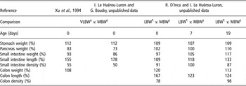
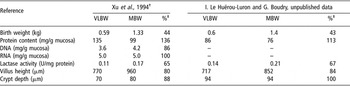
Digestive organs are differently affected by intrauterine growth retardation. In VLBW neonatal piglets, the relative weight of the pancreas is reduced, whereas that of the stomach, small intestine or colon is only slightly affected (Table 2). Moreover, intrauterine growth retardation is associated with a reduction in the wall thickness of the stomach, small intestine and colon and in the density of the small intestine and colon (Table 2; Xu et al., Reference Xu, Mellor, Birtles, Reynolds and Simpson1994; Wang et al., Reference Wang, Huo, Shi, Xu and Hutz2005). Consequently, the intestinal surface area for absorption is highly reduced, the average number of villi per unit area and the height of villi being 15% to 20% lower in VLBW than in MBW piglets (Table 3). Impairment of the intestinal function is also observed in VLBW piglets at birth. As lactase and aminopeptidase N peak at birth or just after birth in MBW piglets (Sangild et al., Reference Sangild, Xu and Trahair2002), the lower lactase and aminopeptidase N activities reported in VLBW piglets (Table 3) indicate a retardation in maturation of the small intestinal function and lowered digestive capacities, as reported in pre-terms (Shulman et al., Reference Shulman, Wong and Smith2005). Differences in digestive organ characteristics between LBW and MBW neonatal piglets are mainly observed in the colon and lessen with increasing age (Table 2). However, functional differences in maturational rate persist when epithelial barrier properties are considered. A recent experiment performed in our laboratory indicates that permeability to macromolecules, determined using Ussing chambers, decreased in the jejunum and increased in the ileum of MBW piglets during the suckling period. In contrast, no change with age was observed in LBW piglets (Boudry et al., Reference Boudry, Morise, Perrier, Sève and Luron2006). In conclusion, gut immaturity is more obvious in VLBW than in LBW neonates. However, some differences observed in the pattern of intestinal maturation between LBW and MBW piglets seem to persist up to the end of the suckling period. The immaturity of the small intestine physiology may enhance risks of developing intestinal diseases and reduce digestive capacities.
Relative weight, length and densityFootnote † of digestive organs of very low or low birth weight piglets compared with median birth weight piglets (expressed as a percentage of median birth weight piglets)

† Relative weight and length of organs was calculated relatively to body weight and relative small intestine and colon density were calculated relatively to organ length.
‡ Mean birth weight of very low birth weight piglets (VLBW) was between 0.55 and 0.63 kg.
§ That of median birth weight (MBW) piglets between 1.3 and 1.4 kg.
¶ That of low birth weight (LBW) piglets was between 0.80 and 1.0 kg.
Structural and functional characteristics of the small intestine in very low (VLBW) and median birth weight (MBW) piglets at birth

†Estimated value from original data.
‡Percentages VLBW of MBW.
Colostrum elicits remarkable growth of the gastrointestinal tract, and especially of the small intestine. Feeding the piglet ad libitum with colostrum during the first 36 h postnatally induces an 80% increase in small intestinal weight (Schober et al., Reference Schober, Simmen, Hadsell and Baumrucker1990; Le Dividich et al., Reference Le Dividich, Tivey, Blum, Strullu and Louat1997). This rapid growth is largely attributed to endocytosis of ingested immunoglobulins, mucosa hyperplasia and protein synthesis (Kelly, Reference Kelly1994; Burrin et al., Reference Burrin, Wester, Davis, Amick and Heath1996; Xu, Reference Xu1996). Therefore, the lower colostrum and milk intake may contribute to the maintenance of the digestive tract immaturity of intrauterine growth-retarded piglets. Besides nutrients, colostrum and, but to a lesser extent, milk contain a variety of bioactive components (Grosvenor et al., Reference Grosvenor, Picciano and Baumrucker1993). Feeding neonates with milk protein-based formula deprived of immunogloblins and growth factors reduces cell turnover and delays cell maturation, as indicated with the lower mitotic index and the higher number and size of vacuolated enterocytes compared with colostrum- and milk-fed 7-day-old piglets (Biernat et al., Reference Biernat, Zabielski, Yao, Marion, Le Huërou-Luron and Le Dividich2001). Long-term effects of foetal and postnatal undernutrition on gut growth and functional maturation are still unknown in pigs and require further investigations.
Regulation of porcine neonatal growth and development by hormones and growth factors
Growth and development are regulated by many hormones and growth factors, produced by the young and/or provided by the colostrum and to a lesser extent by the milk. Among them, growth hormone (GH), insulin, thyroid hormones (TH) and glucocorticoids are the most important. In addition, epidermal growth factor and leptin are of primary importance for intestine and adipose tissue development. All these hormones and growth factors ensure the coordination of growth and energy expenditure in relation to energy supply.
Somatotropic axis
GH and insulin-like growth factor-I (IGF-I) are two of the main regulators of postnatal growth. Deficiencies in GH and IGF-I are associated with prenatal and postnatal growth failure (Ranke, Reference Ranke1987; Baker et al., Reference Baker, Liu, Robertson and Efstratiadis1993). GH acts mainly through IGF-I that induces mitogenesis in target tissues (Louveau and Gondret, Reference Louveau and Gondret2004). In the pig, plasma GH concentrations are very high at birth and decrease sharply during the next 2 to 3 days (Scanes et al., Reference Scanes, Lazarus, Bowen, Buonomo and Gilbreath1987; Carroll et al., Reference Carroll, Veum and Matteri1998). Although the significance of these high levels of plasma GH is not completely understood, GH could contribute to the maintenance of protein accretion in the newborn pig, even in negative energy balance. Plasma IGF-I concentrations increase significantly during the first 3 weeks after birth (Lee et al., Reference Lee, Bazer, Etherton and Simmen1991 and Reference Lee, Chung and Simmen1993; Louveau et al., Reference Louveau, Combes, Cochard and Bonneau1996). GH receptors increase over the first 10 days of life in liver and IGF-I receptors decrease in the skeletal muscle and liver (Breier et al., Reference Breier, Gluckman, Blair and McCutcheon1989; Lee et al., Reference Lee, Chung and Simmen1993; Louveau et al., Reference Louveau, Combes, Cochard and Bonneau1996; Schnoebelen-Combes et al., Reference Schnoebelen-Combes, Louveau, Postel-Vinay and Bonneau1996). The somatotropic axis appears to be functional and responsive to GH administration in neonatal pigs although the responsiveness is reduced compared with older pigs (Harrell et al., Reference Harrell, Thomas, Boyd, Czerwinski, Steele and Bauman1999). Administration of exogenous GH results in a 20% to 30% increase in plasma IGF-I concentration and a 20% decrease in amino acid oxidation in 10-day-old piglets although the magnitude of IGF-I and amino acid responses is of 300% and 60%, respectively, in 123-day-old pigs.
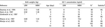
IGFs are also known to be involved in the regulation of adipose tissue growth and development (Louveau and Gondret, Reference Louveau and Gondret2004). It is possible that the lower perirenal fat development observed in LBW involves an alteration of the IGF system as shown in the liver and skeletal muscle of growth-retarded foetuses and neonates (Kampman et al., Reference Kampman, Ramsay and White1993 and Reference Kampman, Ramsay and White1994; Tilley et al., Reference Tilley, McNeil, Ashworth, Page and McArdle2007). In the neonatal sheep, increased adiposity at term has been shown to be associated with increased levels of mRNA for IGF receptors (Symonds et al., Reference Symonds, Pearce, Bispham, Gardner and Stephenson2004). In the neonatal pig, IGF-I infusion results in an increase in weight gain that involves an increased rate of protein and fat accretion (Schoknecht et al., Reference Schoknecht, Ebner, Skottner, Burrin, Davis, Ellis and Pond1997; Dunshea et al., Reference Dunshea, Chung, Owens, Ballard and Walton2002). These effects are more marked in intrauterine growth-retarded piglets, which have a higher protein and fat accretion than control animals. In these animals, IGF-I infusion is able to restore BW and composition to normal (Schoknecht et al., Reference Schoknecht, Ebner, Skottner, Burrin, Davis, Ellis and Pond1997). This higher efficiency of IGF-I infusion in LBW piglets can be explained by their lower plasma IGF-I concentration between birth and 3 days of age (Davis et al., Reference Davis, Fiorotto, Burrin, Pond and Nguyen1997; Ritacco et al., Reference Ritacco, Radecki and Schoknecht1997; Schoknecht et al., Reference Schoknecht, Ebner, Skottner, Burrin, Davis, Ellis and Pond1997; Table 4). A positive correlation between birth weight and plasma IGF-I has been reported in many mammalian studies (Thieriot-Prevost et al., Reference Thieriot-Prevost, Boccara, Francoual, Badoual and Job1988). However, plasma IGF-I concentration of LBW piglets is restored to the level of MBW piglets after the first days of life (Dauncey et al., Reference Dauncey, Burton and Tivey1994; Schoknecht et al., Reference Schoknecht, Ebner, Skottner, Burrin, Davis, Ellis and Pond1997; Mostyn et al., Reference Mostyn, Litten, Perkins, Euden, Corson, Symonds and Clarke2005). Moreover, plasma IGF-I concentration is strongly linked to nutrient bioavailability and difference in concentration may be due to difference in feed intake. Indeed, at 14 days of life, LBW piglets have lower hepatic IGF-I concentration than MBW piglets fed ad libitum but similar to their pair-fed MBW littermates (Dauncey et al., Reference Dauncey, Burton and Tivey1994).
Plasma IGF-I concentrations in low (LBW) and median birth weight (MBW) piglets

†Percentages LBW of MBW.
‡NS, not significantly different (P < 0.05); S, significantly different (P < 0.05).
§Weight at 3 days of age: 1.54 and 1.74 kg for LBW and MBW, respectively.
The developing intestine is a target organ for milk-borne growth factors since functional receptors to these factors are expressed in the apical membranes of the small intestine (Schober et al., Reference Schober, Simmen, Hadsell and Baumrucker1990; Kelly, Reference Kelly1994; Morgan et al., Reference Morgan, Coutts, McFadyen, King and Kelly1996). Moreover, IGF-I present in sow colostrum and milk stimulates gastrointestinal tissue growth and functional maturation in newborn piglets. IGF-I adjunction to milk formulas increases intestinal weight and villosity height (Burrin et al., Reference Burrin, Wester, Davis, Amick and Heath1996), brush border enzyme activities (Houle et al., Reference Houle, Schroeder, Odle and Donovan1997), and the rates of net Na+- and Na+-dependent nutrient absorption (Alexander and Carey, Reference Alexander and Carey1999). On the contrary, GH delivered with an osmotic minipump into the abdominal cavity of newborn piglets failed to improve the absorptive capacity of the small intestine (Fholenhag et al., Reference Fholenhag, Malmlof, Skottner and Nyberg1999). Hormonal regulation of intestinal development seems to be affected by intrauterine growth retardation. Indeed, in association with an altered intestinal morphology, intrauterine growth-retarded piglets have a lower mucosal IGF-I gene expression than controls, whereas they tend to express less GH and insulin receptors than controls (Wang et al., Reference Wang, Huo, Shi, Xu and Hutz2005).
Insulin
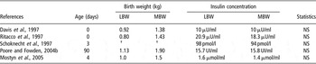
In addition to its well-known acute metabolic actions, insulin plays a role in the control of normal body growth. Child with diabetes exhibits a poor growth, contrasting with the overgrowth of the hyperinsulinemic infant of a diabetic mother (Hill and Milner, Reference Hill and Milner1985). Diabetic pigs have a 50% lower BW than the controls (Romsos et al., Reference Romsos, Leveille and Allee1971a). Administration of insulin restores growth to a rate similar to control pigs (Romsos et al., Reference Romsos, Leveille and Allee1971b). Growth retardation in diabetic rats is associated with a rapid decline in the circulating levels of IGF-I (Maes et al., Reference Maes, Ketelslegers and Underwood1983) that is not restored by GH administration but by insulin only (Phillips and Young, Reference Phillips and Young1976), indicating a modulation of IGF-I release by insulin (Hill and Milner, Reference Hill and Milner1985). In contrast, daily administration of insulin to young pigs does not affect growth rate, feed efficiency, or muscle and adipose tissue mass, suggesting that insulin is not a rate-limiting factor for growth of healthy pigs (Steele and Etherton, Reference Steele and Etherton1983). Similarly, low foetal growth does not seem to be due to low plasma insulin level since the fasting level is similar in LBW and MBW piglets (Table 5). However, even though intrauterine growth retardation does not affect plasma insulin level at birth, it enhances the risk of developing insulin resistance in growing pigs (Poore and Fowden, Reference Poore and Fowden2004b) as in humans (Ong and Dunger, Reference Ong and Dunger2004). Adult pigs of LBW have a poor glucose tolerance (Poore and Fowden, Reference Poore and Fowden2002) that may result from a decrease in insulin sensitivity (Poore and Fowden, Reference Poore and Fowden2004b).
Fasting plasma insulin concentrations in low (LBW) and median (MBW) birth weight piglets

†Weight at 3 days of age: 1.54 and 1.74 kg for LBW and MBW, respectively.
Insulin also plays an important role in the development of tissues, including the intestine and adipose tissue. It is naturally present in colostrum and milk. Insulin concentration is much higher in sow colostrum than in blood plasma: 411 μU/ml v. 5 μU/ml (and this concentration declines after 72 h lactation) (Weström et al., Reference Weström, Ekman, Svendsen, Svendsen and Karlsson1987). There is tangible evidence suggesting that insulin can act locally on the gastrointestinal tract or after being absorbed on peripheral targets (Xu et al., Reference Xu, Wang and Zhang2000; Zabielski et al., Reference Zabielski, Laubitz, Wolinski and Guilloteau2005). Oral insulin (85 mU/ml) enhances the small intestine and mucosal mass and the activity of brush border lactase and maltase in pig neonates (Shulman, Reference Shulman1990; Shulman et al., Reference Shulman, Tivey, Sunitha, Dudley and Henning1992). Interestingly, oral insulin also enhances the expression of its own receptor in small intestine, which may explain the effect of dietary insulin on receptor-mediated postnatal development of the small intestine (Huo et al., Reference Huo, Wang, Xu, Macdonald, Liu and Shi2006). In the adipose tissue of many mammalian species, including pigs, insulin stimulates the anabolic lipid metabolism pathways (Romsos et al., Reference Romsos, Leveille and Allee1971b; Mills, Reference Mills1999). Indeed induction of maternal diabetes results in an increase in lipid deposition in foetuses associated with an elevated plasma insulin and adipose tissue lipogenesis (Hausman and Hausman, Reference Hausman and Hausman1993). It also plays a major role in the regulation of adipogenesis, since in vitro, insulin is required for adipocyte differentiation (Mersmann and Smith, Reference Mersmann and Smith2005).
Thyroid hormones
TH are known to play a major role in the regulation of metabolic adaptations and growth, more particularly of foetal growth and muscle maturity. Pig foetuses from sows fed a high glucosinolate rapeseed diet have low circulating TH and are lighter at the end of gestation (Duchamp et al., Reference Duchamp, Burton, Herpin and Dauncey1994). At birth, TH metabolism of healthy piglets seems to be fully developed. Plasma concentrations of both total and free TH, thyroid gland weights and hepatic 5′-deiodinase activity increase during late gestation (Berthon et al., Reference Berthon, Herpin, Le Dividich and Dauncey1993). Receptors are detected at 80 days of gestation in skeletal muscle, but not in liver, suggesting that porcine muscle can potentially respond to TH much earlier than liver (Duchamp et al., Reference Duchamp, Burton, Herpin and Dauncey1994). During the first 6 h after birth, there is a surge in T3, free T3 and T4 plasma concentrations and apart from a transient decline at 12 h, TH concentrations remain high during the first 2 days and then decline slightly over the next 2 weeks (Slebodzinski, Reference Slebodzinski1981; Berthon et al., Reference Berthon, Herpin, Le Dividich and Dauncey1993 and Reference Berthon, Herpin, Duchamp, Dauncey and Le Dividich1996). The influence of birth weight on TH is controversial in the pig. Although Ritacco et al. (Reference Ritacco, Radecki and Schoknecht1997) found no effect of birth weight on plasma TH concentrations, Bauer et al. (Reference Bauer, Wank, Walter, Blickhan and Zwiener2000b) described an increased plasma T4 concentration in LBW piglets. This was associated with an improved calf muscle blood supply and progressed contractile function. The authors suggested that there was an accelerated muscular development due to intrauterine growth restriction. In the rat, TH have been reported to be involved in the regulation of adipose tissue development (Blennemann et al., Reference Blennemann, Moon and Freake1992) and intestine maturation during weaning (Hodin et al., Reference Hodin, Meng and Chambernain1994). In the pig, available data are consistent with a positive influence of T4 on adipose tissue development in foetuses (Hausman and Hausman, Reference Hausman and Hausman1993; Chen et al., Reference Chen, Hausman and Wright1996; Hausman and Wright, Reference Hausman and Wright1996).
Glucocorticoids
In addition to its well-known role in stress response, cortisol is involved in the regulation of growth during foetal (Bell et al., Reference Bell, Greenwood and Ehrhardt2005) and neonatal life (Lawrence and Fowler, Reference Lawrence and Fowler2002). In the pig, circulating levels of glucocorticoids and catecholamines are very high at birth and dramatically decrease thereafter (Kaciuba-Uscilko, Reference Kaciuba-Uscilko1972; Randall, Reference Randall1983). Cortisol and catecholamines are potent stimulators of catabolism and one can speculate that these high levels induce mobilisation of glycogen stores immediately after birth.
Chronic exposure to elevated glucocorticoids is known to inhibit postnatal growth. Nevertheless, the surge in glucocorticoids levels during the neonatal period may be important for the regulation of postnatal development and growth. The lack of glucocorticoid surge has been shown to be associated with a reduction in piglet growth (Carroll et al., Reference Carroll, Daniel, Keisler and Matteri2000) and a single administration of dexamethasone (1 mg/kg BW) at birth increases the average daily gain during the first 18 days of life (Carroll, Reference Carroll2001). Glucocorticoids can also affect body composition. Indeed, they favour protein catabolism as well as lipogenesis (Lawrence and Fowler, Reference Lawrence and Fowler2002). In young pigs, in vitro studies have shown that glucocorticoids can potentiate IGF-I-stimulated preadipocyte differentiation (Hausman and Hausman, Reference Hausman and Hausman1993). Birth weight does not influence basal plasma cortisol concentrations at 3 or 12 months of age. However, cortisol response to stress is higher in 3-month-old LBW than in HBW piglets. As these LBW pigs also exhibit greater fat depth at 12 months of age, the authors suggest that increased stress responsiveness in early life could play a role in the predisposition of LBW pigs to later fat accumulation (Poore and Fowden, Reference Poore and Fowden2003).
Glucocorticoids exert stimulating effects on intestinal digestive enzymes in the late foetal and early neonatal period when rapid development of the intestinal function takes place (Sangild et al., Reference Sangild, Xu and Trahair2002). The administration of glucocorticoids has been used to stimulate maturation of the small intestine and other organs such as liver, lungs and kidneys in premature babies. However, the reported effects of exogenous glucocorticoids in newborn farm animals are variable and may occur only during a certain developmental period.
Leptin
Leptin is the protein product of the obese (ob) gene and is involved in the regulation of food intake, BW and whole body energy balance in adults (Friedman and Halaas, Reference Friedman and Halaas1998; Barb et al., Reference Barb, Hausman and Houseknecht2001). Its role in the neonate is less documented. In species such as the pig, in which fat is detected before birth, both leptin and leptin receptor mRNA are detected in subcutaneous adipose tissue from 105-day-old foetuses (Chen et al., Reference Chen, Lin, Hausman, Martin, Dean and Hausman2000). Levels of leptin mRNA were much lower in foetuses than in 7-day-old piglets. As in humans or sheep, leptin is detectable in porcine foetal serum. After birth, there is no significant change in the plasma leptin concentration between 3 and 8 days of age in piglets (Litten et al., Reference Litten, Mostyn, Perkins, Corson, Symonds and Clarke2005). Between 40 and 150 days of age, leptin concentration has been reported to increase (Qian et al., Reference Qian, Barb, Compton, Hausman, Azain, Kraeling and Baile1999).
Studies on the effects of exogenous leptin on the young pig are scarce and use different doses (4 to 500 μg/kg BW) and way of leptin administration (intramuscular, intracerebro-ventricular and intravenous, single or chronic injection). Thereby, it is difficult to draw a clear-cut picture on the effects of exogenous leptin. Single injection of high-dose leptin (intra-cerebro-ventricular or in the carotid artery) increases GH secretion and decreases food intake (Barb et al., Reference Barb, Yan, Azain, Kraeling, Rampacek and Ramsay1998). It also induces hypoglycaemia, hypoinsulinaemia and an increased concentration of non-esterified fatty acids (NEFA; Ramsay et al., Reference Ramsay, Bush, McMurtry, Thivierge and Davis2004). Chronically administered leptin (50 μg/kg BW per day) reduces food intake, resulting in decreased growth rates in 27-kg pigs. It also seems to regulate IGF-I liver production in a dose-dependent manner (Ajuwon et al., Reference Ajuwon, Kuske, Ragland, Adeola, Hancock, Anderson and Spurlock2003). In the neonatal pig, intravenous administration of a much lower dose of leptin (4 μg/kg BW per day) increases growth rate and promotes skeletal growth in favour of adipose tissue accretion without any effect on insulinaemia, glycaemia or NEFA concentrations (Litten et al., Reference Litten, Mostyn, Perkins, Corson, Symonds and Clarke2005).
The effects of intrauterine growth retardation on leptinaemia are poorly documented in the pig. Available data indicate that LBW male pigs have similar leptinaemia at 3 months of age, but lower leptinaemia at 12 months of age than HBW pigs. However, leptinaemia is not correlated to current weight or body mass index (BMI) at 12 months of age. The authors suggest that LBW pigs have a deficiency in adipocyte leptin production as adults, although they are not underweight. This low leptin levels, for a given fat mass, may predispose to later obesity, since it characterises a state of perceived energy deficit (Poore and Fowden, Reference Poore and Fowden2004a). In another study, a negative correlation between BW and leptin mRNA abundance in adipose tissue has been established in 2-month-old female pigs (Eckert et al., Reference Eckert, Gatford, Luxford, Campbell and Owens2000). LBW has the opposite effect on leptinaemia in human, but the lack of correlation between fat mass and plasma leptin concentration observed in adults with LBW is also found. In case of humans, LBW adults have higher leptin concentration than individuals at the same BMI, but with a higher BW (Phillips et al., Reference Phillips, Fall, Cooper, Norman, Robinson and Owens1999). Serum-leptin concentrations are low in intrauterine growth-retarded infants at birth, and increase to become higher in these infants at 1 year of age than their normal BW counterparts (Jaquet et al., Reference Jaquet, Leger, Tabone, Czernichow and Levy-Marchal1999). Thus, programming of leptin concentrations by early diet may be one mechanism that links early nutrition to later obesity (Singhal et al., Reference Singhal, Farooqi, O’Rahilly, Cole, Fewtrell and Lucas2002). This is supported by studies in rodents, where rats from undernourished mothers and fed a high fat diet exhibit in adulthood higher leptin, insulin and glucose concentrations and fat pad mass than the control rats (Vickers et al., Reference Vickers, Reddy, Ikenasio and Breier2001); however, these metabolic consequences of maternal undernutrition were reversed by a period of neonatal leptin treatment in female rats (Vickers et al., Reference Vickers, Gluckman, Coveny, Hofman, Cutfield, Gertler, Breier and Harris2005). However, there are many differences between rodent and human adipo-insular axis regulation, and studies in other species are required to conclude on the physiologic role of leptin in the metabolic imprinting.
Like insulin and IGF-I, leptin is also present in sow colostrum and milk (Estienne et al., Reference Estienne, Harper, Barb and Azain2000). Leptin supplementation in milk formulas increases intestinal crypt depth in the upper jejunum, reduces intestinal villi length and the number of vacuolated enterocytes and increases the mitotic index. These results suggest that leptin given into the gastrointestinal tract lumen speeds up the maturation of the small intestine mucosa (Wolinski et al., Reference Wolinski, Biernat, Guilloteau, Weström and Zabielski2003).
Epidermal growth factor (EGF)
EGF is known as a regulator in a wide variety of physiological processes including embryogenesis (Vaughan et al., Reference Vaughan, James, Pascall and Brown1992), growth, tissue repair and regeneration (Zijlstra et al., Reference Zijlstra, Odle, Hall, Petschow, Gelberg and Litov1994). It is produced in various tissues such as brain and the urogenital and gastrointestinal tracts (Kajikawa et al., Reference Kajikawa, Yasui, Sumiyoshi, Yoshida, Nakayama, Ayhan, Yokozaki, Ito and Tahara1991; Peng et al., Reference Peng, Palin, Veronneau, Lebel and Pelletier1997). Sow colostrum and milk contain high concentration of EGF that is involved in the development of intestinal mucosa in newborn pigs. EGF receptor has been identified on the epithelial cells of the gastrointestinal tract, from the oesophagus to the ileum in 1- to 28-day-old pigs (Jaeger and Lamar, Reference Jaeger and Lamar1992). It has been shown in several studies that exogenous EGF influences gut epithelial maturation and function. It increases lactase and sucrase specific activities and protein synthesis rate in jejunal explants and limits gastric acid secretion. In addition, exogenous EGF may facilitate the recovery of traumatised gastric and intestinal tissues (Xu et al., Reference Xu, Wang and Zhang2000).
Data on the effects of EGF on growth and adipose tissue development are scarce and do not concern pigs. Available data in rats are consistent with a role of EGF on adipose tissue development. In newborn rats, daily EGF injections decrease both body and fat pad weight gains with no effects on other organs (Serrero and Mills, Reference Serrero and Mills1991). The number of adipocyte precursors per fat pad is higher in fat pads from EGF-treated animals than in control rats, but triglyceride storage in these tissues is lower. The authors suggest that the increase in the number of adipocyte precursors observed in EGF-treated rats is probably due to a delayed differentiation resulting in the fact that a smaller number of precursors differentiated and moved into the pool of triglyceride-laden adipocyte during the duration of the in vivo experiment.
Conclusion
In pigs, LBW is associated with reduced growth rate throughout the growing period. There is increasing evidence that intrauterine growth retardation causes permanent changes in the developmental processes, resulting in fatter pigs. LBW piglets exhibit delayed maturity of the gastrointestinal tract during neonatal life that may enhance risks of developing intestinal diseases and reduce digestive capacities. Hormonal status is also affected by intrauterine growth retardation at birth and subsequently. However, the mechanisms by which these permanent effects occur and the interrelation between hormones remain unclear. It is also suggested that individuals with LBW may be more susceptible to changes in neonatal nutrition. Thus, the impact of neonatal nutrition on the subsequent physiology and the metabolic status of farm animals warrant further studies and should contribute to sustain research on animal health and meat quality. Such short- and long-term investigations will also find applications in human nutrition, the piglet being used as an animal model of intrauterine growth retardation.