INTRODUCTION
Micrura dellechiajei (Hubrecht, Reference Hubrecht1879) (Nemertea: Pilidiophora: Lineidae) is an elusive ribbon worm, with a conspicuous lack of information regarding its geographic distribution and general natural history. The species seems to have been first recorded from the general region surrounding Naples, Italy by Delle Chiaje (Reference Delle Chiaje1829) who attributed his specimens to a species already known at the time, Cerebratulus bilineatus (Renier, Reference Renier1804). However, the formal description of the species was synthesized fully 50 years later by Hubrecht (Reference Hubrecht1879), based on specimens collected from around Capri, an island south of Naples, in the same vicinity as Delle Chiaje's (Reference Delle Chiaje1829) collection site. These specimens proved to be morphologically compatible with those collected by Delle Chiaje (Reference Delle Chiaje1829). As opposed to Delle Chiaje (Reference Delle Chiaje1829), however, Hubrecht (Reference Hubrecht1879) identified morphological apomorphies possessed by the new specimens and, accordingly, erected a new species, Cerebratulus dellechiajei Hubrecht, Reference Hubrecht1879 to accommodate the specimens. Beyond obvious differences in colour patterns between C. bilineatus and C. dellechiajei, Hubrecht (Reference Hubrecht1879) also noted that, in contrast to C. bilineatus, C. dellechiajei possesses a short caudal cirrus and numerous eyes – attributes that had come to unify species of the genus Micrura Ehrenberg, Reference Ehrenberg, Hemprich and Ehrenberg1831. As a consequence, Bürger (Reference Bürger1895) transferred the species to the genus Micrura and provided extensive illustrations of external and internal anatomy of the species. Although Bürger's illustrations (Reference Bürger1895; plate 4, figures 18, 23, 24, 26, 27, 33) are adequate in allowing for identification of specimens belonging to the species, the aged description by Hubrecht (Reference Hubrecht1879) is insufficient in several regards, and thus in dire need of a complete update, using contemporary data. However, since Bürger's (Reference Bürger1895) treatment of the species about 120 years ago, no specimens have been recorded and, therefore, no data have been available on which to base a redescription. Numerous gracile heteronemertean species possessing a caudal cirrus and cephalic slits have traditionally been included in the genus Micrura but modern molecular phylogenetic studies have revealed an astonishing level of paraphyly within the genus (e.g., Sundberg & Saur, Reference Sundberg and Saur1998; Schwartz, Reference Schwartz2009; Andrade et al., Reference Andrade, Strand, Schwartz, Chen, Kajihara, Von Döhren, Sun, Junoy, Thiel, Norenburg, Turbeville, Giribet and Sundberg2012; Kvist et al., Reference Kvist, Laumer, Junoy and Giribet2014). This indicates that the putative synapomorphies previously considered for the genus were wrong, increasing the need for a modern redescription of M. dellechiajei, as well as a reconsideration of the entire genus.
During September 2011 and July 2012, numerous nemertean samples were collected as part of an oceanographic campaign (the INDEMARES Project) in the Alborán Sea in southern Spain. This collection effort yielded several specimens of Micrura dellechiajei, which had also previously been collected during the 2007/2008 CALMEN oceanographic campaign to the Menorca Channel (Balearic Islands, Spain) – these specimens, however, were poorly preserved and proved refractory to further molecular studies. Importantly, a combination of morphological characters and molecular data seems to be the best approach for inferring species delimitations within Nemertea (Sundberg et al., Reference Sundberg, Thuroczy Vodoti and Strand2010; Sundberg & Strand, Reference Sundberg and Strand2010; Strand & Sundberg, Reference Strand and Sundberg2011; Leasi & Norenburg, Reference Leasi and Norenburg2014; Strand et al., Reference Strand, Herrera-Bachiller, Nygren and Kånneby2014). Therefore, we herein provide a thorough redescription of Micrura dellechiajei, based on traditional histological sections as well as external anatomical observations, of samples from both the Alborán Sea and the Menorca Channel, approximately 1500 km west of its type locality. We also provide a molecular phylogeny of Heteronemertea to estimate the placement of the species within the class and to ensure the monophyly of the sampled specimens, and we briefly discuss these results in the context of the non-monophyly of most genera within Heteronemertea.
MATERIALS AND METHODS
Material examined
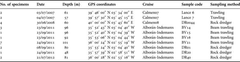
During research cruises CALMEN07, CALMEN08 in the Menorca Channel (Balearic Islands, Spain) in July 2007 and June 2008, as well as ALBORÁN-INDEMARES in the Alborán Sea (Spain) in September 2011 and July 2012 (Figure 1, Table 1), numerous nemertean samples were collected by beam trawler and rock dredger. Depths ranged from 57 to 101 m and samples were generally associated with encrusting red algae, organically enriched sediments (shell sand) and communities of sponges and cnidarians. The following redescription of Micrura dellechiajei is based on observations of 25 living specimens (Table 1) and three sets of histological slides, two of these being from the white morphotype (Figure 1A) and one from the olive-colour morphotype (Figure 1C).
Map showing the collection localities of the sampled specimens of Micrura dellechiajei (Hubrecht, Reference Hubrecht1879) in the Menorca Channel and the Alborán Sea (stars). The circle denotes the type locality of the species off the coast of Naples, Italy.

List of localities of the sampled specimens of Micrura dellechiajei in the Menorca Channel (Balearic Islands) and Alborán Sea (South Spain).
Histology
For the present study, worms were measured and photographed while alive, relaxed in a 7.5% wt/vol solution of MgCl2 mixed 1:1 with filtered seawater, and fixed in Bouin's (cupric picro-formolacetic) solution. Specimens destined for histology were dehydrated in a graded series of ethyl alcohols, cleared with multiple changes of toluene, and embedded in 56°C mp paraffin wax. Three specimens from Alborán Sea were sectioned and studied for histological features. For this purpose, samples were sectioned at 6 μm, mounted on slides, and stained with Mallory triple stain.
Molecular analyses
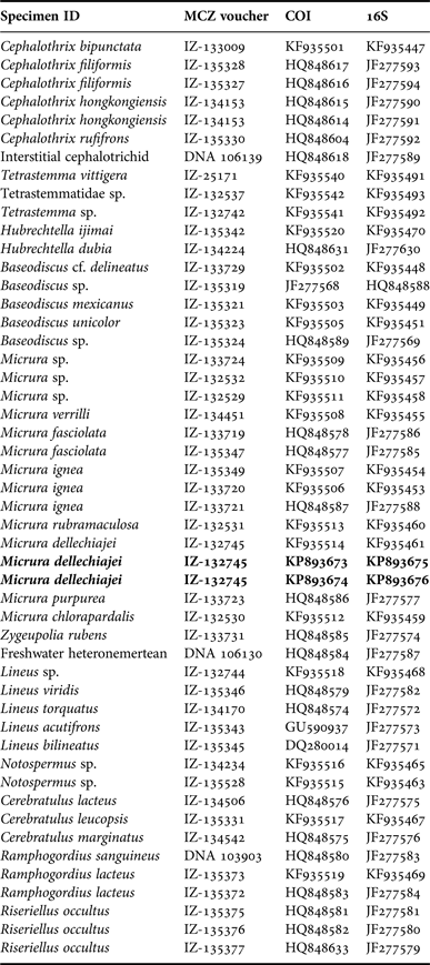
DNA was extracted, amplified, purified and sequenced from three specimens of Micrura dellechiajei (one of them from the white morphotype [Figure 1A] and two of them from the olive-colour morphotype [Figure 1C]) following the exact protocol described by Kvist et al. (Reference Kvist, Laumer, Junoy and Giribet2014), but the present study targeted only the mitochondrial loci cytochrome c oxidase subunit I (COI) and 16S rRNA. These loci were chosen based on their high rate of nucleotide divergence, i.e. their pertinence for teasing out genetic differences between morphologically similar species (see Discussion), coupled with the fact that one of our specimens has been previously sequenced for six loci and placed in a broader phylogenetic context (see Kvist et al., Reference Kvist, Laumer, Junoy and Giribet2014). Subsequently, the newly generated sequences were conjoined with a set of sequences previously compiled for other palaeonemertean, heteronemertean and hoplonemertean taxa (Table 2) and these were jointly aligned using MAFFT ver. 7 (Katoh & Standley, Reference Katoh and Standley2013) employing the L-INS-i strategy and applying default settings. Phylogenetic analyses used both maximum likelihood (ML) and parsimony (MP) methodologies. For ML, RAxML ver. 7.6.3 (Stamatakis, Reference Stamatakis2006) was applied on the CIPRES Science Gateway platform (Miller et al., Reference Miller, Pfeiffer and Schwartz2010) using a partitioned GTR + Γ model of nucleotide evolution, with optimal partition schemes suggested by PartitionFinder ver. 1.1.1 (Lanfear et al., Reference Lanfear, Calcott, Ho and Guindon2012). A heuristic search consisted of 1000 iterations with 25 initial GAMMA rate categories and final optimization with four GAMMA shape categories. For MP, the software TNT (Goloboff et al., Reference Goloboff, Farris and Nixon2008) was used with the following settings: 1000 initial addition sequences, five rounds of ratcheting and three rounds of tree fusing, and requiring that the minimum length tree be found a total of 10 times. For both MP and ML, multiparametric bootstrap support values were calculated from 1000 pseudoreplicates with a different starting tree for each iteration. All trees were rooted at the palaeonemertean Cephalothrix bipunctata Bürger, Reference Bürger1892 following the phylogenetic hypothesis of Kvist et al. (Reference Kvist, Laumer, Junoy and Giribet2014).
List of specimens included in the phylogenetic analyses, with MCZ voucher numbers (more information on each specimen is accessible through http://mczbase.mcz.harvard.edu/), and GenBank accession numbers. Specimens newly sequenced for the present study are denoted in bold font.
TAXONOMY
SYSTEMATICS
Class anopla Schultze, Reference Schultze1851
Subclass heteronemertea
Genus Micrura Ehrenberg, Reference Ehrenberg, Hemprich and Ehrenberg1831
DIAGNOSIS
From Gibson (Reference Gibson1981): ‘Heteronemertea with a single pair of horizontal lateral cephalic slits, posteriorly enlarged to form wide bays; ciliated cerebral canals emerge from ventral wall of cephalic bays; proboscis unbranched, containing two (outer circular, inner longitudinal) or three (outer longitudinal, middle circular, inner longitudinal) muscle layers and none, one or two muscle crosses; rhynchocoel circular musculature not interwoven with body wall muscles; dorsal fibre core of cerebral ganglia bifurcated only at rear into upper and lower branches; nervous system with neither neurochords nor neurochord cells; ganglionic cell layer of brain usually not separated from body wall muscles by neurilemma; foregut with or without splanchnic musculature, if present variably composed of circular and/or longitudinal fibres; dermis variable, mostly with distinct connective tissue layer separating glandular zone from body wall muscles; caudal cirrus present; cephalic glands normally well developed, occasionally weakly formed or absent; frontal organ usually present; eyes present or absent; sexes separate.'
This wide diagnosis accommodates species attributed to Micrura and the validity of this diagnosis is compromised due to the non-monophyletic nature of the genus. However, the specimens used for the present study agree with this diagnosis, as well as those of Hubrecht (Reference Hubrecht1879) and Bürger (Reference Bürger1895), and given the absence of a better system for assigning heteronemerteans to genus, we leave the species in the genus Micrura.
Micrura dellechiajei (Hubrecht, Reference Hubrecht1879)
(Figures 2–4)
Micrura dellechiajei (Hubrecht, Reference Hubrecht1879). (A) photograph of a complete specimen from the Alborán Sea; (B) photograph of a complete specimen from the Menorca channel; (C) photograph of a complete specimen from the Alborán Sea; (D) Bürger's (Reference Bürger1895) original drawings of M. dellechiajei.
Micrura dellechiajei (Hubrecht, Reference Hubrecht1879). (A) macrophotograph of the head of a specimen in dorsal view, showing the characteristic stripe pattern; (B) macrophotograph of the head of a specimen in lateral view showing the cephalic slits and the ocelli (arrowhead); (C) macrophotograph of the posterior end of a specimen in dorsal-lateral view showing the small caudal cirrus (arrowhead); (D) group of specimens for comparison of the differences between the colour nuances of the individuals.
Micrura dellechiajei (Hubrecht, Reference Hubrecht1879). (A) detail of a section through the posterior foregut region showing the lateral nerve cord, the lateral blood lacuna (arrow) and one excretory tubule (arrowhead); (B) detail of a section through the cephalic region showing the dorsal cerebral ganglion with the dorsal (arrowhead) and ventral (arrow) commissures; (C) section through the intestinal region showing the deep lateral intestinal diverticula and the gonads; (D) section through the stomach region showing the moderately convoluted wall; note the rhynchocoelic villus (arrowhead); (E) section through the apical region of the body showing the proboscis pore and a pair of eyes (arrowheads); (F) detail of a section through the cephalic region showing the ciliated canal (arrow) that connects the cephalic slit with the cerebral organ; note the buccal nerves (arrowheads); (G) section of the proboscis showing two pairs of proboscis nerves (arrowheads), the muscle crosses, and the differentiated epithelium. Abbreviations: CB, cerebral ganglion; CS, cephalic slit; CO, cerebral organ; E, epidermis; G, gonad (ovary); I, intestine; LN, lateral nerve cord; MCM, middle circular muscle layer; OLM, outer longitudinal muscle layer; P, proboscis; R, rhynchocoel; S, stomach. Scale bars: 100 μm.
Cerebratulus bilineatus (partim.) (Renier, Reference Renier1804); Cerebratulus delle-chiajei Hubrecht, Reference Hubrecht1879; Meckelia cerebratulus Diesing, Reference Diesing1850; Micrura dellachiajei Bürger, Reference Bürger1895; Micrura delle chiajei Bürger, Reference Bürger1892; Nemertes bilineatus Örsted (Reference Örsted1844); Nemertes bilineata; see Gibson (Reference Gibson1995).
DESCRIPTION
External features. Specimens 4–10 cm long, 3–4 mm wide when fully extended. Body colour varying from very dark to olive or white (Figure 2). Dorsal surface with longitudinal striped pattern alternating symmetrically in colour nuance, with respect to the dorsal midline. Dark specimens with four white dorsal, longitudinal stripes. Olive and white specimens with three dark longitudinal stripes. Some specimens without striped pattern in posterior. Dorsal midline generally orange with dark brown border. Ventral surface pale in all specimens. Head (Figure 3A) ovate, not distinct from rest of body, except for slight constriction at posterior part of cephalic slits. Tip of head obtuse. Lateral horizontal slits long, open and shallow, reaching behind cerebral ganglia (Figure 3B). When relaxed, body round in cross section, but anterior end dorsoventrally flattened. Posterior end tapering, bearing minute caudal cirrus, sometimes difficult to observe (Figure 3C). Mouth composed of thin but elongate slit, slightly posterior to end of lateral slits. Black ocelli in rows throughout anterior portion of slits (Figure 3B). Gonads visible through body wall in intestinal region, forming rows on both sides of ventral midline. Specimens contract when exposed to light and when disturbed, coiling in a spiral, releasing mucus and usually fragmenting.
Body wall, musculature, and parenchyma. Epidermis ciliated, 35–50 μm thick, dominated by serous goblet cells. Ciliated cells separating globet cells, predominating in cephalic slits. Mouth with slightly longer epithelial cells. Epidermis resting upon basal lamina. Dermal gland zone as thick (35–50 μm) as epidermis, placed under the basal lamina. Dermal gland zone consisting of hard-to-detect circular dermal musculature, very thin (15–20 μm, depending on contraction) longitudinal dermal musculature, and with layer of gland cells embedded on dermal connective tissue stratum. Well-developed (80–150 μm, depending on contraction) body wall with outer longitudinal musculature (OLM) (Figure 4A, D) below dermal zone. Thin peripheral neural sheath with nerve cords embedded, present under OLM. Well-developed (25 μm) middle circular musculature (MCM) followed by an inner longitudinal musculature (ILM), 30 μm thick, present under peripheral neural sheath. Precerebral cephalic musculature present as framework of outer longitudinal muscle fibres mixed with radial and oblique muscle fibres, with interspersed gland cells. Circular muscles around the rhynchodaeal blood lacuna absent. MCM starts at middle of cerebral region, at point of connection between cerebral organ canals and cephalic slits. Dorsoventral muscles absent in the anterior intestinal region.
Proboscis apparatus. Proboscis pore ciliated, opening subterminally and ventrally near tip of head (Figure 4E). Proboscis reaches rhynchodaeum as tubular chamber with ciliated epithelium; globet cells absent. Thin basal lamina surrounding pore lumen. Rhynchodaeum musculature consisting of thin dermal circular layer and discrete longitudinal layer. Rhynchocoel extends for most of body length. Circular muscle fibres of rhynchocoel wall very thick throughout cerebral region; these do not intermingle with muscles of body wall. Rhynchocoel CM and body wall MCM in contact dorsally throughout most of length of body, always with fine coat of ILM as separator. Rhynchocoel with circular musculature in mouth region, intertwined with longitudinal musculature of foregut ventrally and throughout part of length of villus. Villus with spongy appearance. Rhynchocoel wall consisting almost exclusively of thin circular muscular coat in midgut region. Proboscis insertion point just anterior to cerebral ganglia, with two proboscis nerves entering from cephalic nerve region. Proboscis significantly longer than body length, unbranched, possessing five distinct layers: thin endothelial lining, inner longitudinal muscle zone, circular muscle layer, neural plexus with two pairs of proboscis nerves discernable (Figure 4G), and outer glandular epithelium. Epithelium of proboscis differentiated into two regions, one anterior and one posterior; one forming a ridge bearing glandular, cup-shaped structures with apical arrays of small rhabditiform granules, and other with wide folds bearing gland cells lacking such granules. Two muscle crosses distinguishable, running from circular muscle layer to endothelium of proboscis, situated perpendicular to opposing pairs of proboscis nerves.
Alimentary canal. Mouth present as ventral slit posterior to cephalic slits, 1 mm long. Interior margin with deep ridges and thick epithelium covered with glandular cells. Foregut wall moderately convoluted, with distinct lining of cilia and microvilli. Subepithelial gland-cell zone ensheathing the stomach at foregut (Figure 4D). Posterior part of foregut marked by change in wall thickness, becoming thinner. Intestinal region with convoluted wall with numerous large spherical glandular cells and bearing deep lateral diverticula for most of length, alternating with gonads in mature specimens (Figure 4C).
Circulatory system. Blood lacuna dorsal to the proboscis pore in head region. Rhynchodaeum dividing blood lacuna into two thin-walled cephalic blood vessels. Posterior to proboscis insertion, at level of brain commissures, lateral vessels anastomose to form U-shaped vessel cradling ventral portion of rhynchocoel. In half region of brain, vessel bifurcating, giving rise medially to mid dorsal blood vessel, forming long rhynchocoelic villus, approximately 2–2.5 mm long (Figure 4D). U-shaped vessel widening at level of cerebral organs, progressively becoming bisected by connective tissue. Lateral branches expand forming thin-walled lateral lacunae, bathing posterior portions of cerebral organs, then extending further posteriorly, adjacent to rhynchocoel. Lacunae eventually giving rise to ventrolateral vascular plexus passing backwards towards margins of buccal cavity and anterior part of foregut, extending to excretory tubules. At end of foregut, upper branches of vascular plexus joining dorsal lacunae to form pair of main dorsolateral blood vessels. In intestinal region, blood supply consisting of paired ventrolateral (running alongside intestine) and dorsolateral (close to rhynchocoel) vessels, and single small mid-dorsal vessel.
Nervous system. Brain well developed, with paired dorsal cerebral ganglia larger than ventral ones. Cerebral ganglia pink, visible through dorsal and ventral body wall. Dorsal cerebral commissure shorter and narrower than ventral (25 and 35 μm in diameter, respectively) (Figure 4B). Both dorsal and ventral ganglia enclosed by connective tissue. Outer and inner neurilemma absent around ganglia and lateral nerve cords. Dorsal ganglia bifurcated posteriorly, forming upper and lower neuropil. Ventral ganglia more elongate, extending slightly past dorsal lobes. Ventral ganglia confluent with lateral nerve cords (Figure 4A, D). Neurochords and neurochord cells not distinguished in nervous system. Numerous dorsal cephalic nerves leading forward from anterior borders of brain lobes, innervating cephalic gland region. Two buccal nerves arising from near rear of ventral ganglia (from inner lateral margin), and extending into foregut wall (Figure 4F). Dorsal nerve arising from dorsal commissure, extending posteriorly adjacent to body wall MCM. Peripheral neural sheath encircles the body wall MCM.
Sensory organs. Eyes 25–30 μm in diameter, consisting of rudimentary reddish pigment-cup ocelli (Figure 4E). Ocelli (N = 20–25) irregularly but longitudinally distributed along lateral cephalic margins, almost in rows, below and above cephalic slits. Two apical sense organs observed on anterior tip of head at level of proboscis pore, appearing as small ciliated pits opening independently, lacking epidermal glandular cells. Pits surrounded by glandular cells interspersed between cephalic musculature fibres in anterior part of head. Lateral horizontal cephalic slits beginning at tip of head and reaching posterior part of cerebral organs, at posterior brain region. Neuroglandular cerebral organs ovoid (maximum 230–250 μm in diameter), with typical lineid structure, attaching to rear of dorsal cerebral ganglia. Cerebral organ canal opening into small chamber of cephalic slit anterior to end (Figure 4F). Epithelium of cerebral organ canal surrounded by nerve cells, bearing cilia, lacking glandular cells. Glandular cells surrounding blind terminus of canal.
Excretory system. Well developed, confined to foregut region of body, consisting of several thick-walled longitudinal collecting tubes (20–30 μm in diameter) running close to blood lacunae, extending anteriorly and posteriorly and opening as two lateral nephridiopores near posterior part of foregut region (Figure 4A).
Reproductive system. All specimens mature. Only females sectioned, sexes are assumed separate. Gonads distributed in lateral rows on each side of intestine (Figure 4C), distributed between diverticula from dorsal to ventrolateral margins. All ovaries bearing up to 10 or more oocytes (170–230 μm diameter), with nucleus (30–40 μm diameter). No oviducts observed.
ECOLOGY
Specimens were found on maërl beds, constituted mainly by Lithophyllum racemus (Lamarck) Foslie, 1901, Lithothamnion philippii Foslie, 1897, and Phymatolithon calcareum (Pallas) Adey & McKibbin, 1970, and in organically enriched sediments consisting mainly of molluscan shells, with sessile animal communities including species of Porifera (Spongia Linneaus, 1759, Faciespongia Burton, 1934 and Axinella Schmidt, 1862) and Cnidaria (Alcyonium Pallas, 1766, Epizoanthus Gray, 1867, Eunicella Verrill, 1869, Paramuricea Koelliker, 1865 and Leptogorgia Milne-Edwards, 1857). Among the more conspicuous and numerous species associated with the samples containing M. dellechiajei were the echinoderms Parastichopus regalis (Cuvier, 1817) and Hacelia attenuata Gray, 1840, as well as the polychaete Hyalinoecia tubicola (O.F. Müller, 1776). Specimens were sexually mature between June and September.
PHYLOGENY
The maximum likelihood (ML) analysis resulted in a tree with a final GAMMA-based lnL score of −21869.649765 (Figure 5) and the parsimony analysis (MP) resulted in two equally parsimonious trees at 5392 steps and a compound retention index and consistency index of 0.508 and 0.255, respectively (the strict consensus tree is presented in Figure 6). Except for some minor rearrangements, mainly within Palaeonemertea, the ML and MP trees are fully congruent but the ML tree receives slightly higher overall bootstrap support. Both ML and MP trees based on the sequences included in this dataset have previously been discussed at length (Kvist et al., Reference Kvist, Laumer, Junoy and Giribet2014) and, therefore, we here focus mainly on the phylogenetic placement of the included specimens of Micrura dellechiajei. In both analyses, the included specimens form a monophyletic group with full support. However, in the ML tree, this clade forms the sister group of two specimens of Notospermus Huschke, 1829 (Figure 5), whereas the clade is the sister group of Cerebratulus leucopsis (Coe, 1901) in the MP tree. Neither of these positions receive resampling support in the analyses, but the M. dellechiajei + C. leucopsis clade recovered by the MP analysis is the sister group of the same specimens of Notospermus, suggesting that there is some affiliation between these species and Micrura dellechiajei. As is typical for a Sanger-based approach (Andrade et al., Reference Andrade, Strand, Schwartz, Chen, Kajihara, Von Döhren, Sun, Junoy, Thiel, Norenburg, Turbeville, Giribet and Sundberg2012; Kvist et al., Reference Kvist, Laumer, Junoy and Giribet2014), most of the genera within Heteronemertea, including Micrura, Cerebratulus Renier, Reference Renier1804 and Lineus Sowerby, 1806, are found to be paraphyletic. It should be noted that Lineus bilineatus (discussed below) is not affiliated closely with M. dellechiajei, as it places in a remote region of the tree – these species were probably confused by Delle Chiaje (Reference Delle Chiaje1829).
Best scoring tree from the maximum likelihood analysis of COI + 16S rRNA dataset (lnL = −21869.649765). Likelihood bootstrap values >50% are shown to the left and above each node. Specimens sequenced for the present study are denoted in bold font and are encircled by a yellow background, and IZ numbers refer to the morphological voucher ID deposited in the Department of Invertebrate Zoology collection of the MCZ.
Strict consensus of two equally parsimonious trees (length: 5392 steps; consistency index: 0.255; retention index: 0.508) produced by TNT for the COI+16S rRNA dataset. Likelihood bootstrap values >50% are shown to the left and above each node. Specimens sequenced for the present study are denoted in bold font and are encircled by a yellow background, and IZ numbers refer to the morphological voucher ID deposited in the Department of Invertebrate Zoology collection of the MCZ.
DISCUSSION
The taxonomic history of Micrura dellechiajei (Hubrecht, Reference Hubrecht1879) is complex, much like other nemerteans described in the 19th century. Delle Chiaje (Reference Delle Chiaje1829, Reference Delle Chiaje1841) recorded two species with longitudinal stripe patterns: Cerebratulus bilineatus (now Lineus bilineatus, Renier, Reference Renier1804) and Polia bilineata, Delle Chiaje, Reference Delle Chiaje1841. The latter species was referred to as Nemertes bilineata by Örsted (Reference Örsted1844), Meckelia cerebratulus by Diesing (Reference Diesing1850), and Cerebratulus dellechiajei by Hubrecht (Reference Hubrecht1879). However, Hubrecht (Reference Hubrecht1879: 214) also mentioned in his description of C. dellechiajei that ‘A small caudal appendage similar to that of other species which have formerly been united in the genus Micrura is present in most of the specimens’, which ultimately lead Bürger (Reference Bürger1895) to provide an extensive description and finally cementing the placement of the species within the genus Micrura, under the currently valid (see Norenburg & Gibson, Reference Norenburg and Gibson2013) specific epithet Micrura dellechiajei.
Some of the taxonomic confusion surrounding this species may stem from the stark variation in colour portrayed by its specimens. As evident in Bürger's (Reference Bürger1895) drawings (see Figure 2D of the present paper), as well as in the photographs of the specimens collected in Spanish waters (Figure 2A–C), the colouration ranges from very dark (Menorca Channel), to olive or white (Alborán Sea). In addition, Hubrecht (Reference Hubrecht1879) mentioned that the eyes are disposed in four series on the lateral margins of the head, and recorded a maximum of 120 eyes. Our newly studied Spanish specimens also show this disposition, but the number of eyes is much lower (N = 30), which could potentially serve as a further point of taxonomic confusion between M. dellechiajei and other heteronemertean taxa. Importantly, nephridia were not observed by Bürger (Reference Bürger1895), whereas we here show that a well-developed excretory system is present for this species (Figure 4A). A caveat of our study may be the fact that neither type material nor material from the type locality were available for study (histological or molecular) and, therefore, there is a risk that Bürger (Reference Bürger1895) referred to a different species. However, no specimens of this rare animal have been reported from or near Capri in 120 years, and thus our study can serve to fix a name to well-studied specimens, although we take no nomenclatorial action.
Out of the nearly 500 species of Pilidiophora (Gibson, Reference Gibson1995; Kajihara et al., Reference Kajihara, Chernyshev, Sun, Sundberg and Crandall2008), four genera contain an overwhelming majority of the diversity, namely Cerebratulus, Lineus, Micrura and Baseodiscus Diesing, Reference Diesing1850. Cladistic analyses based only on morphological characters have been ineffective in resolving the phylogenetic relationships among the genera of Pilidiophora, due to an extensive amount of homoplasy (Schwartz & Norenburg, Reference Schwartz and Norenburg2001; Schwartz, Reference Schwartz2009; Strand et al., Reference Strand, Herrera-Bachiller, Nygren and Kånneby2014). Inadequate descriptions of new species currently presents one of the main challenges to nemertean taxonomy and systematics. Molecular studies on the phylogenetic relationships of nemerteans (Sundberg & Saur, Reference Sundberg and Saur1998; Sundberg et al., Reference Sundberg, Turbeville and Lindh2001; Thollesson & Norenburg, Reference Thollesson and Norenburg2003; Strand et al., Reference Strand, Hjelmgren and Sundberg2005; Andrade et al., Reference Andrade, Strand, Schwartz, Chen, Kajihara, Von Döhren, Sun, Junoy, Thiel, Norenburg, Turbeville, Giribet and Sundberg2012, Reference Andrade, Montenegro, Strand, Schwartz, Kajihara, Norenburg, Turbeville, Sundberg and Giribet2014; Kvist et al., Reference Kvist, Laumer, Junoy and Giribet2014) have tried to overcome this issue, but have instead found that a staggering amount of genera are non-monophyletic – however, most of these studies agree that nemertean orders (Palaeonemertea, Heteronemertea, Hoplonemertea) and suborders (Monostilifera, Polystilifera) are natural (i.e. monophyletic) groups. The trees shown for the present study corroborate this finding (see Figures 5 & 6), even when based solely on mitochondrial genes. The choice of genes was guided by the fact that these loci often show higher rates of nucleotide evolution, as compared with the commonly used 18S rRNA and 28S rRNA, such that they hold the potential to more readily tease out potential cryptic differences between the sequenced specimens (e.g. Chen et al., Reference Chen, Strand, Norenburg, Sun, Kajihara, Chernyshev, Maslakova and Sundberg2010). In addition, whereas we here use phylogenetic inference to, principally, test the monophyletic status and amount of nucleotide changes between our specimens, M. dellechiajei has previously been robustly placed in a phylogenetic context (see Kvist et al., Reference Kvist, Laumer, Junoy and Giribet2014) using six molecular loci – both nuclear and mitochondrial. The single specimen used in that study (MCZ IZ-132745) is also represented herein. Seeing as the three specimens included here show zero nucleotide variation in the relatively fast-evolving COI and 16S rRNA, this warrants the assumption that all three specimens used herein would form a clade, as the sister group to specific members of the genus Notospermus (see Figure 3 in Kvist et al., Reference Kvist, Laumer, Junoy and Giribet2014). Importantly, the use of only COI and 16S rRNA by the present study also recovers the species as sister group to a clade of Notospermus. Despite the above, it remains that poorly defined genera, such as Lineus, Micrura and Cerebratulus are in dire need of a taxonomic revamp and restructured classification. Currently, the best approach to delimiting and diagnosing nemertean taxa seems to be a combination of morphological characters (including traditional histology) and modern molecular techniques (e.g. Schwartz, Reference Schwartz2009; Junoy et al., Reference Junoy, Andrade and Giribet2010; Puerta et al., Reference Puerta, Andrade and Junoy2010; Kajihara et al., Reference Kajihara, Olympia, Kobayashi, Katoh, Chen, Strand and Sundberg2011). It is now crucial that future studies use rigorous taxonomic protocols and focus their efforts on resolving the paraphyletic nature of these genera, under the umbrella of molecular phylogenetics and next-generation sequencing, if we are to fully understand the natural history of these enigmatic and charismatic groups.
ACKNOWLEDGEMENTS
The authors thank scientists and technicians participating in the cruise in the Alborán-INDEMARES Oceanographic Campaign, supported by the European Project LIFE + Indemares. CALMEN07 and CALMEN08 expeditions were supported by the Secretaría General de Pesca Marítima (SGPM) of Spain. We are deeply grateful to all participants in the oceanographic campaigns on board RV ‘Emma Bardán’ especially to the captains and crew of the vessel, who fully contributed to the high-quality collection of the samples, as well as to the chief scientist of the two cruises, Nuria Ortega. In addition, SK thanks the Wenner–Gren Foundations and Olle Engkvist Byggmästare's Foundation for support. This research has benefited from funds of a grant from the Project ‘Ayudas a la Investigación 2013’, from the Instituto de Estudios Ceutíes, Ceuta, Spain, and from internal funds from the Museum of Comparative Zoology. Two anonymous reviewers provided comments that improved this paper.