Introduction
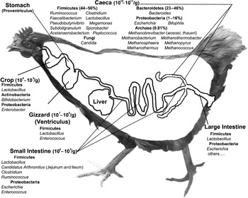
The gastrointestinal tracts (GITs) of chickens harbor microbial communities, or microbiomes, that play important roles in: growth and development, including the production of energy-rich short chain fatty acids (SCFA; Dunkley et al. , Reference Dunkley, Dunkley, Njongmeta, Callaway, Hume, Kubena, Nisbet and Ricke2007); promotion of GIT villus and crypt morphology (Shakouri et al. , Reference Shakouri, Lji, Mikkelsen and Cowieson2009); nutrient utilization, including reduction in luminal viscosity (Shakouri et al. , Reference Shakouri, Lji, Mikkelsen and Cowieson2009), the deconstruction of dietary polysaccharides (Beckmann et al. , Reference Beckmann, Simon and Vahjen2006; Qu et al. , Reference Qu, Brulc, Wilson, Law, Theoret, Joens, Konkel, Angly, Dinsdale, Edwards, Nelson and White2008); nutrient absorption (Cole and Boyd, Reference Cole and Boyd1967); and well-being of their chicken hosts, including detoxification (Hai et al. , Reference Hai, Zhou, Gong, Young, Su, Li, Zhu, Tsao and Yang2010). The chicken GIT is inhabited by various bacteria (Qu et al. , Reference Qu, Brulc, Wilson, Law, Theoret, Joens, Konkel, Angly, Dinsdale, Edwards, Nelson and White2008), methanogenic archaea (Saengkerdsub et al. , Reference Saengkerdsub, Anderson, Wilkinson, Kim, Nisbet and Ricke2007a, Reference Saengkerdsub, Herrera, Woodward, Anderson, Nisbet and Ricke2007b), fungi (Okulewicz and Zlotorzycka, Reference Okulewicz and Zlotorzycka1985), and viruses (Qu et al. , Reference Qu, Brulc, Wilson, Law, Theoret, Joens, Konkel, Angly, Dinsdale, Edwards, Nelson and White2008). Protists are more sparsely distributed (Okulewicz and Zlotorzycka, Reference Okulewicz and Zlotorzycka1985) and are generally regarded as pathogens. The composition of the GIT microbiome reflects co-evolution among the inhabiting microbes, genetic, immune, and metabolic interactions with the host, and environmental influences (Yeoman et al. , Reference Yeoman, Chia, Yildirim, Berg Miller, Stumpf, Leigh, Kent, Nelson, White and Wilson2011). Microbes are found across the entire length of the GIT, where they show spatial variation in community composition biogeographically (Fig. 1; Gong et al. , Reference Gong, Si, Forster, Huang, Hai, Yulong, Yang and Han2007) as well as between luminal and mucosa-associated populations (Gong et al. , Reference Gong, Forster, Yu, Chambers, Sabour, Wheatcroft and Chen2002). Microbial diversity and abundance are most evident in the ceca (Gong et al. , Reference Gong, Si, Forster, Huang, Hai, Yulong, Yang and Han2007), where more than 2200 operational taxonomic units (OTUs; 95% sequence ID; Danzeisen et al. , Reference Danzeisen, Kim, Isaacson, Tu and Johnson2011) and as many as 3500 genotypes (Qu et al. , Reference Qu, Brulc, Wilson, Law, Theoret, Joens, Konkel, Angly, Dinsdale, Edwards, Nelson and White2008) have been predicted. Consistently, microbial fermentation is most active in this section of the GIT.
Major taxa surveyed along the chicken GIT. Data on taxa and their spatial distribution are taken from Qu et al. (Reference Qu, Brulc, Wilson, Law, Theoret, Joens, Konkel, Angly, Dinsdale, Edwards, Nelson and White2008), Saengkerdsub et al. (Reference Saengkerdsub, Anderson, Wilkinson, Kim, Nisbet and Ricke2007a, Reference Saengkerdsub, Herrera, Woodward, Anderson, Nisbet and Ricke2007b), and Gong et al. (Reference Gong, Forster, Yu, Chambers, Sabour, Wheatcroft and Chen2002). Virus and phage populations are not presented or adequately sampled and listed cecal colonists are limited to the most common and abundant taxa. Numerous other taxa have been described in the chicken ceca.

As a result of issues that relate to zoonoses, food safety, animal nutrition, and health, the composition and function of the chicken GIT microbiome has received significant attention from researchers for almost 40 years. The original study of chicken GIT community composition by Salanitro et al. (Reference Salanitro, Blake and Muirhead1974) looked at 325 strains isolated by culture from the cecum of 5-week-old broiler hens. These strains were reported to represent up to 81% of the cultivable microbes from the chicken ceca. However, by the early 1990s it was recognized that the richness of species in all microbiomes, and indeed the Earth's biosphere, had been significantly underestimated by conventional microbial culturing methodologies (Amann et al. , Reference Amann, Ludwig and Schleifer1995). In fact, the majority of microbial species colonizing the chicken GIT have not been cultivated. More recently, culture-independent methods have been developed to overcome cultivation biases and allow more complete and detailed information on microbial community diversity, composition, and function.
16S rRNA gene-directed microbiome composition
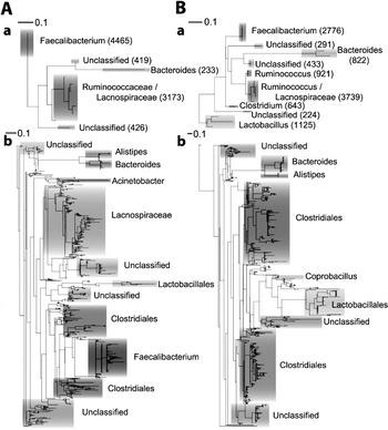
The use of the 16S rRNA gene as a phylogenetic marker to study bacterial and archaeal diversity and composition across various environments has resulted in tremendous quantities of information about microbial community dynamics. In particular, the increasing affordability and capability of second and subsequent-generation high-throughput sequencing platforms have made it possible to explore microbiomes at unprecedented phylogenetic depth. These surveys have uncovered the fine-grained structure of microbial communities occupying these ecosystems, exposing important features such as the existence of a rare biosphere, whose low-abundance populations dominate ecosystem diversity (Fig. 2; Dethlefsen et al. , Reference Dethlefsen, Huse, Sogin and Relman2008; Huse et al. , Reference Huse, Dethlefsen, Huber, Mark Welch, Relman and Sogin2008; Turnbaugh et al. , Reference Turnbaugh, Hamady, Yatsunenko, Cantarel, Duncan, Ley, Sogin, Jones, Roe, Affourtit, Egholm, Henrissat, Heath, Knight and Gordon2008). Although outnumbered by an order of magnitude by bacteriophage (Rodriguez-Valera et al. , Reference Rodriguez-Valera, Martin-Cuadrado, Rodriguez-Brito, Pasic, Thingstad, Rohwer and Mira2009), bacteria are the most abundant and diverse domain of life in the chicken GIT.
Maximum likelihood trees of abundant (a) and rare (b) OTUs. Trees are built from sequence data generated by Qu et al. (Reference Qu, Brulc, Wilson, Law, Theoret, Joens, Konkel, Angly, Dinsdale, Edwards, Nelson and White2008) for Chick1 (Panel A) and Chick94 (Panel B) with detailed taxonomic assignments. Unclassified refers to a taxonomic classification of less than 70% confidence by RDP at Order. Taxonomic clades are shaded according to a shared taxonomic designation. The number of sequence tags in each of the OTUs in the modal biosphere is given in parentheses following their taxonomic classification.

Microbial density and diversity are greatest in the cecum where longer digesta transit times permit more substantial microbial fermentation (Rehman et al. , Reference Rehman, Vahjen, Awad and Zentek2007). In the cecal pouches, bacteria are present at concentrations of 1010–1011 cells/g cecal material, encoding more than 95% of the genetic information present (Qu et al. , Reference Qu, Brulc, Wilson, Law, Theoret, Joens, Konkel, Angly, Dinsdale, Edwards, Nelson and White2008; Danzeisen et al. , Reference Danzeisen, Kim, Isaacson, Tu and Johnson2011). Consistent with other host-associated microbiomes (Ley et al. , Reference Ley, Hamady, Lozuone, Turnbaugh, Ramey, Bircher, Schlegel, Tucker, Schrenzel, Knight and Gordon2008), the bacterial phylum Firmicutes is the predominant phylum in the chicken crop, gizzard, small intestine, and cecum (Rehman et al. , Reference Rehman, Vahjen, Awad and Zentek2007; Qu et al. , Reference Qu, Brulc, Wilson, Law, Theoret, Joens, Konkel, Angly, Dinsdale, Edwards, Nelson and White2008; Danzeisen et al. , Reference Danzeisen, Kim, Isaacson, Tu and Johnson2011). Firmicutes represent 50–90% of all taxa in the cecum (Qu et al. , Reference Qu, Brulc, Wilson, Law, Theoret, Joens, Konkel, Angly, Dinsdale, Edwards, Nelson and White2008; Danzeisen et al. , Reference Danzeisen, Kim, Isaacson, Tu and Johnson2011), while culture-dependent and -independent approaches indicate the proportion of Firmicutes (principally in the form of Lactobacilli) is greater than 90% in other GIT locations (Gong et al. , Reference Gong, Si, Forster, Huang, Hai, Yulong, Yang and Han2007; Rehman et al. , Reference Rehman, Vahjen, Awad and Zentek2007). Archaea are less abundant, being present at concentrations of 105–107 cells per gram of cecal material (Saengkerdsub et al. , Reference Saengkerdsub, Anderson, Wilkinson, Kim, Nisbet and Ricke2007a) and encoding around 1–2% of the genetic information present in the ceca (Qu et al. , Reference Qu, Brulc, Wilson, Law, Theoret, Joens, Konkel, Angly, Dinsdale, Edwards, Nelson and White2008; Danzeisen et al. , Reference Danzeisen, Kim, Isaacson, Tu and Johnson2011). Methanobrevibacter is the predominant archaeal genus in the chicken ceca, with taxa similar to Methanobrevibacter woesei being the most prolific of this domain (Fig. 1; Saengkerdsub et al. , Reference Saengkerdsub, Anderson, Wilkinson, Kim, Nisbet and Ricke2007a). Other archaeal taxa exist, and consistent with other GIT environments, all archaea appear to be involved in the methanogenic dissipation of hydrogen produced during fermentation (Saengkerdsub et al. , Reference Saengkerdsub, Anderson, Wilkinson, Kim, Nisbet and Ricke2007a, Reference Saengkerdsub, Herrera, Woodward, Anderson, Nisbet and Ricke2007b).
Analyses of rarefaction curves and diversity indexes indicate that microbial richness and diversity increase with age (Danzeisen et al. , Reference Danzeisen, Kim, Isaacson, Tu and Johnson2011). In the work by Danzeisen et al. (Reference Danzeisen, Kim, Isaacson, Tu and Johnson2011), OTUs (95% sequence ID) corresponding to Roseburia, Coprococcus, Butyricoccus, Papillibacter (all Firmicutes), and Escherichia (Proteobacteria) were found to be abundant constituents of the chicken ceca, but were not detected before 14 days of age, while other OTUs classified as Fastidiospila, Hespellia, Lactobacillus, and Coprococcus (all Firmicutes) were not detected before 35 days of age. Methanobacteriales were detected in the fecal samples of 25% of chickens as early as 3 days of age and found in 100% of chickens tested from 5 days of age (Saengkerdsub et al. , Reference Saengkerdsub, Herrera, Woodward, Anderson, Nisbet and Ricke2007b).
Although 16S rRNA gene surveys provide taxonomic information, they fail to provide information related to microbial function. While these can be uncovered for isolated microbes in culture using a variety of directed-assays, metagenomic, metatranscriptomic, and metabolomic analyses offer the ability to understand these physiological roles for individual species (including those that have not yet been cultured), in situ, and in the context of the entire microbiome.
Shotgun metagenomic analyses
Gene-based metagenomic surveys provide a measure of the metabolic capabilities of a microbiome. To date, two shotgun metagenomic surveys have been performed in chickens, both focusing on the ceca. The first, performed by Qu et al. (Reference Qu, Brulc, Wilson, Law, Theoret, Joens, Konkel, Angly, Dinsdale, Edwards, Nelson and White2008) determined the distribution of ~200,000 genes present in healthy chickens and in chickens experimentally infected with Campylobacter jejuni. In the second, Danzeisen et al. (Reference Danzeisen, Kim, Isaacson, Tu and Johnson2011) looked at differences in genes between control chickens and those fed sub-therapeutic levels of antibiotics for growth enhancement. These studies uncovered a large amount of information relating to the prevalence of mobile elements, and genes involved in nutrition, virulence, and antibiotic resistance, which will be discussed below. They also provide an unbiased look at the diversity and distribution of all types of microbes, including bacteria, archaea, viruses, and eukaryotic microbes.
To date, no studies have integrated the global gene expression patterns or metabolite profiles from the chicken GIT to a metagenomic backbone. Therefore, currently our knowledge is limited to the metabolic potential of the chicken GIT microbiome. Several studies have investigated the transcriptional dynamics of microbial isolates of the chicken GIT, including comparisons of in vitro and in vivo grown Salmonella enterica serovar Enteritidis PT4 and Salmonella enteric serovar Typhimurium (Dhawi et al. , Reference Dhawi, Elazomi, Jones, Lovell, Li, Emes and Barrow2011; Harvey et al. , Reference Harvey, Watson, Hulme, Jones, Lovell, Berchieri, Young, Bumstead and Barrow2011), respectively. These studies revealed significant metabolic differences between in vitro and in vivo grown cells, as well as striking differences in the expression of important virulence factors (Dhawi et al. , Reference Dhawi, Elazomi, Jones, Lovell, Li, Emes and Barrow2011; Harvey et al. , Reference Harvey, Watson, Hulme, Jones, Lovell, Berchieri, Young, Bumstead and Barrow2011). One of the studies (Harvey et al. , Reference Harvey, Watson, Hulme, Jones, Lovell, Berchieri, Young, Bumstead and Barrow2011) also revealed significant differences in the growth rate and motility of Salmonella Typhimurium. These studies highlight the stark contrasts between evidence obtained at the laboratory bench and the functional reality of microbes occupying GIT environments, arguing strongly for a systems biology understanding. Even these experiments are one step removed from biological reality as they were performed in gnotobiotic hatchlings; it will therefore be interesting to determine the significance of these findings in situ alongside fully developed microbial ecosystems.
This is not an argument for a complete switch to meta-omic techniques. These techniques provide the opportunity to survey the system-wide dynamics of a microbiome, but meta-omic techniques need to be integrated with genomic and transcriptomic information from isolated organisms so they are correctly interpreted and contextualized, leading to a more complete understanding of the ecology and evolution of the microbiome.
Genomic analyses of microbes isolated from the chicken GIT
Most genome-sequencing projects focusing on chicken isolates have been directed toward pathogenic viruses (e.g. Barbosa et al. , Reference Barbosa, Zavala, Cheng and Villegas2007; Linde et al. , Reference Linde, Munir, Zohari, Stáhl, Baule, Renström and Berg2010; Qiu et al. , Reference Qiu, Sun, Wu, Dong, Hu, Meng, Wu and Liu2011; Abro et al. , Reference Abro, Renström, Ullman, Belák and Baule2012; Diel et al. , Reference Diel, Susta, Cardenas Garcia, Killian, Brown, Miller and Afonso2012). Those projects focusing on autonomous microbial life forms have almost universally targeted zoonotic or host pathogens (Johnson et al. , Reference Johnson, Kariyawasam, Wannemuehler, Mangiamele, Johnson, Doetkott, Skyberg, Lynne, Johnson and Nolan2007; Ahir et al. , Reference Ahir, Roy, Jhala, Bhanderi, Mathakiya, Bhatt, Padiya, Jakhesara, Koringa and Joshi2011), including GIT isolates (Pearson et al. , Reference Pearson, Gaskin, Segers, Wells, Nuijten and van Vliet2007; Thomson et al. , Reference Thomson, Clayton, Windhorst, Vernikos, Davidson, Churcher, Quail, Stevens, Jones, Watson, Barron, Layton, Pickard, Kingsley, Bignell, Clark, Harris, Ormond, Abdellah, Brooks, Cherevach, Chillingworth, Woodward, Norberczak, Lord, Arrowsmith, Jagels, Moule, Mungall, Sanders, Whitehead, Chabalgoity, Maskell, Humphrey, Roberts, Barrow, Dougan and Parkhill2008; Cooper et al. , Reference Cooper, Cooper, Zuccolo, Law and Joens2011; Feng et al. , Reference Feng, Xu, Li, Zhang, Wang, Zhu, Cao, Li, Johnston, Zhou, Liu and Liu2012) or opportunistic pathogens (Johnson et al. , Reference Johnson, Fernandez-Alarcon, Bojesen, Nolan, Trampel and Seemann2011). A handful of bacteria have been isolated and sequenced from the chicken GIT without a clear zoonotic link, including Bacteroides salanitronis BL78, Lactobacillus crispatus ST1 and Lactobacillus salivarius NIAS840. The B. salanitronis and Lactobaillus genomes were all reported in the past few years and describe bacteria of potential importance to GIT health (Ojala et al. , Reference Ojala, Kuparinen, Koskinen, Alatalo, Holm, Auvinen, Edelman, Westerlund-Wikström, Korhonen, Paulin and Kankainen2010; Gronow et al. , Reference Gronow, Held, Lucas, Lapidus, Del Rio, Nolan, Tice, Deshpande, Cheng, Pitluck, Liolios, Pagani, Ivanova, Mavromatis, Pati, Tapia, Han, Goodwin, Chen, Palaniappen, Land, Hauser, Chang, Jefferies, Brambilla, Rohde, Göker, Detter, Woyke, Bristow, Markowitz, Hugenholtz, Krypides, Klenk and Eisen2011; Ham et al. , Reference Ham, Kim, Seol, Jang, Jeong, Oh, Kim, Kang, Kim and Cha2011). In addition to bacterial and viral genomes (Thomson et al. , Reference Thomson, Clayton, Windhorst, Vernikos, Davidson, Churcher, Quail, Stevens, Jones, Watson, Barron, Layton, Pickard, Kingsley, Bignell, Clark, Harris, Ormond, Abdellah, Brooks, Cherevach, Chillingworth, Woodward, Norberczak, Lord, Arrowsmith, Jagels, Moule, Mungall, Sanders, Whitehead, Chabalgoity, Maskell, Humphrey, Roberts, Barrow, Dougan and Parkhill2008), genome-sequencing efforts have also successfully targeted a Siphoviridae-family bacteriophage, SPN3UB (Lee et al. , Reference Lee, Shin and Ryu2012) and a Podoviridae-family phage, ΦCPV1 (Volozhantsev et al. , Reference Volozhantsev, Verevkin, Bannov, Krasilnikova, Myakinina, Zhilenkov, Svetoch, Stern, Oakley and Seal2011). Bacteriophage SPN3UB was isolated from chicken feces (Lee et al. , Reference Lee, Shin and Ryu2012), while ΦCPV1 was isolated from chicken intestinal contents. These phages are infectious to important zoonotic pathogens and are being explored as alternatives to antibiotics for the control of Salmonella Typhimurium and Clostridium perfringens, respectively.
Although the molecular interrogation of the chicken GIT microbiome is only in its adolescence, its contributions to our understanding of growth, health, and development of the chicken host have been significant, and may lead to new methods for the mitigation of zoonotic diseases that use chickens as a vector.
The chicken GIT microbiome's role in host nutrition
The chicken GIT microbiome produces enzymes enabling the deconstruction of dietary polysaccharides (Beckmann et al. , Reference Beckmann, Simon and Vahjen2006). These enzymes are critical to host nutrition because chickens, like most animals, lack the genes for glycoside hydrolase (GH), polysaccharide lyase (PL), and carbohydrate esterase (CE) enzymes that are necessary to facilitate this process (Morris, Reference Morris2003). Metagenomic analyses have illustrated the significance of the cecal microbiome's contribution to carbohydrate metabolism. Genes encoding GHs, PLs, CEs, and other proteins involved in carbohydrate metabolism (transporters and those involved in central carbohydrate metabolism) have been shown to be more abundant than any other category of gene in this environment (~20% of genes; Qu et al. , Reference Qu, Brulc, Wilson, Law, Theoret, Joens, Konkel, Angly, Dinsdale, Edwards, Nelson and White2008; Danzeisen et al. , Reference Danzeisen, Kim, Isaacson, Tu and Johnson2011).
During the deconstruction of dietary polysaccharides, GIT bacteria produce SCFAs (Topping and Clifton, Reference Topping and Clifton2001; Dunkley et al. , Reference Dunkley, Dunkley, Njongmeta, Callaway, Hume, Kubena, Nisbet and Ricke2007). The composition and proportions of these SCFAs vary depending on microbial composition, which is to some degree adaptable, and fine-tuned by the composition and structure of the fiber component of the chicken's diet (Topping and Clifton, Reference Topping and Clifton2001). Acetate is the primary SCFA produced in most GIT environments, including the chicken, followed by propionate and butyrate (Topping and Clifton, Reference Topping and Clifton2001; Dunkley et al. , Reference Dunkley, Dunkley, Njongmeta, Callaway, Hume, Kubena, Nisbet and Ricke2007). Other SCFAs such as valerate, isobutyrate, and isovalerate are also produced in trace amounts (Dunkley et al. , Reference Dunkley, Dunkley, Njongmeta, Callaway, Hume, Kubena, Nisbet and Ricke2007). Concentrations of butyrate are of particular physiological significance, as this SCFA is the primary energy source of colonic epithelia and has been shown to be essential to homeostasis of colonocytes and development of GIT villus morphology (Panda et al. , Reference Panda, Rama Rao, Raju and Shyam Sunder2009; Donohoe et al. , Reference Donohoe, Garge, Zhang, Sun, O'Connell, Bunger and Bultman2011). The three major SCFAs (acetate, propionate, and butyrate) all appear important to colonic musculature and vasculature in the GIT (Topping and Clifton, Reference Topping and Clifton2001). These SCFAs are also of critical importance to host energetics and hydration. SCFAs stimulate fluid and electrolyte uptake and are absorbed transepithelially as a source of energy that contributes between 10% (humans) and up to 70% (ruminants) of the host's daily energy requirements (McNeil, Reference McNeil1984; Topping and Clifton, Reference Topping and Clifton2001; Flint and Bayer, Reference Flint and Bayer2008). Although their exact contribution in chickens has yet to be determined, the SCFA butyrate has been shown to improve growth performance and carcass quality characteristics in chickens (Panda et al. , Reference Panda, Rama Rao, Raju and Shyam Sunder2009).
The GIT microbiome also contributes to nitrogen metabolism. Genes involved in the metabolism of protein (9–10% of genes), amino acids (8–9%), and nitrogen (1%) have all been shown to be abundant (Qu et al. , Reference Qu, Brulc, Wilson, Law, Theoret, Joens, Konkel, Angly, Dinsdale, Edwards, Nelson and White2008; Danzeisen et al. , Reference Danzeisen, Kim, Isaacson, Tu and Johnson2011). The relative proportions of genes dedicated to the metabolism of these three nitrogen sources are consistent with protein being the major source of nitrogen and depicting the major direction of nitrogen flux (protein – amino acids – nitrogenous compounds) in the GIT. The microbial metabolism of dietary protein that escaped host metabolism earlier in the GIT provides further amino acids for egg production, maintenance, and growth (Latshaw and Zhao, 2011). However, subsequent metabolic processing to ammonia or urea is of no nutritive value to the host and approximately half of the available dietary nitrogen is excreted, mostly as ammonia in chickens (Latshaw and Zhao, 2011). This hyper-production of ammonia and subsequent excretion is not only nutritionally inefficient but also underpins negative effects on performance, health, and mortality in poultry houses, and is a major environmental and public health concern (McCubbin et al. , Reference McCubbin, Apelberg, Roe and Divita2002; Xin et al. , Reference Xin, Gates, Green, Mitloehner, Moore and Wathes2011).
Genes dedicated to fatty acid and lipid metabolism are also detected (1–2%; Qu et al. , Reference Qu, Brulc, Wilson, Law, Theoret, Joens, Konkel, Angly, Dinsdale, Edwards, Nelson and White2008), suggesting microbial modulation of lipid profiles as has been described in other livestock (Dhiman et al. , Reference Dhiman, Nam and Ure2005). Conjugated linoleic acid (CLA) is one of the best-studied microbially produced fatty acid and is produced by certain microbes as an intermediate during the biohydrogenation of the polyunsaturated linoleic acid (Palmquist et al. , Reference Palmquist, Lock, Shingfield and Bauman2005). CLA has been found to naturally occur in chicken meat (Dhiman et al. , Reference Dhiman, Nam and Ure2005). Dietary supplementation of CLA has been shown to increase lean body mass in chickens and to be incorporated into tissue lipids (Simon et al. , Reference Simon, Männer, Schäfer, Sagredos and Eder2000). It should also be noted, however, that dietary CLA also appears to affect yolk quality and embryo mortality in laying hens by altering yolk fatty acid composition and albumen and yolk pH, a feature that can be overcome by the co-supplementation of olive oil (Aydin et al. , Reference Aydin, Pariza and Cook2001).
The incorporation of a molecular understanding of the microbiome with nutritional science therefore paves the way for new research that should seek to optimize the composition of the chicken GIT microbiome. Such research could provide new opportunities to enhance SCFA production, reduce nitrogen losses or optimize fatty acid profiles (which may vary between broiler and laying hens). Such benefits could lead to significant improvements in poultry production and the associated economics. SCFA and CLA production also have been linked to host health (Badinga and Greene, Reference Badinga and Greene2006; Wong et al. , Reference Wong, de Souza, Kendall, Emam and Jenkins2006) and may provide additional benefit in reducing disease.
The role of the chicken GIT microbiome in reducing pathogen loads
For over 100 years chickens have been recognized as an important source of zoonotic infection (Higgins, Reference Higgins1898), a feature that has long plagued the poultry industry. This may be exacerbated by less active innate and humoral immune systems in chickens that are permissive to colonization by pathogenic bacteria such as species of Salmonella and Campylobacter (Toth and Siegel, Reference Toth and Siegel1986; Jeurissen et al. , Reference Jeurissen, Janse, van Rooijen and Claassen1998) and numerous host-specific or host-promiscuous viruses. Metagenomic analyses have shown us that genes associated with virulence are abundant within the chicken microbiome (~8% of all genes; Qu et al. , Reference Qu, Brulc, Wilson, Law, Theoret, Joens, Konkel, Angly, Dinsdale, Edwards, Nelson and White2008; Danzeisen et al. , Reference Danzeisen, Kim, Isaacson, Tu and Johnson2011). These mostly include genes for antibiotic resistance (>55% of virulence-related genes), and iron scavenging (13%), but also include genes involved in types III and IV secretion (>2%), adhesion (>1%), invasion and intracellular resistance (1%; Qu et al. , Reference Qu, Brulc, Wilson, Law, Theoret, Joens, Konkel, Angly, Dinsdale, Edwards, Nelson and White2008), lipid A biosynthesis (not quantified), and type I pilus formation (not quantified; Danzeisen et al. , Reference Danzeisen, Kim, Isaacson, Tu and Johnson2011). Many of these virulence genes, including those involved in type IV secretion, type I pilus formation, lipid A biosynthesis, and iron scavenging were found to represent a significantly larger portion of the total genes identified in chickens subjected to various sub-therapeutic antibiotic treatments (STAT; Danzeisen et al. , Reference Danzeisen, Kim, Isaacson, Tu and Johnson2011).
The composition of the chicken GIT maintains a fine balance; disruptions to key species can enable the dramatic proliferation of pathogenic microbes (Kimura et al. , Reference Kimura, Mimura, Nishida and Kobayashi1976; Morishita and Mitsuoka, Reference Morishita and Mitsuoka1976) and dramatic increases in the proportion of virulence genes (Danzeisen et al. , Reference Danzeisen, Kim, Isaacson, Tu and Johnson2011). Conversely, a stable and healthy GIT microbiome can limit the colonization of zoonotic pathogens, such as Salmonella (Hudault et al. , Reference Hudault, Bewa, Bridonneau and Raibaud1985) and Campylobacter species (Soerjadi-Liem et al. , Reference Soerjadi-Liem, Snoeyenbos and Weinack1984) as well as transform clinically significant fungal mycotoxins periodically found in feed to non-toxic derivatives (Hai et al. , Reference Hai, Zhou, Gong, Young, Su, Li, Zhu, Tsao and Yang2010). Although Enterococcus faecium and some Lactobacillus isolates have been suggested to limit the colonizing potential of some major pathogens through direct competitive interactions (Jin et al. , Reference Jin, Ho, Abdullah, Ali and Jalaludin1996; Carina Audisio et al. Reference Carina Audisio, Oliver and Apella2000), relative exclusion is strongly correlated to increasing species complexity (Hudault et al. , Reference Hudault, Bewa, Bridonneau and Raibaud1985; Fukata et al. , Reference Fukata, Hadate, Baba and Arakawa1991;Schoeni and Wong, Reference Schoeni and Wong1994). The pre-establishment of the microbiome prior to infection is an important precursor of resistance (Hudault et al. , Reference Hudault, Bewa, Bridonneau and Raibaud1985), perhaps relating to the order and complexity of epithelial adherence. It is therefore clear that maintaining chicken GIT health is one key to limiting pathogen loads and increasing food safety in the poultry industry. Microbial diversity in the chicken GIT is sensitive to a number of perturbing agents, including parasitic infection with Ascaridia galli (Okulewicz and Zlotorzycka Reference Okulewicz and Zlotorzycka1985) or protozoal infection with Eimeria tanella (Kimura et al. , Reference Kimura, Mimura, Nishida and Kobayashi1976), as well as human interventions such as the provision of antibiotics (Danzeisen et al. , Reference Danzeisen, Kim, Isaacson, Tu and Johnson2011).
Antibiotics are used therapeutically to treat disease in humans and domestic animals. In the late 1940s, it was recognized that sub-therapeutic levels of antibiotics (STAT) could be used to expedite and enhance the growth of chickens (Stokstad and Jukes, Reference Stokstad and Jukes1950) and other livestock (Gustafson and Bowen, Reference Gustafson and Bowen1997). The serendipitous finding came from chickens fed fermentation waste from cyclotetracycline production as an inexpensive source of vitamin B12 (Stokstad and Jukes Reference Stokstad and Jukes1950). In addition, STAT was recognized as a tool to reduce pathogen loads and decrease the risks of zoonotic transmission (Gustafson and Bowen, Reference Gustafson and Bowen1997). The mechanisms that link STATs to animal productivity have not been established but because of their growth promoting activity STATs became widely used in production facilities. Today STATs are still used in the USA and several other countries around the world as they have been for more than 50 years (Collignon et al. , Reference Collignon, Powers, Chiller, Aidara-Kane and Aarestrup2009; Chapman et al. , Reference Chapman, Jeffers and Williams2010).
Molecular methods have established that STATs act non-specifically affecting a broad range of microbial taxa. Monensin in combination with either virginiamycin or tylosin has been shown to significantly decrease bacteria of the major phylum Firmicutes, including Roseburia, Enterococcus, Lactobacillus and Blautia, and increase Proteobacteria such as Escherichia and Ruminococcaceae such as Anaerotruncus, Subdoligranulum, and Sedimentibacter (Danzeisen et al. , Reference Danzeisen, Kim, Isaacson, Tu and Johnson2011).
In the 1960s, it was suggested that STATs could lead to proliferation of the pool of antibiotic resistance genes and allow their transfer to human pathogens (Swann, Reference Swann1969). Evidence suggests a number of pathogens can colonize both the human and chicken GIT (Johnson et al. , Reference Johnson, Wannemuehler, Johnson, Stell, Doetkott, Johnson, Kim, Spanjaard and Nolan2008, Reference Johnson, Logue, Wannemuehler, Kariyawasam, Doetkott, DebRoy, White and Nolan2009; Gipp et al. , Reference Gipp, Hlahla, Didelot, Kops, Maurischat, Tedin, Alter, Ellerbroek, Schreiber, Schomburg, Janssen, Batholomäus, Hofreuter, Woltemate, Uhr, Brenneke, Grüning, Gerlach, Wieler, Suerbaum and Josenhans2011), providing opportunities for gene exchange. Gene-directed metagenomic surveys have provided clear evidence that microbes colonizing the chicken GIT are an abundant source of antibiotic resistance genes (Qu et al. , Reference Qu, Brulc, Wilson, Law, Theoret, Joens, Konkel, Angly, Dinsdale, Edwards, Nelson and White2008; Zhou et al. , Reference Zhou, Wang and Lin2012). Most prolific are genes encoding resistances to fluoroquinolones, tetracyclines, cobalt, zinc, cadmium, and common antibiotics used in poultry production (Qu et al. , Reference Qu, Brulc, Wilson, Law, Theoret, Joens, Konkel, Angly, Dinsdale, Edwards, Nelson and White2008; Danzeisen et al. , Reference Danzeisen, Kim, Isaacson, Tu and Johnson2011). Methicillin (Qu et al. , Reference Qu, Brulc, Wilson, Law, Theoret, Joens, Konkel, Angly, Dinsdale, Edwards, Nelson and White2008) and beta-lactam (Qu et al. , Reference Qu, Brulc, Wilson, Law, Theoret, Joens, Konkel, Angly, Dinsdale, Edwards, Nelson and White2008; Danzeisen et al. , Reference Danzeisen, Kim, Isaacson, Tu and Johnson2011) resistance genes are also common. This abundance of antibiotic resistance genes has been contrasted with the near absence of these genes in animals with no historical exposure to antibiotics and minimal interactions with humans or other animals from areas where antibiotics are frequently used (Thaller et al. , Reference Thaller, Migliore, Marquez, Tapia, Cedeño, Rossolini and Gentile2010). However, certain antibiotic resistances are ancient properties of microbes (D'Costa et al. , Reference D'Costa, King, Kalan, Morar, Sung, Schwarz, Froese, Zazula, Calmels, Debruyne, Golding, Poinar and Wright2011). Genes encoding ampicillin and spectinomycin resistance have been detected in free-range chickens not routinely subjected to STATs (Zhou et al. Reference Zhou, Wang and Lin2012), though in the same study antibiotic-resistance genes were found to be almost four times more prevalent in conventionally raised (STAT-treated) chickens (Zhou et al. Reference Zhou, Wang and Lin2012). Danzeisen et al. (Reference Danzeisen, Kim, Isaacson, Tu and Johnson2011) found that antibiotic resistance genes were not enriched by controlled short-term STAT application. This could suggest that the low dosages of STATs have limited bactericidal action and therefore elicit a limited selection pressure, but may also reflect the long-term adaptation of the chicken GIT microbiome to STAT use.
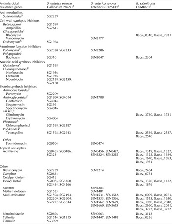
Perhaps the most disturbing aspect of this abundance of antibiotic resistance genes in the chicken ceca is that they are co-occurring in an environment that also has a high abundance of mobile DNA elements, as detected in shotgun metagenomic surveys (Qu et al. , Reference Qu, Brulc, Wilson, Law, Theoret, Joens, Konkel, Angly, Dinsdale, Edwards, Nelson and White2008; Danzeisen et al. , Reference Danzeisen, Kim, Isaacson, Tu and Johnson2011). The genome sequences of chicken isolates Salmonella Enteritidis P125109 and Salmonella enterica serovar Gallinarum 287/91 carry potentially mobile genomic islands (Thomson et al. , Reference Thomson, Clayton, Windhorst, Vernikos, Davidson, Churcher, Quail, Stevens, Jones, Watson, Barron, Layton, Pickard, Kingsley, Bignell, Clark, Harris, Ormond, Abdellah, Brooks, Cherevach, Chillingworth, Woodward, Norberczak, Lord, Arrowsmith, Jagels, Moule, Mungall, Sanders, Whitehead, Chabalgoity, Maskell, Humphrey, Roberts, Barrow, Dougan and Parkhill2008). Plasmids carrying virulence genes have been observed in potentially zoonotic Escherichia coli strains of chicken origin (Johnson et al. , Reference Johnson, Wannemuehler, Johnson, Stell, Doetkott, Johnson, Kim, Spanjaard and Nolan2008). Even the avirulent L. salivarius NIAS840 and B. salanitronis Bl78 isolates carry three plasmids each (Gronow et al. , Reference Gronow, Held, Lucas, Lapidus, Del Rio, Nolan, Tice, Deshpande, Cheng, Pitluck, Liolios, Pagani, Ivanova, Mavromatis, Pati, Tapia, Han, Goodwin, Chen, Palaniappen, Land, Hauser, Chang, Jefferies, Brambilla, Rohde, Göker, Detter, Woyke, Bristow, Markowitz, Hugenholtz, Krypides, Klenk and Eisen2011; Ham et al. , Reference Ham, Kim, Seol, Jang, Jeong, Oh, Kim, Kang, Kim and Cha2011). A quick survey of the gene contents of these plasmids shows the largest of the B. salanitronis plasmids, pBACSA01, encodes apparatus necessary for conjugative and type IV DNA transfer, while all three B. salanitronis plasmids carry genes for mobilization, indicating that all three B. salanitronis plasmids could be moved between bacterial hosts. Mobile elements have different host ranges but are well described for their ability to move among very disparate microbial hosts. Staphylococcus aureus’ recent adaptation to the chicken GIT (during the STAT era) included the acquisition of novel mobile genetic elements (Lowder et al. , Reference Lowder, Guinane, Ben Zakour, Weinert, Conway-Morris, Cartwright, Simpson, Rambaut, Nübel and Fitzgerald2009). Although antibiotic resistance genes were largely absent from plasmids that pre-date the antibiotic era (Hughes and Datta, Reference Hughes and Datta1983), they are commonplace among today's GIT microbes (Schultsz and Geerlings, Reference Schultsz and Geerlings2012). These genes are functional across multiple host species as exemplified by Zhou et al. (Reference Zhou, Wang and Lin2012), who demonstrated that antibiotic-resistance genes identified from a metagenomic clone library could be introduced into a strain of C. jejuni and be functionally active. Therefore mobile elements may provide a vehicle permitting the transfer of genes that facilitate antibiotic resistance and virulence to initially antibiotic-sensitive, avirulent microbes, potentially including those of clinical importance (Gyles, Reference Gyles2008). Consistently, surveys have demonstrated erythromycin resistance is prevalent in C. jejuni strains (Ladely et al. , Reference Ladely, Harrison, Fedorka-Cray, Berrang, Englen and Meinersmann2007) and multiple antibiotic resistances and virulence traits are prevalent in strains of E. coli (Johnson et al. Reference Johnson, Wannemuehler, Johnson, Stell, Doetkott, Johnson, Kim, Spanjaard and Nolan2008; Glenn et al. , Reference Glenn, Englen, Lindsey, Frank, Turpin, Berrang, Meinersmann, Fedorka-Cray and Frye2012) isolated from STAT chickens. A quick catalogue of the antimicrobial resistance genes present in the genomes of chicken GIT isolates S. Gallinarum 287/91 and S. Enteritidis P125109 shows these organisms each carry an impressive arsenal of genes potentially enabling increased resistance to multiple antimicrobials (Table 1). Even the avirulent chicken isolate B. salanitronis DSM1870 carries an assortment of antibiotic resistance genes, perhaps chronicling a strong anthropogenically imposed selection pressure of this environment.
Antimicrobial resistance genes in the genomes of some chicken GIT isolates

1 Locus tags of genes in this genome potentially increasing resistance to antimicrobial; 2Macrolides, Lincosamides, Streptogramins, Ketolides; CBactericidal; SBacteriostatic.
Collectively, these studies clearly illustrate a mechanism through which the history of STAT use could have contributed to the proliferation of microbial antibiotic resistances. This is further supported by a number of indirect pieces of evidence. Yet, conclusive evidence has for a long time been lacking in this argument. Recent work in pigs has provided much clearer evidence that zoonotic transfer between humans and STAT-treated animals can lead to microbes obtaining antibiotic resistances (Price et al. , Reference Price, Stegger, Hasman, Aziz, Larsen, Andersen, Pearson, Waters, Foster, Schupp, Gillece, Driebe, Liu, Springer, Zdovc, Battisti, Franco, Zmudzki, Schwarz, Butaye, Jouy, Pomba, Porrero, Ruimy, Smith, Robinson, Weese, Arriola, Yu, Laurent, Keim, Skov and Aarestrup2012). Although the area remains contentious, legislative steps are already in place to illuminate STAT use in USA farming practices. A mechanistic understanding of STAT-mediated growth promotion could lead to the identification of new and more broadly accepted agents to facilitate this process. Optimizing GIT microbial communities through microbiome-directed nutrition or probiotics could provide new opportunities to limit pathogen colonization. Further research into alternatives to antibiotics that are target-specific, such as lytic phages could attenuate the transmission of zoonotic pathogens without negatively impacting production.
Conclusions
Molecular interrogation of the chicken GIT microbiome has given us a new level of understanding of its composition and spatial structure. Surveys of phylogenetic markers such as the 16S rRNA gene have allowed researchers to overcome the roadblocks associated with culture-based surveys. In tandem with modern sequencing technologies, these surveys have also allowed us to describe more than the most abundant few organisms. Gene-directed metagenomic surveys have described the functional content of the microbiome illustrating its contribution to host nutrition. These surveys, along with microbial genomic analyses, have also provided clearer evidence of the abundance of antimicrobial and pathogenicity traits circulating in the chicken GIT. The prevalence of mobile elements further raises concerns about the role of poultry in exacerbating the virulence and resistance of zoonotic human pathogens. Transcriptional analyses have shown the disparity between in vitro and in vivo experimentation, and the deployment of metatranscriptomics is needed to place this information in an in situ and physiologically relevant context. The field of GIT microbiology is moving toward an integrated systems level understanding. Early meta-omic analyses have enabled those with an interest in the chicken GIT to also venture down this road. An improved and integrated understanding of the role of nutrition and the microbiome in mitigating disease and promoting animal growth and productivity may lead next-generation farming practices to a level that exceeds that currently achieved through STAT provision.