Implications
There has been no evidence suggesting the effects of ovine serum immunoglobulin (Ig) on animal immunomodulation. The results of this study clearly demonstrate an effect of ovine serum Ig on immunomodulation in the growing rat. The proliferation of lymphocytes, phagocytic activity and rat digesta antibodies were all significantly enhanced. The results now need to be confirmed in production animals, but ovine Ig may offer a novel means of enhancing immunity.
Introduction
The development of immunomodulators from natural sources for dietary supplementation in both animals and humans is an active area of research. Recently, immunonutrition has been applied to improve the clinical course of recovery for severely sick or surgical patients who often need an exogenous supply of nutrients delivered through the parental or enteral routes (Calder, Reference Calder2007).
Immunoglobulins (Igs) are the primary anti-infective component of plasma, colostrum and breast milk. These specialized proteins protect the body from harmful bacteria, viruses and other environmental pathogens by either binding to them or by forming an encapsulating barrier (Kraehenbuhl and Neutra, Reference Kraehenbuhl and Neutra1992). Human Ig has been used as a prophylactic treatment for children born prematurely with necrotizing enterocolitis (Eibl et al., Reference Eibl, Wolf, Furnkranz and Rosenkranz1988). To date, Ig has been used therapeutically for intervention in Campylobacter jejuni (Hammarstrom et al., Reference Hammarstrom, Smith and Hammarstrom1993), Clostridium difficile (Tjellstrom et al., Reference Tjellstrom, Stenhammar, Eriksson and Magnusson1993), Helicobacter pylori, rotavirus and cryptosporidial infections.
Several studies carried out in production animals at weaning (Kats et al., Reference Kats, Nelssen, Tokach, Goodband, Hansen and Laurin1994; Torrallardona et al., Reference Torrallardona, Conde, Badiola, Polo and Brufau2003; Pierce et al., Reference Pierce, Cromwell, Lindemann, Russell and Weaver2005) have demonstrated beneficial effects from the ingestion of spray-dried animal plasma (SDAP) containing Ig, such as increased intestinal tissue growth rate, increased feed intake and higher body weight (BW) gain. Furthermore, the beneficial effects of feeding SDAP are more pronounced with higher pathogen challenge, as when pigs were housed in an on-farm conventional nursery, which was exposed to a greater load of subclinical pathogenic organisms, the animals exhibited an enhanced growth rate and feed intake compared to those reared in the off-site experimental (cleaner) settings (Coffey and Cromwell, Reference Coffey and Cromwell1995). A study by Bosi et al. (Reference Bosi, Casini, Finamore, Cremokolini, Merialdi, Trevisi, Nobili and Mengheri2004) suggests that the Ig present in SDAP appears to elicit its effect by binding to the pathogenic bacteria, thereby preventing their attachment to the mucosa in the lumen of the gastrointestinal tract (GIT).
An important consideration with orally administered Ig is whether the proteins become denatured during their passage through the GIT, thus losing activity. Several reports have shown that orally administered Ig retains activity in the GIT (Tjellstrom et al., Reference Tjellstrom, Stenhammar, Eriksson and Magnusson1993; Bogstedt et al., Reference Bogstedt, Johansen, Hatta, Kim, Casswall, Svensson and Hammarstrom1996; Casswall et al., Reference Casswall, Hammarstrom, Veress, Nord, Bogstedt, Brockstedt and Dahlstrom1996), and partially digested or undigested Ig with opsonic activity has been found in the faeces of low BW infants who had been treated orally with Ig (Blum et al., Reference Blum, Phelps, Ank, Krantman and Stiehm1981).
Recently, we have reported that freeze-dried ovine serum Ig (FDOI) selectively improves growth performance, organ growth and gut morphology in rats when compared with a control diet and with inactivated ovine Ig (IOI; Balan et al., Reference Balan, Han, Rutherfurd, Singh and Moughan2009). There have been no reports, however, regarding the effects of ovine Ig on immunomodulation in animals.
The objective of this study was to ascertain whether orally administered ovine serum Ig selectively modulates immune function in the growing rat. A number of indices of immune function were studied including phagocytic activity, lymphocyte proliferation of spleen cells, cytokine production and IgA and IgG concentrations in intestinal digesta and plasma. Comparison was made among a diet containing FDOI, a casein-based control diet (BD, no Ig) and a negative control diet (IOI-containing diet).
Material and methods
Preparation and quantitation of ovine serum Ig
An ovine Ig fraction was prepared by ammonium sulphate precipitation of fresh lamb’s blood as described previously (Balan et al., Reference Balan, Han, Rutherfurd, Singh and Moughan2009). The final product was freeze-dried, a portion of which was inactivated by dry heating (121°C, 15 min) to produce the IOI. The inactivation of Ig in the IOI material was confirmed by circular dichroism (Chirascan, Applied Photophysics, Surrey, UK) based on the method of Vermeer and Norde (Reference Vermeer and Norde2000).
The IgG content in the ovine Ig fraction was analysed by direct ELISA using a rabbit anti-sheep IgG (AbD Serotec, Oxford, UK) according to the method as reported by Balan et al. (Reference Balan, Han, Rutherfurd, Singh and Moughan2009). The ELISA was performed in quadruplicate for each sample and the data were averaged.
Animal study
The study was approved by the Massey University Animal Ethics Committee (06/132) and procedures complied with the New Zealand Code of Recommendations and Minimum Standards for the Care and Use of Animals for Scientific purposes (New Zealand Animal Welfare Advisory Committee, 1995). Forty-five male Sprague–Dawley rats (140 to 160 g BW) were housed singly in stainless steel cages with free access to water in a room maintained at 22 ± 2°C with a 12 h light/dark cycle.
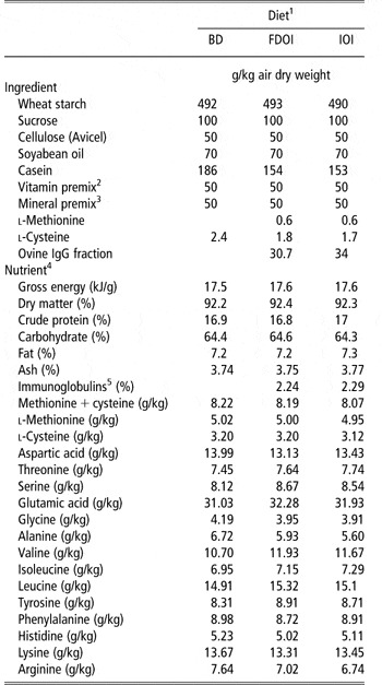
The rats were given 1 week to acclimatize, during which they consumed the basal diet ad libitum. After acclimatization, the rats were randomly allocated to one of three diets (Table 1; n = 15 per diet) for the 3-week study.
Ingredient composition and determined nutrient and energy contents of the control and test diets

BD = basal diet; FDOI = freeze-dried ovine immunoglobulin; IOI = inactivated ovine immunoglobulin; Ig = immunoglobulin.
1All diets were formulated to meet or exceed the nutrient requirements of growing rats.
2The mixture supplied: (mg/kg diet): retinol acetate 5.0, DL-tocopheryl acetate 200, menadione 3.0, thiamin hydrochloride 5.0, riboflavin 7.0, pyridoxine hydrochloride 8.0, D-pantothenic acid 20, folic acid 2.0, nicotinic acid 20, D-biotin 1.0, myo-inositol 200, choline chloride 1500; (μg/kg diet): ergocalciferol 25, cyanocobalamin 50.
3The mixture supplied: (g/kg diet): Calcium hydrogen phosphate 6.29, magnesium oxide 1.06, potassium chloride 5.24, sodium chloride 1.97; (mg/kg diet) chromic potassium sulphate 1.97, cupric carbonate 10.7, ferric citrate 424, manganese sulphate 78.0, zinc oxide 48.2; (μg/kg diet): cobalt chloride 29.0, potassium iodate 151, ammonium molybdate152, sodium selenite 151.
4values for nutrients were from chemical analysis except for carbohydrate, which was calculated by difference; methionine and cysteine were calculated values.
5Estimated Ig concentration in each diet.
The diets were supplemented with synthetic methionine and cysteine to meet or exceed the National Research Council (NRC, 1995) recommendations for the growing rat, as methionine plus cysteine was the first limiting amino acid (Table 1). The diets included a BD, a test diet containing FDOI and a negative control diet, IOI. The ovine Ig fractions that consist of IgG at a concentration of 73% were included in the diets (FDOI and IOI) at a concentration approximately equal to the amount of SDPP IgG used in previous studies (Pierce et al., Reference Pierce, Cromwell, Lindemann, Russell and Weaver2005; Balan et al., Reference Balan, Han, Rutherfurd, Singh and Moughan2009; Table 1). Food was provided ad libitum in powdered form.
Chemical analysis
Dry matter, ash, crude protein (CP), total fat and gross energy were determined according to the methods described by the Association of Official Analytical Chemists (AOAC; 2003). Carbohydrates were determined by difference using the formula (100 – (moisture + ether extract + ash + CP) = carbohydrate). The amino acid compositions of the diets (with the exception of methionine and cysteine that were calculated) were determined in duplicate after hydrolysis (6 mol/l HCl for 24 h at 110°C) using ion exchange HPLC with ninhydrin post-column derivatization.
Post-mortem procedure
At day 21 of the experiment, rats were anaesthetized by the intra-peritoneal injection (0.1 ml/100 g live weight) of a mixture containing two parts acepromazine maleate BP (0.2%, w/v, ACP), five parts ketamine (10%, w/v), one part xylazine (10%, w/v) and two parts sterile water. Blood sample was collected by cardiac puncture and a portion was immediately transferred to a heparinized vacutainer tube (Becton Dickinson, NJ, USA) to isolate plasma for measurement of plasma IgA and IgG. The remaining heparinized blood was used for assessing the phagocytic activity of peripheral blood leukocytes (PBLs). The spleen was removed aseptically for the lymphocyte proliferation assay. The jejunum (middle of the intestine) and ileum (anterior to ileocaecal valve) were collected and washed intra-luminally with 5 ml of phosphate buffer saline (PBS) containing 1% protease inhibitor (Sigma, Auckland, New Zealand). The suspensions were then centrifuged (HERAEUS, Fresco 17, Thermo Electron Corporation, MA, USA) at 3000 g for 10 min to remove debris and the clear supernatant was stored at −20°C until the measurement of intestinal IgA and IgG.
Assessment of phagocytosis
Assessment of the phagocytic activity of PBLs by flow cytometry was based on the method of Wan et al. (Reference Wan, Park and Lau1993) with some modifications. Briefly, 5 μl fluorescein isothiocyanate-labelled Escherichia coli (1 × 10⁹/ml, Molecular Probes Incorporated, OR, USA) and 100 μl blood were mixed in glass tubes on ice and incubated at 37°C for 30 min. The PBLs were fixed for 1 min with 100 μl paraformaldehyde (8%, v/v) and erythrocytes lysed by the addition of 1 ml ice-cold water. Following centrifugation, the resulting pellets were resuspended in 0.5 ml of PBS, thoroughly mixed and transferred to an FACS tube containing 50 μl trypan blue (0.4% in PBS, w/v) to quench extraneous fluorescence. The level of phagocytic activity was determined using an FACS-Calibur flow cytometer (Becton Dickinson, NJ, USA).
Preparation of splenocytes
Single-cell suspensions were prepared from spleen in complete RPMI-1640 medium (RPMI-1640 medium supplemented with 10% foetal calf serum, 10 mmol HEPES, 2 mmol-l-glutamine, 100 U/ml penicillin, 100 mg/ml streptomycin sulphate and 50 mmol 2-mercaptoethanol; all reagents from Gibco, CA, USA). The spleen tissue was mechanically disrupted by cutting into small pieces using scissors and sucking up and down with a sterile 1 ml syringe. The resulting spleen cell suspension was transferred to a 15-ml tube containing 5 ml RPMI-1640 and centrifuged at 1000 r.p.m. for 10 min. Supernatants were then discarded. Erythrocytes were lysed using 5 ml ACK lysis buffer (0.15 M NH4Cl, 10 mmol KHCO3 and 0.1 mmol EDTA) with occasional mixing during a 5-min incubation period. The remaining leukocyte suspensions were washed twice in complete RPMI-1640 medium and then resuspended in 3 ml RPMI-1640 medium, from which 100 μl of suspended cells were transferred to a flow cytometer tube containing 400 μl of PBS and 2 μl of propidium iodide. Viable lymphocytes were identified and counted using Becton Dickinson Immunocytometry Systems ‘Cell Quest’ software on an FACS Calibur flow cytometer (Becton Dickinson) by gating around the mixed lymphocyte cell population. Spleen lymphocyte cell numbers were adjusted to a final concentration of 2 × 10⁶ lymphocyte-like cells/ml in complete RPMI-1640 immediately before use.
Lymphocyte proliferation assay
Aliquots of 105 spleen lymphocyte cells in 50 μl of complete RMPI-1640 medium were added in triplicate to the wells of a 96-well, flat-bottomed tissue culture plate (Nunc, MO, USA) and cultured in the presence of either 2.5 μg/ml concanavalin A (Con A; Sigma), 5 μg/ml lipopolysaccharide (LPS; Sigma), diluted (1 :49 ) phytohaemagglutinin (PHA; Gibco) or complete RPMI-1640 in place of the mitogen (control wells). The cells were then cultured for 48 h at 37°C in a 5% humidified CO2-air atmosphere, before being pulsed for 18 h with 0.5 mCi methyl-3H-thymidine (Amersham Biosciences, NJ, USA) per well. Each plate was then harvested onto a 96-well glass fibre mat using a Tomtek cell harvester 96 (Hamden, CT, USA) and counted using a Wallac MicroBeta Trilux 1450 liquid scintillation and luminescence counter (PerkinElmer Life Sciences, MA, USA). Stimulation index was calculated as counts per minute (cpm) in wells with mitogen divided by cpm in wells without mitogen.
Analysis of cytokines
In all, 2 ml of the spleen lymphocyte cell suspensions (4 × 10⁶ cells) were added to each well of a 24-well plate (Costar, MO, USA) and cultured in the presence and absence of ConA (1 mg/ml; Sigma) for 48 h at 37°C. Cell-free supernatant fractions were harvested and stored at −20°C until assayed.
The presence of interferon-γ (IFN-γ) and interleukin-4 (IL-4) in the culture supernatants was determined by sandwich ELISA using IFN-γ and IL-4 cytokine DuoSet ELISA kits (R&D systems, DuoSet®, Abingdon, Oxford, UK) according to the manufacturer’s instruction.
Quantitative analysis of IgA and IgG
A sandwich rat IgA and IgG ELISA was used to quantify the IgA and IgG concentrations in plasma and intestinal digesta (jejunum and ileum); all antibodies were obtained from AbD Serotec, Oxford, UK. Briefly, for IgA quantitation, an ELISA plate was coated with mouse anti-rat IgA antibody (PRP01), and incubated for 1 h at room temperature. After washing, purified rat IgA kappa standards (MCA191) or suitably diluted samples were added to designated wells. Following further washings, horseradish peroxidase-conjugated mouse anti-rat IgA antibody (STAR111P) was added to each well as a detection antibody. After an incubation period, TMB enzyme substrate (3,30,5,50-tetramethyl benzidine) was added to each well. After 20 min, the reaction was stopped with 2 mol/l H2SO4 and the absorbance was determined at 450 nm. The ELISA procedure for IgG was similar to that described for IgA quantitation, using purified rat IgG standards (PRP12), but the antibodies used were goat anti-rat IgG (STAR71) and horseradish peroxidase-conjugated sheep anti-rat IgG (AARP10P).
Statistical analysis
Results are expressed as mean ± s.e.m. Data were analysed using a one-way ANOVA using the GLM procedure of SAS/PROC GLM (SAS, version 9.1, SAS Institute Inc., Cary, NC, USA) and means were compared using Tukey’s test. Statistical significance was accepted at P < 0.05.
Results
PBL phagocytosis
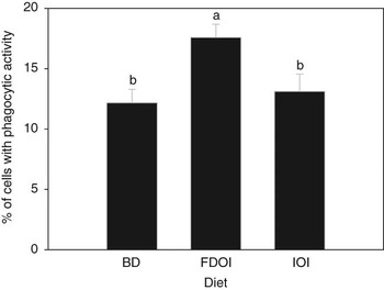
The phagocytic activities of PBLs from rats fed BD, FDOI and IOI diets for 3 weeks are shown in Figure 1. Levels of phagocytic activity were greater (P = 0.002) in rats receiving the FDOI diet than in rats fed either the IOI or the BD diet. There was no difference (P > 0.05) between rats fed the BD and IOI diets.
Phagocytic activity of peripheral blood leukocytes in rats fed a diet containing ovine Ig for 21 days. Values are means ± s.e. (n = 15); a,bMeans with superscripts without a common letter differ, P < 0.05.

Lymphocyte proliferation
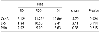
ConA-induced lymphocyte proliferative responses (stimulation index) of spleen cells in rats fed the FDOI diet were greater (P = 0.024) when compared to rats fed either the BD or the IOI diet (Table 2). Feeding the rats with different diets (BD, FDOI and IOI) did not result in any difference (P > 0.05) in spleen lymphocyte proliferative responses to LPS or PHA.
Spleen lymphocyte proliferative responses (stimulation index) to ConA, LPS and PHA for rats fed a diet containing ovine Ig for 21 daysFootnote 1

ConA = concanavalin A; LPS = lipopolysaccharide; PHA = phytohemagglutin; Ig = immunoglobulin; BD = basal diet; FDOI = freeze-dried ovine immunoglobulin; IOI = inactivated ovine immunoglobulin.
1 Values are means ± s.e.m. (n = 15), means in a row with superscripts without a common letter differ, P < 0.05.
Cytokine analysis
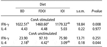
IFN-γ and IL-4 productions in ConA stimulated and unstimulated (in vitro) spleen cell cultures are shown in Table 3. In stimulated spleen cells, IFN-γ was higher (P = 0.008) in rats fed the FDOI diet than in rats fed the BD diet. The level of IL-4 production was not affected by the diets (BD, FDOI and IOI). In unstimulated spleen cells, IFN-γ production was not affected (P > 0.05) by the dietary intake of BD, FDOI and IOI, but the IL-4 concentrations were found to be higher (P = 0.044) in rats fed the FDOI diet than in rats receiving the BD diet.
INF-γ and IL-4 productions (pg/ml) by spleen lymphocyte cells from rats fed a diet containing ovine Ig for 21 daysFootnote 1 with and without ConA stimulation

INF-γ = interferon-γ; IL = interleukin; Ig = immunoglobulin; ConA = concanavalin A; BD = basal diet; FDOI = freeze-dried ovine immunoglobulin; IOI = inactivated ovine immunoglobulin.
1 Values are means ± s.e.m. (n = 15), means in a row with superscripts without a common letter differ, P < 0.05.
Rat IgA and IgG concentrations in intestinal digesta and plasma
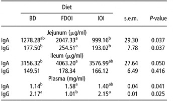
The IgA concentrations in the jejunal digesta were higher (P = 0.037) in rats receiving the FDOI diet than in rats fed the IOI diet. In the ileal digesta and plasma, IgA concentrations were higher (P = 0.05 and P = 0.041, respectively) in rats fed the FDOI diet than in rats receiving the BD diet (Table 4).
IgA and IgG concentrations of intestinal digesta and plasma for rats fed a diet containing ovine Ig for 21 daysFootnote 1

BD = BASAL diet; FDOI = freeze-dried ovine immunoglobulin; IOI = inactivated ovine immunoglobulin.
1 Values are means ± s.e.m. (n = 15), means in a row with superscripts without a common letter differ, P < 0.05.
The IgG concentrations in the jejunal digesta were higher (P = 0.037) in rats receiving the FDOI diet than in rats fed either the BD or the IOI diet. Ileal IgG concentrations were not affected (P > 0.05) by the dietary consumption of BD, FDOI and IOI. Plasma IgG levels were higher (P = 0.025) in rats receiving either the BD or the IOI diet than in rats fed the FDOI diet (Table 4).
Discussion
Phagocytic cells (polymorphonuclear leukocytes, monocytes and macrophages) and natural killer cells are the major effectors of innate immunity. These are considered as the first line of defense against foreign pathogens (Watzl et al., Reference Watzl, Girrbach and Roller2005). Oral administration of bovine colostrum is known to stimulate host non-specific immunity by increasing the systemic immune response or by modulating immunocompetent cell function (Sugisawa et al., Reference Sugisawa, Itou and Sakai2001). In this study, the phagocytic activity of PBLs was enhanced in rats fed the FDOI diet compared to rats fed either the BD or the IOI diet. Therefore, we can suggest that feeding the rats with FDOI may improve their innate immunity via enhancement of phagocytic activity of PBLs. Further study is required to determine a definitive mechanism of action. However, it is possible that it may be similar to that proposed by Rutherfurd-Markwick et al. (Reference Rutherfurd-Markwick, Johnson, Cross and Gill2005) who reported that mice fed whey protein concentrate (WPC) exhibited significant increases in both phagocytosis by blood leukocytes and ConA-induced proliferation of spleen lymphocytes, compared to those from mice receiving a milk protein-free diet. They suggested that constituent cysteine residues of WPCs may provide a substrate for glutathione synthesis and that this serves to potentiate antioxidant activity and membrane function of immune system cells sufficient to promote increased cellular activity (Bounous et al., Reference Bounous, Batist and Gold1989).
Lymphocyte proliferative responses to mitogens (i.e. stimulation index) are widely used to ascertain T- (ConA is a T-cell mitogen) and B (LPS is a B-cell mitogen)-cell function (Gill et al., Reference Gill, Watson and Brandon1992). In this study, rats fed the FDOI diet had a higher stimulation index in ConA-stimulated spleen lymphocytes compared to rats receiving the BD and IOI diets. This result may indicate that the FDOI diet enhanced T-cell function.
Naive T cells recognize an antigen using the major histocompatibility complex molecules via the T-cell receptor complex, and can be developed into three major subsets: Th1, Th2 and Treg/Th3 based on the pattern of cytokines they secrete. The key Th1 cytokines such as IFN-γ, IL-2, IL-3, IL-12 and TNF-α are involved predominantly in cell-mediated immune response and confer immunity against harmful intracellular agents such as bacteria, viruses and tumour cells, and also downregulate intestinal IgA concentrations. They also activate and differentiate T and B lymphocytes as well as macrophages, whereas cytokines such as IL-4, IL-5 and IL-6, which are secreted by Th2 cells, confer humoral responses, activate B lymphocytes, upregulate mucosal immunity and mediate responses against extracellular parasites, as well as upregulating IgA concentration. Treg/Th3 cells secrete IL-10 and TGF-β cytokines. A balance between Th1 and Th2 cytokines may be necessary to maintain a normal IgA immune response (Kramer et al., Reference Kramer, Sutherland, Bao and Husband1995; Reiner and Seder, Reference Reiner and Seder1995; DiPiro, Reference DiPiro1997).
In this study, since the spleen lymphocytes from the rats fed the FDOI diet showed a greater stimulation index in the presence of ConA, we analysed cell culture supernatants for their cytokine pattern (IFN-γ and IL-4) to ascertain T-cell polarity. In FDOI-fed rats, the pattern of cytokine expression indicates that there might have been a selective activation of Th1 cells in the presence of ConA and selective activation of Th2 cells (to a lesser extent than Th1 cells) in the absence of the mitogens. A balance between Th1 and Th2 cytokines is necessary for normal immune function because if the diet influences persistent Th1 polarity, it may lead to cell-mediated immunity and cytotoxic T-lymphocyte induction, resulting in inflammatory responses, whereas persistent Th2 polarity leads to more IgE production, resulting in allergy and hypersensitivity reactions (Thyphronitis et al., Reference Thyphronitis, Tsokos, June, Levine and Finkelmen1989). In this study, we found no significant differences in the plasma IgE levels of rats fed BD, FDOI and IOI (data not shown), suggesting that the rats fed the FDOI diet had not developed any allergic type reaction. Our findings are similar to that of Boudry et al. (Reference Boudry, Buldgen, Portetelle, Gianello, Thewis, Leterme and Dehoux2007) who reported an immunomodulatory effect of bovine colostrum on the gut associated lymphoid tissue of weaned piglets, which responded by producing both Th1 cytokines and Th2 cytokines at different levels. They concluded that Th1/Th2 bipolar response protects the piglets from both allergic (food) and infectious (pathogens) diseases.
When Ig present in the FDOI diet is delivered orally, the gut-associated lymphoid tissue may be its primary target, resulting in stimulation of IgA secretion in the intestine. The immunological defense against bacteria and various potential pathogens in the gut is carried out by intestinal IgA in a process known as immune exclusion where intestinal IgA binds to the bacteria, pathogen or its antigen, thereby preventing their translocation (Amin et al., Reference Amin, Diebel and Liberati2007). Rats consuming the FDOI diet in this study had greater amounts of secretory IgA (sIg) in both jejunal and ileal digesta that might play a major role in protecting the intestine from invading pathogens (Mayer, Reference Mayer1997) and suppressing inflammatory processes (Parlesak et al., Reference Parlesak, Schäfer and Bode2002). Rats fed the FDOI diet also had greater amounts of rat jejunal IgG. Taken together, the elevated amounts of intestinal digesta IgA and IgG of rats fed the FDOI diet indicate that the animals receiving this diet may be better able to preserve a functional gut compared to rats fed the other diets, by preventing the adverse effects of possible pathogens in the GIT. This is in line with our previous observations that villus length, crypt depth, villus/crypt ratio and villus surface area of the small intestine were greater (P < 0.001 to 0.703) in rats receiving the FDOI diet than in rats fed either the BD or the IOI diet (Balan et al., Reference Balan, Han, Rutherfurd, Singh and Moughan2009).
Plasma IgA plays an important role in inhibiting allergen absorption (Benyacoub et al., Reference Benyacoub, Czarnecki-Maulden, Cavadini, Sauthier, Anderson, Schiffrin and von der Weid2003). In this study, plasma IgA concentrations were also greater in rats fed the FDOI diet than in rats fed the BD diet. This is similar to the results from a study (Puri et al., Reference Puri, Rattan, Bijlani, Mahapatra and Nath1996) in which diets supplemented with Enterococcus faecium (SF68) stimulated immune function in dogs by increasing the concentrations of intestinal and plasma IgA. Intestinal IgA, produced by B lymphocytes in the intestinal cells, may cross the mucosal barrier and enter the bloodstream influencing the plasma IgA concentration (Puri et al., Reference Puri, Rattan, Bijlani, Mahapatra and Nath1996).
In this study, rats fed the FDOI diet had lower plasma IgG levels when compared to rats fed the BD and IOI diets. A possible explanation for this could be that the ovine Ig present in the FDOI may remove pathogens or microorganisms in the GIT, resulting in less pathogenic antigens being transported across the gut to blood vessels resulting in a lower production of rat plasma IgG. In our previous study, we found that lactobacillus species were upregulated in ileal and colonic digesta in the rats fed the FDOI diet when compared to the rats fed the BD diet (Balan et al., Reference Balan, Han, Rutherfurd, Singh and Moughan2009). Livingston et al. (Reference Livingston, Loach, Wilson, Tannock and Baird2010) reported that the percentage of Foxp3+ T cells from mice colonized by Lactobacillus species was found to be significantly higher than that from Lactobacillus-free mice in both mesenteric lymph nodes and spleen cell populations, clearly showing that mice colonized with Lactobacillus species developed more tolerance of the immune system in gut-associated lymphoid tissue than mice without Lactobacillus. This information suggests that rats fed the FDOI diet may develop tolerance by upregulation of the Lactobacillus species in the gut.
Igs are at least partially resistant to digestion in the upper GIT. Rodriguez et al. (Reference Rodriguez, Blanch, Romano, Saborido, Rodenas and Polo2007) recently reported that porcine Ig partially resisted the digestion process in the GIT of adult dogs and cats fed diets containing SDAP, or concentrated Ig derived from pig blood. Indeed, reports have shown that human serum Ig and bovine Ig resist digestion in the upper GIT of humans and retain toxin binding and neutralizing activity (Losonsky et al., Reference Losonsky, Johnson, Winkelstein and Yolken1985; Warny et al., Reference Warny, Fatimi, Bostwick, Laine, Lebel, LaMont, Pothoulakis and Kelly1999). Given that circular dichroism analysis (data not shown) confirmed that the Ig in the IOI treatment was inactivated, our results suggest that active (undenatured) Ig in the FDOI diet was the active ingredient responsible for the observed immune responses.
In conclusion, orally administered FDOI selectively modulated immune function in the growing rat through increasing phagocytic activity of PBLs and lymphocyte proliferation, and by influencing the balance between Th1 and Th2 cytokine productions, and the sIgA and sIgG in the intestinal contents and plasma. Altogether, these effects may result in the animal having increased resistance to infection. To our knowledge, this is the first report of the immunomodulatory effects of dietary ovine serum Igs in an animal species.