Idiopathic chylopericardium is the accumulation of chylous fluid in the pericardial cavity without any known pre-disposing factors.Reference Wang, Hung, Cherng, Yen, Ng and Lee1 Currently, the development of advanced diagnostic tools, including magnetic resonance lymphangiography, enables the identification of the mechanism of chylopericardium.Reference Krishnamurthy, Hernandez, Kavuk, Annam and Pimpalwar2 We report an unique case of a 29-month-old girl who presented with chronic massive pericardial effusion and was diagnosed with lymphatic anomalies that were successfully treated with thoracic duct embolisation.
Case Report
A 29-month-old, previously healthy girl visited an external referring hospital because of fever, cough, and sputum production for 2 weeks prior to referral. Cardiomegaly was observed on chest X-ray. Echocardiography revealed a large pericardial effusion. Pericardiocentesis was performed and 200 ml of pericardial effusion was drained daily via percutaneous catheter drainage. The patient was transferred to our hospital because the massive pericardial effusion persisted for >2 weeks.
Her chest X-ray showed cardiomegaly with diffuse interstitial thickening in both lungs at the time of the visit to our hospital. Echocardiography showed a massive pericardial effusion.
Her effusion was serosanguinous and exudative: albumin, 2.8 g/dl (serum albumin, 3.4 g/dl). On analysis of pericardial fluid, the cell profile revealed a lymphocyte-dominant (96%) cell count, negative fluid cultures, and no malignant cells. During her usual diet without any specific restrictions, the triglyceride (100–115 mg/dl) and total cholesterol levels (93–133 mg/dl) were mildly elevated, with cholesterol/triglyceride ratio of 0.9:1.2.
The pericardial effusion resolved spontaneously 5 weeks after the initial detection of the effusion and the patient was discharged. However, massive pericardial effusion recurred repeatedly, 3 months and 9 months after the first episode.
Although the effusion did not have prominent chylous features (triglyceride, 106 mg/dl; total cholesterol, 132 mg/dl in the pericardial effusion and serum triglyceride, 58 mg/dl; total serum cholesterol, 154 mg/dl), lymphoscintigraphy using Technetium-99m antimony trisulfide colloid was performed due to chronic and recurrent pericardial effusion and pulmonary lymphangiectasia, which was suspected based on the findings of a previous CT. As lymphoscintigraphy revealed mild radioactivity around the heart, contrast enhanced magnetic resonance lymphangiography was performed by injecting contrast medium (Dotarem/normal saline 1:1 mixture 10 ml) through right inguinal lymph nodes.
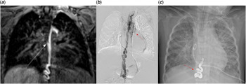
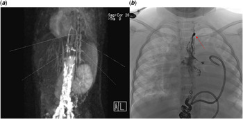
The magnetic resonance lymphangiography showed a tortuously dilated thoracic duct in the mediastinum with suspected chylous reflux to the pericardial space at the level of the aortic arch (Fig 1a). We presumed that the pericardial effusion was caused by chylous reflux. On invasive, percutaneous fluoroscopic contrast enhanced lymphangiography using the retrograde catheter through the femoral vein, superior vena cava, innominate vein, lymphovenous junction, and abdominal thoracic duct, abnormal communication of prominent lymphatic collaterals to the pericardial cavity was more prominently visualised (Fig 1b), because this catheter unexpectedly blocked lymphatic drainage into the innominate vein at the lymphovenous junction, which led to the abrupt aggravation of pericardial effusion. From this finding, we could confirmed that chylous reflux from thoracic duct caused massive chronic pericardial effusion. Thoracic duct embolisation was performed using multiple microcoils and 1:2 glue/lipiodol mixture at the T11 level (Fig 1c). The patient could have been discharged after resolution of pericardial effusion. However, a large amount of pericardial effusion recurred after 5 months. The second magnetic resonance lymphangiography using undiluted Dotarem solution 10 ml through bilateral inguinal lymph nodes showed enhancement of the periaortic structures, suggestive of persistent lymphatic fluid inflow to the pericardium through the remnant upper part of the thoracic duct (Fig 2a). A second lymphatic embolisation was performed to completely occlude the remaining upper part of the thoracic duct and abnormal lymphatic vessels alleged to distribute over not only the pericardium but also bilateral lung hila. Because the conventional antegrade access to the thoracic duct through cisterna chyli was not possible due to prior thoracic duct embolisation, two microcatheters were retrogradely inserted from the common femoral venous access via the junction between the left subclavian vein and thoracic duct at the venous angle. First, the outflow of the thoracic duct was occluded using multiple microcoils to prevent pulmonary embolism of the embolic agent. Then, the 1:5 volume mixture of n-BCA Histoacryl-tissue adhesive (B. Braun Melsungen AG W. Germany) and Lipiodol (Lipiodol Ultrafluide, Laboratoire Guerbet, Aulnay-Sous-Bois, France) was injected through the other microcatheter to occlude abnormal lymphatic vessels in the mediastinum as well as the remaining part of the thoracic duct (Fig 2b). After the second embolisation, the patient has been doing well without recurrence of pericardial effusion for 12 months until now.
(a) Dynamic T1-weighted magnetic resonance lymphangiography obtained after intranodal injection of gadolinium contrast medium to the inguinal lymph node shows the tortuously dilated thoracic duct connected to the pericardium through an anomalous mediastinal lymphatic vessel (arrow). (b) Direct catheter lymphangiography with the tip in the central conducting duct at the abdomen confirmed a reflux from the thoracic duct into dysplastic lymphatic channels in the mediastinum and left pericardial space (arrow). Note the pigtail catheter in the pericardial space. (c) Chest radiograph obtained after thoracic duct embolisation shows multiple microcoils (arrow) and glue cast at the lower thoracic spine level.

(a) The major collateral vessel around the occluded thoracic duct was observed on magnetic resonance lymphangiography. (b) Thoracic duct embolisation was performed at the cervical–thoracic level with 1:3 glue after coil embolisation (arrow) to prevent venous reflux of glue.

Discussion
Idiopathic chylopericardium is a rare disease.Reference Itkin, Swe, Shapiro and Shrager3,Reference Svedjeholm, Jansson and Olin4 Milky white or pink fluid on pericardial fluid analysis is the usual finding in chylopericardium, with elevated triglyceride level in the effusion (181–4299 mg/dl).Reference Yu, Jia, Ye, Zhou and Liu5 However, pericardial fluid was serosanguinous, and the triglyceride level was only mildly elevated (100–110 mg/dl) in this patient. Hence, chylopericardium was not suspected initially. Recurrent massive pericardial effusion without a definite cause and peribronchovascular and interstitial thickening of both lungs on chest CT led us to suspect the presence of lymphatic abnormalities, which was proven by magnetic resonance lymphangiography and lipiodol lymphangiography.
Recently, percutaneous intervention was developed as a minimally invasive alternative treatment to surgical treatment for chylothorax or chylopericardium.Reference Nadolski and Itkin6,Reference Cope and Kaiser7 Some studies have reported that thoracic duct embolisation is effective as a treatment for non-traumatic chylothorax or chylopericardium.Reference Nadolski and Itkin6,Reference Chen and Itkin8 Even though the specific method of thoracic duct embolisation has not yet been unified, some researchers have argued that embolisation in the proximal region (rather than in the outflow or abnormal vessels) may be sufficient.Reference Chen and Itkin8 However, in our case, pericardial effusion recurred after initially successful thoracic duct embolisation at proximal level of the thoracic duct. This is probably because the chylous lymphatic fluid from the abdomen could bypass the occluded segment of the proximal thoracic duct via collateral vessels to reach the remnant upper part of thoracic duct and finally the abnormal lymphatic vessels distributing over the lung hilum and pericardium (Fig 2a). Pericardial effusion finally resolved after the second thoracic duct embolisation occluding the remnant upper part of the thoracic duct as well as the abnormal lymphatic vessels in the mediastinum.
Thoracic duct embolisation can theoretically cause lower extremity oedema and gastrointestinal symptoms.Reference Chen and Itkin8,Reference Laslett, Trerotola and Itkin9 Although the number of cases is small, the incidence of associated complications after thoracic duct embolisation was reported to be approximately 10% in these reports. These studies indicate that thoracic duct embolisation is a relatively safe and effective procedure.Reference Chen and Itkin8,Reference Laslett, Trerotola and Itkin9 However, further studies are needed for safety in children. Complications may be rare, but this patient requires long-term follow-up for an assessment of problems associated with thoracic duct embolisation. In the current era, lymphoscintigraphy is not always necessary and could be replaced by MRI lymphangiography as a primary diagnostic imaging tool. MRI lymphangiography is also useful as it allows a radiation-free comprehensive map of the central lymphatics (both abdominal and thoracic) to help select patients best suited to thoracic duct interventions.
Conclusion
In a patient with idiopathic chronic pericardial effusion, lymphatic abnormality is one of the causes that should be considered. Magnetic resonance lymphangiography was useful in the diagnosis in a patient with chylopericardium. Thoracic duct embolisation might prove to be a successful mode of treatment for massive and recurrent chylopericardium.
Acknowledgements
None.
Financial Support
This study was not supported by any funding.
Conflict of Interest
The authors declare that they have no conflict of interest.
Ethical Standards
For this type of study, formal consent is not required.
Informed Consent
Informed consent was obtained from all individual participants included in the study.
Consent for Publication
Consent for publication was obtained for every individual person’s data included in the study.