Introduction
In freshwater fish species, such as zebrafish (Danio rerio), embryos have to be kept in a culture media with an osmolarity value of approximately 35 mOsm (Westerfield, Reference Westerfield2003) for their in vitro culture and micromanipulation, whereas cultured cells must be kept at 315 mOsm (Calvi and Maisse, Reference Calvi and Maisse1998). This may involve certain problems of osmolar compatibility when experimental techniques, such as embryonic chimaerism using in vitro cultured cells, are practised. This technique is very important in the reproduction biotechnology of fish species because of its commercial interest. Osmolarity can also involve some technical difficulties in studies of cell de-differentiation when, as has already been proposed in mammals, cells and early embryos are co-cultured to change the differentiation patterns of those cells by epigenetic effects, before being used as donor nuclei in somatic cloning experiments (Burnside and Collas, Reference Burnside and Collas2002; Cervera and García-Ximénez, Reference Cervera and García-Ximénez2004a and Reference Cervera and García-Ximénez2004b).
In fish, although the effects of salinity have been extensively examined in marine and estuarine fish embryos and larvae (Van Der Wal, Reference Van Der Wal1985; Young and Dueñas, Reference Young and Dueñas1993; Hart and Purser, Reference Hart and Purser1995; Haddy and Pankhurst, Reference Haddy and Pankhurst2000), they remain largely untested in freshwater fish species. To our knowledge, the effects of salinity on zebrafish embryos have been tested only recently (Sawant et al., Reference Sawant, Zhang and Li2001; Adams et al., Reference Adams, Zhang and Rawson2005), but not often those due to the culture media osmolarity or composition.
In preliminary assays, we observed that intact and dechorionated zebrafish embryos could survive and develop to the pharyngula stage with normal developmental timings in L-15. However, after that, they curved their tails, paralysed their movements and stopped their development (results not published). The above-mentioned effects could be due to the culture media composition (L-15) (Westerfield, Reference Westerfield2003) or, although not exclusively, due to its osmolarity (315 mOsm). Thus, these effects were evaluated in the present work, in an attempt to distinguish those due to hyperosmolarity from those caused by the composition of the culture media. These results are an important first step in future works on chimaerism and co-culture of somatic cells with stopped early embryos.
Material and methods
Wild zebrafish (Danio rerio) dechorionated embryos from 1000-cell stage (midblastula transition, MBT) to Oblong stage (3 to 4 h post fertilisation at 28.5°C) were used. They were collected from adults kept in a 6 : 3 proportion (females/males) by siphoning fertilised eggs from the bottom of the tank. Granular food was supplemented with recently defrosted hen egg yolk in place of live food (daphnia or Artemia nauplius), which is usually recommended for egg production. All chemical products and culture media were from Sigma-Aldrich (St Louis, MO, USA).
After washing with conventional tap water on a nylon mesh, egg embryos were selected under a microscope and washed again with tap water. No bleaching treatment was applied, but sterility conditions of media and pipettes after dechorionation were strict.
Dechorionation was carried out by pronase treatment (1.5 mg/ml in Hanks’ 10%) followed by immersion twice in Hanks’ buffered salt solution diluted 10% (v/v) in distilled water (H-10; 35 mOsm). The embryos were kept for 1 h in H-10. Next, partially or totally damaged embryos (lysed or blastoderm–yolk disconnection) due to previous manipulation were removed, and finally intact embryos were assigned to experimental groups (see below).
Dechorionated eggs were cultured in 35-mm petri dishes (corning) for 6 days at 28.5°C in the following culture media: Hanks’ buffered salt solution (CH), L-15 with 20% of foetal bovine serum (FBS) (L-15) and Hanks’ buffered salt solution diluted 10% (v/v) (H-10; control group).
Osmolarity was measured using a cryoscopic osmometer (Osmomat 030; Gonotec, Berlin, Germany): CH and L-15 osmolarity was around 315 mOsm, whereas H-10 osmolarity was approximately 35 mOsm.
L-15 was included in the experimental design because it is needed to co-culture cells and early embryos for carrying out de-differentiation studies like those proposed in mammals.
Two consecutive experiments were carried out. Embryonic survival and development effects were evaluated when midblastula transition (MBT) embryos were exposed to hyperosmolar culture media during the whole culture period (6 days) or only for a limited time (1 h), maximum time after the MBT stage in which the chimaerism can be performed. In the first experiment, embryos were kept throughout their development (6 days) in the two assayed culture media (CH and L-15) with the aim of studying the long-term hyperosmolar effects on embryos, important in future experiments of co-culture stopped embryos with somatic cells. In the second experiment, MBT embryos were only kept in both culture media for 1 h and cultured in H-10 later. In this case, we studied the hyperosmolar effects on embryos when they were exposed during the minimum time required to carry out the chimaerism.
The following parameters were evaluated in both experiments.
Survival rate and structural integrity (blastoderm–yolk disconnection and lysis) were evaluated at 10 h post fertilisation (100% epiboly). Survival rate was also evaluated by tail motility and/or heartbeat at 28 h (pharyngula period). Survival rate (motility and/or heartbeat) was again evaluated at 48 h (hatching period). In addition, alteration of timing of the pattern of development was established by eye pigmentation, as well as by developmental abnormalities such as motility loss (corporal paralysis) or tail deformation (curled-up shape and/or atrophy). Finally, survival was again evaluated at 6 days by corporal motility and/or by heartbeat. At the same time, the presence of a functional swim bladder was tested.
At least three replicates were performed in all experimental groups. These repeats varied from three to six depending on the experimental group. The χ 2 tests were used to analyse effects of treatments on the various developmental parameters. The Yates correction for continuity was used when a single degree of freedom was involved.
Results
Experiment 1
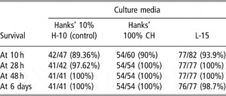
Related to survival rate, blastoderm–yolk disconnections are attributed to previous manipulation (washing and dechorionation), because they were produced before embryos were assigned to the different experimental groups. Damaged embryos (by lysis and blastoderm–yolk disconnections) were observed in all three groups until 10 h post fertilisation. They were still detected in the high-osmolarity groups (CH and L-15) at 28 h post fertilisation, whereas they were not observed in the control group (H-10) (Table 1). However, the greater effect of osmolarity on the survival rate was detected later, over the 48 h period in which survival decreased strongly in high osmolarity (Table 1), being even lower in the L-15 group (6 days: L-15: 45.1% v. CH: 72.34% v. H-10: 100%; P< 0.05). It must be emphasised that survival rates (evaluated by beating heart) in both high-osmolarity groups were zero beyond 6 days.
Experiment 1: survival rate of midblastula embryos long-term cultured (6 days) in high-osmolarity media

Between columns, data with different superscript letters are significantly different (P < 0.05).
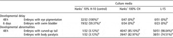
As was expected, developmental timing was fully normal in H-10, whereas a delay in eye pigmentation was observed at 48 h in the high-osmolarity groups (CH and L-15) (Table 2). Pigmentation was not complete in the L-15 group. This delay in the developmental timing was also shown at 6 days by the non-functionality of the swim bladder.
Experiment 1: alterations of developmental patterns of midblastula embryos long-term cultured (6 days) in high-osmolarity media

Between columns, data with different superscript letters are significantly different (P < 0.05).
Some delay in body pigmentation patterns was detected at 28 h in high-osmolarity groups, but results were not included in tables because objective quantification was difficult. On the other hand, embryos cultured in high-osmolarity media (CH and L-15) slowed their developmental timing, and finally stopped and remained in the pharyngula stage until their death (6 days). Embryos that were cultured in H-10 developed normally until the fry stage.
Hyperosmolarity conditions also brought about certain developmental abnormalities at 28 h, although they were not fully evident until 48 h. These embryos showed tail deformation (curly) as well as body paralysis at 48 h when they showed tail movements at 28 h. Differences in the rate of abnormal specimens between high-osmolarity groups reached levels of significance (CH: 85.10% v. L-15: 98.04%; P < 0.05) (Table 2). In fact, most of the embryos that were cultured in high-osmolarity conditions progressively slowed their heartbeat rhythm, enlarged their pericardium and heart, and stopped their heartbeat at 6 days, but finally all embryos lost their heartbeat 2 to 3 days later at maximum. The above-mentioned loss of heartbeat occurred earlier in the L-15 group than in the CH group. In this way, it may be highlighted that no embryos in L-15 or CH showed heartbeat after a few days.
Experiment 2
Differential effects in the survival rate were not observed (Table 3), even at 6 days, when midblastula embryos were cultured for 1 h in H-10, CH or L-15, even though a 10% loss was observed in the three groups until the first evaluation (at 10 h post fecundation).
Experiment 2: survival rate of midblastula embryos cultured 1 h in high osmolarity media

Between columns, data with different superscript letters are significantly different (P< 0.05).
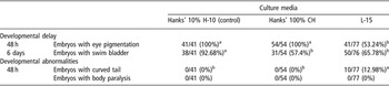
Regarding teratogenic effects, curved tails were only observed in 13% of the embryos cultured in L-15. This difference even reached significant levels with regard to CH (L-15: 12.98% v. CH: 0%; P < 0.05) (Table 4). Loss of motility was not detected in any group at 48 h.
Experiment 2: alterations of developmental patterns of midblastula embryos cultured 1 h in high-osmolarity media

Between columns, data with different superscript letters are significantly different (P< 0.05).
With respect to alterations of developmental timing, modification in eye pigmentation patterns were only observed in those embryos exposed to L-15 medium (50% with fully pigmented eyes at 6 days of development) (Table 4), whereas the swim bladder was evident later in L-15 (66% at 6 days) and CH (57% at 6 days) groups than in the control group (H-10: 93% at 6 days) (Table 4).
Discussion
Results of the first experiment show that the negative effects on development and survival of midblastula embryos cultured in L-15 are essentially due to high osmolarity and not due to media composition, because both L-15 and CH caused the same unfavourable final effects on embryos (at 6 days). In any case, the differential response to these media in some parameters, such as eye pigmentation (Experiment 2: CH 100% v. L-15 53.24%) or tail curvature (Experiment 1: L-15 98.04% v. CH 85.10%; Experiment 2: L-15 12.98% v. HC 0%) rates, may be due to an additional toxic effect exerted by some component of L-15 culture medium. Surprisingly, this toxic effect did not affect embryo cell survival during in vitro culture. Some negative effects of components of the culture media on embryos have been described in mammals, such as those due to phosphates (Escribá et al., Reference Escribá, Silvestre, Saeed and García-Ximénez2001) or phenol red, although the latter is recommended precisely as a tracer for some substances in intraembryonic microinjection experiments in zebrafish (Gilmour et al., Reference Gilmour, Jessen and Lin2002).
Although Sawant et al. (Reference Sawant, Zhang and Li2001) indicate that higher salinity impaired the nuclear division of the embryonic cells in zebrafish embryos, in our case L-15 medium, which is usually used in fish cell culture, can support embryonic development, even early larvae, although that medium finally caused motility loss and developmental inhibition.
It may be that hyperosmolarity may not affect early embryos, either because of the low hydraulic permeability of their outer cell membranes (Adams et al., Reference Adams, Zhang and Rawson2005), or because there are mechanisms that make up for the osmolarity change in that embryonic stage.
On the other hand, but not exclusively, results from the second experiment suggest that damage caused due to the exposure to high-osmolarity media for a limited time in the MBT stage can be compensated and/or repaired later either by cell functional replacement (Sekirina and Neganova, Reference Sekirina and Neganova1995; Boiani et al., Reference Boiani, Eckardt, Leu, Schöler and McLaughlin2003) or by maternal effects. The existence of these compensating mechanisms would also explain the observed delay in developmental timing.
All the above-mentioned effects would be present in embryos that bear the osmolarity change at the midblastula stage. Adams et al. (Reference Adams, Zhang and Rawson2005) detected an effect on survival in zebrafish when early embryos were exposed since fecundation to osmolarity values around 105 mOsm, which indicates that embryo survival depends on the embryonic stage and on the time of exposure for which embryos are in hyperosmolar medium.
Whatever the case, results of the present work indicate that L-15 cell culture medium supported early embryo development, which involves the possibility of carrying out embryonic–somatic cells co-culture experiments.
The optimum osmolar condition for cultured cells and also embryonic cells (certainly MBT cells immediately lysed at 35 mOsm after their extraction from the embryos, data not published) are around 315 mOsm and so, during chimaerism experiments (usually practised at MBT stage), the present results indicate that midblastula embryos can acceptably bear the effects caused by 315 mOsm (CH) for 1 h, even though this involves a certain delay in developmental timing. Otherwise, this cannot be said for L-15 medium because, as pointed out before, it seems to cause additional and non-reversible negative effects. According to our knowledge, in chimaerism experiments with freshwater species, such osmolar effects were not taken into account until now.
Acknowledgements
The authors thank Mr Neil Macowan for revising the English version of the manuscript.