INTRODUCTION
Pycnogonids (sea spiders) are one of the most intriguing groups of arthropods. These exclusively marine animals found from shorelines to abyssal depths (>1320 spp.) show unique morphological features (e.g. prominent external proboscis, extremely reduced abdomen, reproductive and digestive systems extending into walking legs) that make them difficult to relate to any other arthropod group. Their sister relationship to Chelicerata, the most accepted phylogenetic hypothesis, is yet to be fully understood (Dunlop & Arango, Reference Dunlop and Arango2005; Brenneis et al., Reference Brenneis, Ungerer and Scholtz2008; Regier et al., Reference Regier, Shultz, Zwick, Hussey, Ball, Wetzer, Martin and Cunningham2010), and has propelled several recent works on pycnogonid evolution and novel morphological data (Vilpoux & Waloszek, Reference Vilpoux and Waloszek2003; Brenneis et al., Reference Brenneis, Ungerer and Scholtz2008; Ungerer & Scholtz, Reference Ungerer and Scholtz2009). However, many aspects of pycnogonid biology remain understudied. Since Arnaud & Bamber's (Reference Arnaud and Bamber1987) comprehensive review, few papers on reproduction (Tomaschko et al., Reference Tomaschko, Wilhelm and Bückmann1997; Barreto & Avise, Reference Barreto and Avise2008; Bilinski et al., Reference Bilinski, Szymanska and Miyazaki2008) and feeding biology (Bain, Reference Bain1991; Imandeh & King, Reference Imandeh and King2001; Heß & Melzer, Reference Heß and Melzer2003) have been published. A common highlight of these and previous studies though, is the uniqueness of different aspects of the biology of Pycnogonida when compared to other arthropods and their digestive system is no exception.
The digestive system seems to be similar within the group (Richards & Fry, Reference Richards and Fry1978), but, unique among the arthropods. The proboscis is perhaps the most prominent feature and has been studied in detail by Dohrn (Reference Dohrn1881), Hoek (Reference Hoek1881), Wirén (Reference Wirén1918) and Fry (Reference Fry1965), and more recently at the ultrastructural level in Fahrenbach & Arango (Reference Fahrenbach and Arango2007). The midgut, which is the gut section where intracellular digestion occurs, comprises the medial trunk and reaches the leg processes at different lengths (Richards & Fry, Reference Richards and Fry1978). In pycnogonids, contrary to what occurs in most arthropods (crustaceans, insects and some Chelicerata), the midgut cannot be divided into functional portions, that is anterior and posterior, but the whole length of the midgut shows the same type of multifunctional epithelial cells (Richards & Fry, Reference Richards and Fry1978). The hindgut is reduced within the small abdomen, and is the gut section where faecal pellets congregate to be evacuated through the anal opening.
Previous studies, based on the analysis of feeding behaviour (Wyer & King, Reference Wyer and King1974; Stock, Reference Stock1978; Bain, Reference Bain1991), showed pycnogonids feeding on phytodetritus and seaweeds, as well as being predators of usually sessile items such as hydroids (Prell, Reference Prell1909; Fry, Reference Fry1965; Russel & Hedgpeth, Reference Russel and Hedgpeth1990; Bain, Reference Bain1991; Tomaschko et al., Reference Tomaschko, Wilhelm and Bückmann1997; Heß & Melzer, Reference Heß and Melzer2003), Anthozoa (Bamber, Reference Bamber1985; Tomaschko et al., Reference Tomaschko, Wilhelm and Bückmann1997; Arango, Reference Arango2001; Braby et al., Reference Braby, Pearse, Bain and Vrijenhoek2009), and Bryozoa (Fry, Reference Fry1965); in some cases they have also been observed feeding on mobile prey such as polychaetes (Arnaud & Bamber, Reference Arnaud and Bamber1987; Soler-Membrives et al., Reference Soler-Membrives, Rossi and Munilla2011), copepods (Lotz, Reference Lotz1968) and molluscs, bivalves and nudibranchs (Lotz, Reference Lotz1968; Rogers et al., Reference Rogers, De Nys and Steinberg2000; Arango & Brodie, Reference Arango and Brodie2003), among others, generally by sucking their prey (Arnaud & Bamber, Reference Arnaud and Bamber1987). However, it is not clear how the digestive system and the processes involved may vary between taxa and according to the different feeding behaviours. Fahrenbach & Arango (Reference Fahrenbach and Arango2007) briefly related the feeding habits of several species from different families to the external proboscis structure. Here, we investigate the digestive system of two Mediterranean species Ammothella longipes (Hodge, 1864) and Endeis spinosa (Montagu, 1808) with different diets and feeding behaviours. The former, closely related to the predator Ammothella appendiculata (see Fahrenbach & Arango, Reference Fahrenbach and Arango2007), is a seasonal opportunist, carnivorous during spring and detritivorous during autumn and winter while both carnivory and detritivory seem to occur in summer (Soler-Membrives et al., Reference Soler-Membrives, Rossi and Munilla2011). According to its morphology and general behaviour (Wyer & King, Reference Wyer and King1974; Arnaud & Bamber, Reference Arnaud and Bamber1987; Fahrenbach & Arango, Reference Fahrenbach and Arango2007), Endeis spinosa is considered a surface grazer or detritus feeder. In spite of the studies conducted in this species, there is no information about the seasonal changes in its feeding behaviour.
The major goal of this study then, is to provide a detailed examination of the digestive system of Ammothella longipes and Endeis spinosa using light, scanning and transmission electron microscopes, as well as to discuss behavioural and morphological data relating them to food preferences. Moreover, the relevance of this study falls on to the discussion of the digestive process in Pycnogonida and its possible evolutionary and ecological implications.
MATERIALS AND METHODS
Collecting of live pycnogonids took place between summer and autumn of 2007 and 2008 on a north-western Mediterranean beach (41°40′37″N 2°48′29″E) in Blanes, Catalonia (Spain). Each batch consisted of a sample of Stypocaulon spp. community (Stypocaulon scoparia, Halopteris filicina, or a mix of them) which was carefully bagged in situ and extracted using SCUBA diving in 7–10 m depth where the species are commonly distributed (Soler-Membrives et al., Reference Soler-Membrives, Rossi and Munilla2011). Batches were transported in a cooler filled with ice (10–12°C) and then left in separate trays maintaining constant cold water temperature, to avoid digestive tract degradation. Pycnogonids were sorted and identified to species level. Individuals of A. longipes and E. spinosa were chosen and fixed either for histology or for electron microscopy.
Live animals destined for histology were fixed in 4% formalin in seawater, and completely dehydrated through a graded ethanol series. Some individuals were embedded in paraffin and serial sections (from 4 to 10 µm thick) were obtained and then stained with haematoxylin and eosin. Other individuals were embedded in Technovit 7100 resin (Kulzer, Heraeus, Germany), and semi-thin seriated sections (2 µm) were stained with toluidine blue. Samples were examined under a Leica DM5000B light microscope, and digital images were captured by a Prog-Res C3 camera (resolution 3.3 mpixels).
Live animals destined for electron microscopy were fixed for 24 hours in 1% glutaraldehyde with 3% NaCl in a 0.1 M sodium cacodylate buffer adjusted to pH 7.2. Most specimens were processed intact but proboscis of some specimens, from both species, were sectioned longitudinally with a surgical blade under a dissecting microscope. Samples were washed several times with the same buffer, postfixed in 1% cacodylic osmium tetroxide at 4°C for 2 hours and dehydrated through a graded ethanol series. Some fixed specimens were critical-point dried, mounted on stubs, coated with gold-palladium, and observed using a Hitachi S-570 scanning electron microscope. Specimens to be observed by transmission electron microscope were embedded in Spurr's resin. The sections were stained with uranyl acetate for 10 minutes and lead citrate for 18 minutes, and examined in Philips 300 and Hitachi H7000 transmission electron microscopes operating at 75 kV.
Food circulation along the trunk and legs was observed in 50 and 10 selected live specimens for A. longipes and E. spinosa respectively that showed food in their digestive systems, under an optical microscope (Olympus BX50). To avoid detritus adhered on the external side of the pycnogonids cuticle pycnogonids were sonicated in a Selecta Ultrasons bath during 5 minutes at 50 Hz. Afterwards, they were transferred onto glass slides rinsed with 0.22 µm filtered seawater. The proboscis and body cuticle were carefully pulled apart with dissecting needles to expose the intestinal tract, and midgut contents were extracted carefully under a dissecting microscope by gently inserting a dissecting needle into the midgut lumen and cutting the midgut by pressing against the needle with a syringe needle. The midguts were then spread out on a glass slide. Groups of 20 individuals of A. longipes at five different dates throughout a year and a total of 10 specimens of E. spinosa were selected for the behavioural feeding experiments. Observations were carried out 4 times every four hours during 5 consecutive days with live animals maintained at room temperature in small aquaria; the pycnogonids were observed by means of a stereoscopic microscope (Leica S8APO) and the light covered with a transparent dark paper filter, to reduce the light intensity.
RESULTS
Morphological structure of the digestive system
The digestive system of the pycnogonids under study is divisible into three regions, i.e. foregut, midgut, and hindgut, exhibiting particular characteristics for each of the species studied.
FOREGUT
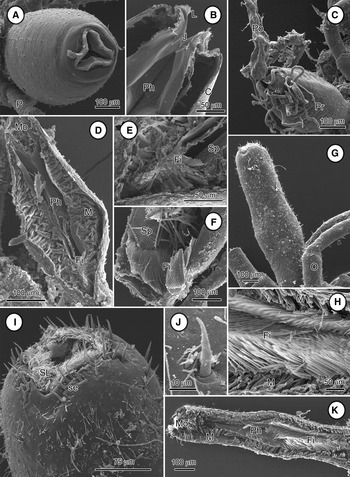
The mouth of Ammothella longipes is positioned at the tip of the fusiform proboscis (Figure 1A–D). The mouth is formed by the typical triradiate symmetry of most pycnogonids and show three external folded lips fringed by setae and sharp trident jaws (see Figures 1A, B & 2A). In contrast, Endeis spinosa has a long and straight proboscis, the apical mouth has three densely setose lips and jaws are not conspicuous (see Figure 1G, I).
Scanning electron microscopy images of proboscis of Ammothella longipes (A–F) and Endeis spinosa (G–K). (A) The lips are continuous and their flutings are seen around the periphery of the mouth. Note the fusiform proboscis of this species; (B) frontal section showing the half distal portion of the proboscis. Note the sharp tips of the jaws (J) and the peripheral lips (L); (C) a specimen of this species with a polychaete (Po) into its mouth, revealing its capacity to predate mobile prey. Some inner structures from the polychaete are visible around the mouth; (D) longitudinal half portion of the proboscis. Pharyngeal filter (Fi) is restricted to the posterior section, and musculature (M) is well developed; (E) detail of the anterior, showing the section where the pharyngeal filter (Fi) starts, with some spines (Sp); (F) pharyngeal filter (Fi) opened showing its few setae and some spines (Sp); (G) lateral view of the long and straight proboscis; (H) pharyngeal filter (Fi) crammed full of dense setae; (I) antero-lateral view of the mouth. Large setae (se), presumably all sensory, are seen surrounding the mouth, and densely setose lips (SL) inside; (J) insertion detail of a long seta surrounding the mouth; (K) longitudinal half portion of the proboscis. The pharyngeal filter (Fi) is fully visible in this plane of section, which occupies half of the proboscis length; cuticle (C), mouth (Mo), oviger (O), pharynx (Ph), pharynx muscle fibres (M), palp (P), proboscis (Pr).
The foregut in both species starts continuously from the mouth and extends through the proboscis. The anterior foregut—also called ‘pharynx’ by some authors (Helfer & Schlottke, Reference Helfer, Schlottke and Bronns1935; Sanchez, Reference Sanchez1959; King, Reference King1973)—has a trifoliate lumen in cross-section followed by a large sac, both layered by a thin cuticle. In A. longipes, this inner cuticle seems to be smooth up to the posterior section of the proboscis where the pharyngeal filter starts—also called ‘oyster basket sieve’ (Schlottke, Reference Schlottke1933)—(Figure 1D–F). In E. spinosa, the filter is much denser and occupies half of the proboscis length (see Figures 1H, K & 2C).
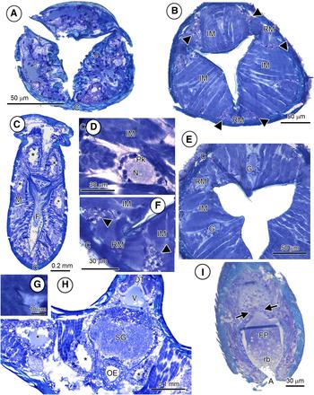
Both species have approximately 100 muscle fibres in the interradial zones and about 50 muscles in the radial zones (see Figure 2B, E). Proboscis musculature is more developed in the fusiform proboscis of A. longipes than in the straight proboscis of E. spinosa (Figure 1D, K). No recognizable salivary glands were found in either A. longipes or E. spinosa, but both species showed secretion tissue on both sides of each lip vertex in the apical region of the proboscis (see Figure 2B, F).
Histological sections of the proboscis of Ammothella longipes (A, B, D–G) and Endeis spinosa (C, H), and hindgut of Endeis spinosa (I). (A) Apical proboscis transverse section showing the sharply pointed trident jaws (J); (B) proboscis transverse section showing the triradiate symmetric secretion tissues (arrowheads) situated on both sides of each lip vertex; (C) proboscis horizontal section. Pharyngeal filter (F) is fully visible in this plane of section, which is extended throughout the pharynx sac. Note the midgut tract (*) extending towards the proboscis in this species; (D) detail of a proboscis nervous ganglion in parasagittal section. Note that ganglia are formed by perikarya (Pk) surrounding a neuropil (N), layered peripherally by the connective lamina; (E) proboscis transverse section. Two of three triradiate symmetric nervous ganglia (G) are visible; (F) detail of a lip vertex showing the secretory tissues (arrowheads) at both sides of the lip ridge; (G) detail of the tripartite valve between foregut and midgut; (H) cross-section at level of oesophagus and the first pair of legs. Ocular tubercle (OT) is shown at the top of the image as well as the dorsal median vessel (V), and the supraoesophageal ganglion (SG) underneath. Note the oesophagus (OE) with different sections of the midgut tract (*) laterally; (I) hindgut parasagittal section, showing the hindgut totally filled with faecal pellet (FP). The valve that separates the midgut from the hindgut is arrowed. Note the residual bodies (rb) that form the faecal pellet gathered near the anal opening (A); cuticle (C), lips (L), interradial muscle fibres (IM), proboscis musculature (M), radial muscle fibres (RM).
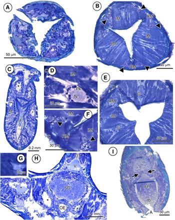
The nervous system observed in the foregut is similar in both species. There were three nervous structures in dorsal and ventrolateral positions which consisted of an amorphous mass surrounded by some cell bodies which in turn were wrapped in a layer of connective lamina (see Figure 2D, E). The short oesophagus, located after the pharyngeal filter, is a thin muscular tube covered by a thin cuticle and presents a single layered epithelium of 10–15 µm thick (see Figure 2H). This structure is resembled within the taxon. No functional digestive cells are observed in the oesophagus. The tripartite valve, which delimits the end of foregut, is of ectodermal origin and consists of an extension of foregut epithelial cells with a slight thickening of the lining cuticle (Figure 2G).
MIDGUT
In pycnogonids, the midgut has diverticula (caeca) into the walking legs. These caeca extend to the end of the tibiae in A. longipes and towards the propodus in E. spinosa. Moreover, in contrast with other pycnogonids, the midgut of E. spinosa extends forward into the proboscis with a pair of caeca (Figure 2C, H). Otherwise, there seem to be few differences in the microscopic structure and ultrastructure of the midgut tracts between A. longipes and E. spinosa. Neither structural nor functional differences have been observed between trunk midgut sections and pedal caeca sections of both species.
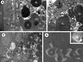
The midgut epithelium is located inwards the following tissues: the haemocoel, a thin cell layer, and some thin muscle fibres (Figure 3A). A thin basal lamina of 0.2 µm thick was observed between each tissue layer. The midgut epithelial cells show marked differences in their appearance depending on the phase in the digestion cycle. During resting periods, epithelial cells (resting cells) have a diameter of about 10–20 µm and contain a basal nucleus, dense basophilic cytoplasm, small rounded mitochondria, well-developed rough endoplasmatic reticulum (rER) and secretion granules of about 2 µm diameter (Figure 3A). During digestion, midgut cells can reach up to 35 µm high and their cytoplasm becomes acidophilic. Large secondary lysosomes which harbour large electron-dense inclusions, glycogen granules, and other cellular organelles such as rER, and electro-dense granules can be observed within epithelial cells (Figure 3A–C). Additionally, some concentric rings of about 1–2 µm diameter, hereafter called residual bodies, appear in the epithelial cells during digestion (Figure 3D). At the end of the digestion process, residual bodies, autophagic vacuoles and some isolated lipid vacuoles are the only organelles observed in the epithelial cells. Digestion ends with the migration and release of these organelles to the midgut lumen. No brush border is seen at the apical distal region of the epithelial cells facing the midgut lumen.
Ultrastructure of the midgut epithelium of Ammothella longipes (A–C) and Endeis spinosa (D). (A) Basal part of midgut epithelium close to integument, showing two midgut epithelial cells at different stages of digestion. The cuticle (C) lies against a border of microvilli (MV) provided by the epithelium, which in turns faces the epidermis (E) showing groups of glycogen granules (g). The haemocoel (H) lies between the epidermis and the thin quasi-endothelium (EN) of the midgut tract, both lined by a basal lamina (arrows). Some thin muscle fibres (MF) externally surround the midgut epithelium. Note two digestive cells that interdigitate, below a digestive cell just before digestion (DC1) with some large zymogen droplets (Z), organized endoplasmatic reticulum (er) and mitochondria (m), and above a digestive cell at the end of the digestive process (DC2) with large secondary lysosomes (SL) and an acidophilic cytoplasm; (B) basal portion of a digestive cell at a late stage of digestion with large secondary lysosomes (SL) and elements of endoplasmatic reticulum (ER). The cells are layered by a basal lamina (arrows), facing to the blood space; (C) basal portion of a digestive cell at the end of digestion surrounded by a basal lamina (arrows), with large secondary lysosomes (SL), glycogen granules (g) and some mitochondria (m); some nuclei (n) externally surround the midgut epithelium; (D) several residual bodies (RB) surrounding a lipid vacuole (L). Inset: detail of the electron-dense inclusions of a concentric residual body (RB). Scale bar of inset: 500 nm.
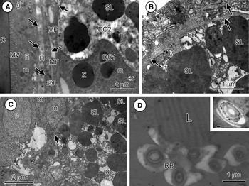
Digestive cells are found one beside the other at different stages of development, so that there are no midgut zones with epithelial cells at the same digestive stage. The membrane of these cells interdigitates in the region of contact between two consecutive cells (Figure 3A).
HINDGUT
The hindgut is located inside the peg-shaped abdomen ending on a terminal anus. It is separated from the midgut by a tripartite valve (Figure 2I), where a thin layer of cuticle starts. Hindgut epidermal cells are surrounded apically by the thin cuticle, which faces the hindgut lumen. A thin basal lamina separates the hindgut epidermal cells from the proctodeal muscle fibres. The terminal anal opening is about 10–15 µm wide when it is opened to excrete. The hindgut does not seem to have absorptive, secretory or excretory functions and only serves for the convection of indigestible residues.
Food circulation along the trunk and legs
No discernible pieces have been found in the gut contents analysed for both species.
Once the food passes through the pharyngeal filter and oesophagus, the following distribution of food along the body is equal in both carnivorous and detritivorous species. Small food particles are retained just before the valve between the foregut and midgut where about each ten of them are grouped into a morula-like food bolus. Then, when the valve opens, some food boli go further into the midgut. The food boli move forward and backwards through trunk and legs midgut aided by the intestinal fluid pressured by the peristaltic movements of the midgut wall (see supplementary material; video). During these movements the food boli may attach to the midgut epithelium, and some of the food vacuoles composing the food bolus may break and remain attached to the epithelium to be digested. Meanwhile, the rest of the food boli continue moving until they re-attach. There is no evidence of a peritrophic membrane wrapping the food bolus. The waste products are discharged by the epithelial cells to the midgut lumen and may form a new bolus or may attach to other boli to move forward and backwards until reaching the hindgut. There, the boli conformed by waste products (i.e. the faecal pellets) move until they reach the valve between the midgut and hindgut. When the hindgut lumen is full, the faecal pellet—a dense matrix, which is completely filled with residual bodies—is seen. Faecal pellets pile up until expelled through the anus. The composition of the food vacuoles forming the morula-like food boli is still unknown, though different size and compacting appearances can be noticed.
Feeding behaviour
Behavioural feeding patterns were studied from the selected specimens for the behaviour observations. Pycnogonids were observed showing a feeding behaviour in 21% of the occasions.
The predation rate was 34% of the feeding events. Only two predations were observed on nematodes and two other specimens were observed firmly retaining caprellids, though it was not possible to discern whether they were conducting predation. The 98% of predations were on polychaetes, and in 10 out of 23 specimens of A. longipes found predating a polychaete, the prey was still alive and shaking vigorously. When polychaetes were used as prey, the feeding behaviour of A. longipes could be categorized into a pattern: A. longipes catches the prey with its legs and moves it closer to the mouth. Then, A. longipes breaks the cuticle and the epidermis of the prey with its jaws and sucks its liquid (Figure 1C). It seems that only liquid is absorbed, as no recognizable parts of the prey can be seen under light microscopes immediately after ingestion. The prey remained into the pycnogonids proboscis between 24 and 48 hours. The 64% of the feeding events were categorized into a pattern of sucking detritus: this species can be seen placing their proboscis on their legs or on algae branches, possibly sucking the detritus deposited on them, as no specimens were found feeding directly on algae. Ammothella longipes could survive without prey for long periods (up to three months).
The feeding rates of E. spinosa were 15% during the behaviour observations. No evidence of predation has been found for this species. In regard to the feeding pattern observed, E. spinosa places the proboscis against the algae branches or the mass of detritus screening and moving it laterally until small pieces can be taken. They are capable of inserting their proboscis into the narrow cracks or between crowded algae branches and suck the small pieces of detritus trapped in there.
DISCUSSION
The general organization of the digestive system in Ammothella longipes and Endeis spinosa is similar; however, the main differences appear in proboscis structure. These differences not only are in accordance with the taxonomic classification of the species within the group but also reflect concordance to their alimentary lifestyle. Despite the differences noticed in the feeding behaviour and the diet of the two species, the midgut structure presents almost no differences, also when compared to that of other ammotheid species (Fahrenbach & Arango, Reference Fahrenbach and Arango2007). Therefore, the feeding structures observed in the proboscis—the sharp trident-shaped jaws and the well-developed musculature in A. longipes, and the densely setose lips and the large pharyngeal filter in E. spinosa—and oral secretions seem critical to achieve the crumbled state of the ingested food. The food pulp reaches the midgut at subcellular size where it is then ready to be digested by the epithelial cells. Our observations highlight the importance of the proboscis' anatomical structure and clarify the structures and processes involved during feeding and digestion in pycnogonids.
Relation between foregut and feeding behaviour
Ammothella longipes is a small-sized pycnogonid of about 6 mm leg span that belongs to the family Ammotheidae. This family is characterized by having functional palps but chelifores with atrophied or absent chelae (except for a few species) in adult forms. Ammothella longipes has a proboscis adequate to its predatory behaviour. The sharp pointed jaws and the setae on the lips of A. longipes correspond with the feeding behaviour observed for this species. Ammothella longipes is considered an omnivorous species which during spring periods seem to be carnivorous, as they were often observed preying on polychaetes (see Figure 1C; Soler-Membrives et al., Reference Soler-Membrives, Rossi and Munilla2011). For this reason, they seem to need aggressive jaws to retain their prey as well as to cut the thin cuticle and epidermis of the prey; then, its internal fluids are exposed and can be sucked. The interradial muscle fibres of the proboscis are responsible for the opening of the pharynx lumen when contracted, while muscle fibres located at radial ridges cause the closing of the foregut lumen when contracted (Dencker, Reference Dencker1974). The pumping of the food through the mouth directly into the filter is caused by the combination of both muscle fibres. Thus, the developed musculature found in A. longipes proboscis is in accordance with their carnivorous diet, needed to retain while sucking their prey fluids. Moreover, this species has a compact, oval-shaped body, with relatively short but tough robust, curved, bizarre legs that help the palps in catching and retaining prey, even if it is alive and shaking vigorously (e.g. nereid polychaetes). The ornamented setose body and legs may facilitate detritus deposition on the body surface, which seems to be another substantial food source during periods in which prey are not easily available (Soler-Membrives et al., Reference Soler-Membrives, Rossi and Munilla2011).
In contrast, Endeis spinosa, a larger-size pycnogonid (about 15 mm leg span) compared to other Mediterranean species is characterized by complete absence of chelifores and palps in adults. Endeis spinosa has very long thin legs with some scattered setae. These well-separated long legs, which are sometimes three times longer than the body, are not capable of retaining moving prey. In contrast to A. longipes, E. spinosa has a proboscis long, straight and movable, adequate to find and select detritus of an appropriate size. This species has weaker jaws with densely setose lips and several setae scattered on the tip surface of the proboscis that probably have a sensorial function; as they do not have palps, the distal setae may detect available food and help to select it. The musculature of E. spinosa within the straight proboscis is less developed compared to A. longipes. The long proboscis allows them to have an extended filter in the pharynx that prevents large debris passing towards the midgut; a similar extended filter is found in the terrestrial spiders that also feed by sucking up fluids (Felgenhauer, Reference Felgenhauer, Harrison and Kohn1999). Generally, the function of the pharyngeal filter is to screen the food before passing it further down. As E. spinosa ingests debris, the large filter with multiple long setae removes any substantial object that could damage its digestive tract. These anatomical features can be expected in detritus-feeders, such as most Endeis species (King, Reference King1973; Fahrenbach & Arango, Reference Fahrenbach and Arango2007).
The morphologically similar species Endeis mollis Carpenter, 1904, a tropical species, is known to be carnivorous, feeding on sessile invertebrates like hydrozoan corals and zoanthid polyps (Arango, Reference Arango2003). Although E. mollis presents carnivorous feeding habits, both endeids have similar proboscis morphology. That may be due to the fact that E. mollis is a predator of soft and sessile prey and they only need to pierce the tissue of prey, insert their proboscis and suck fluids out. On the contrary to some predators of restless prey (as in Ammothella longipes), it seems that endeids do not need a strong proboscis to maintain their prey fastened.
In this study we found six masses of secretory tissue in both species. It could not be determined if these tissues are structured in glands, but according to the location which is the same as that described in Fahrenbach & Arango (Reference Fahrenbach and Arango2007) for salivary glands, we suggest the tissue observed in our species may correspond to salivary secretory tissues. The salivary secretion product in the proboscis of pycnogonids serves for the oral digestion of food, as do salivary glands in the majority of Chelicerata (Harrison & Foelix, Reference Harrison, Foelix, Harrison and Foelix1999; Filimonova, Reference Filimonova2009). These secretions may be more important in those species which ingest not only liquids but small pieces of food, which have to be completely crumbled prior to its intracellular digestion in the midgut as in A. longipes.
The triradiate sucking pharynx and oesophagus leading to the Y-shaped lumen in transverse section is probably the most evident plesiomorphic condition of sea spiders. This condition has been found among ecdysozoans including nematodes, tardigrades, onychophorans and some euarthropods such as Acari and Amblypygi (Schmidt-Rhaesa, Reference Schmidt-Rhaesa2007). At present, it is not clear whether the scattered distribution of the characteristic has evolved convergently by means of some functional requirements, or the shape of the sea spider foregut lumen is a symplesiomorphy (Miyazaki, Reference Miyazaki2002).
Midgut and the digestive process
The general structure of the midgut of A. longipes and E. spinosa corresponds to that described in other pycnogonids, but differs from the structure described in other marine arthropods (e.g. crustaceans; Harrisson & Humes, Reference Harrison, Humes, Harrison and Foelix1992). Pycnogonids body is extremely reduced, the need for increasing the digestive surface has developed to extend the midgut tract into the legs, an autapomorphic feature of this group.
In most chelicerates the midgut consists of anterior and posterior sections, and shows two different types of cells in the intestinal epithelium (secretion cells and absorptive cells) (Harrison & Foelix, Reference Harrison, Foelix, Harrison and Foelix1999). By contrast, in pycnogonids no differentiation of anterior and posterior regions of the midgut are observed, as happens in most Tardigrada (Dewel et al., Reference Dewel, Nelson, Dewel, Harrison and Kohn1993; Schmidt-Rhaesa, Reference Schmidt-Rhaesa2007), supporting the plesiomorphy of this character.
The interpretation of digestion in pycnogonids is controversial, and the outline for digestion has been misinterpreted and re-interpreted again mainly by Schlottke (Reference Schlottke1933), Sanchez (Reference Sanchez1959) King (Reference King1973) and Richards & Fry (Reference Richards and Fry1978). The present work supports the Richards & Fry (Reference Richards and Fry1978) hypothesis stating that only one type of cell exists with both secretion and absorption functions and that it presents a variable morphology depending on the stage of the digestive process, although with some specifications. Many invertebrates have midgut glandular cells producing various protein-rich secretions like glycoproteins and enzymes or their precursors (e.g. Voltzow, Reference Voltzow, Harrison and Kohn1994 for molluscs; Harrison & Foelix, Reference Harrison, Foelix, Harrison and Foelix1999 for chelicerates; Storch et al., Reference Storch, Strus and Brandt2002 for crustaceans). These cells are not observed in the digestive tract of the specimens studied. The electron dense vacuoles observed inside digestive cells at ultrastructural level, and which show high affinity to eosin and toluidine blue under light microscope are interpreted as vacuoles containing hydrolytic enzymes and, consequently, the large vacuoles observed in the digestive cells at the beginning of digestion are interpreted as secretion granules probably containing zymogen. Thus, we suggest that digestive cells are multifunctional, and subsequently are involved in zymogen secretion, absorption, intracellular digestion, and excretion of waste products, each process prevailing at different stages of the cell cycle, excluding the previous hypothesis of two specialized cell types (gland and digestive) (Schlottke, Reference Schlottke1933; Sanchez, Reference Sanchez1959; King, Reference King1973).
Further chemical studies are needed to understand the composition of the food boli observed in the pycnogonids midgut tract (i.e. lipids, proteins or carbohydrates), and whether there is differential attachment to the midgut epithelium depending on their nutrient content or waste product content.
CONCLUSIONS
The two Mediterranean pycnogonids studied here present differences in the foregut structure. The shape and musculature of proboscis, lips and jaws of the mouth and the pharyngeal filter, seem to be the main structures relating to the type of diet (a predator species against a detritus-feeder) while the midgut and hindgut structurally show a similar form in the different lineages. Our study provides new evidence on the relation between morphological characteristics of the foregut and the feeding behaviour of unrelated species of pycnogonids, adding relevant data for comparative studies in the understudied fields of anatomy and feeding ecology of Pycnogonida.
ACKNOWLEDGEMENTS
We are grateful to Sílvia Crespo and all the members of the Animal Biology (Veterinary Faculty), Universitat Autònoma de Barcelona, for their help in the microscopic and ultrastructure analyses and to the staff of the Electron Microscopy Services of the Universitat Autònoma de Barcelona. We also thank Emilio Valbuena for his help in collecting and sorting and John Healy for his comments which helped improve the manuscript. This work was funded by the CGL 2007-66876 project: Fauna Ibérica, Chelicerata, Pycnogonida (Ministerio de Ciencia e Innovación).
Supplementary materials and methods
The supplementary material referred to in this article can be found online at journals.cambridge.org/mbi.