Introduction
Parasite infections decrease food intake (Sykes et al., Reference Sykes, Poppi and Elliot1988), and increases the loss of nitrogen (N) from the body either as endogenous protein in the gastrointestinal tract (e.g. mucus, plasma leakage or sloughed cells; Kimambo et al., Reference Kimambo, MacRae, Walker, Watt and Coop1988) and/or as N excretion in urine and faeces (Sykes and Coop, Reference Sykes and Coop1976). These effects may, in part, explain the decreased growth performance (Sykes and Coop, Reference Sykes and Coop1976; van Houtert et al., Reference van Houtert, Barger, Steel, Windon and Emery1995) and production (Steel et al., Reference Steel, Symons and Jones1980 and Reference Steel, Jones and Symons1982) that is observed during parasite infections in lambs.
Intestinal parasites, including Trichostrongylus colubriformis, cause atrophy of the brush-border membrane (Coop and Angus, Reference Coop and Angus1975), and this may result in a decreased ability to absorb amino acids (AA) and other nutrients. This is likely to be the cause of the increased fractional protein synthesis in the small intestine (Symons and Jones, Reference Symons and Jones1975; Bermingham et al., Reference Bermingham, McNabb, Sutherland, Waghorn, Sinclair, Treloar and Roy2006) and leucine oxidation and protein turnover in the gastrointestinal tract (Yu et al., Reference Yu, Bruce, Calder, Milne, Coop, Jackson, Horgan and MacRae2000) that have been observed during parasitic infection.
The gastrointestinal tract preferentially uses AA from arterial supply (MacRae et al., Reference MacRae, Bruce, Brown and Calder1997a), and it is therefore possible that a reduction in the absorption of AA may increase the requirement for arterial AA in the gastrointestinal tract. The already high metabolic requirement of the gastrointestinal tract and liver is, therefore, likely to increase further with infection, resulting in a shift in nutrient partitioning by tissues within the body to meet the demands of the affected tissues, with priority placed on the maintenance of body protein (including repair of damaged tissues; Coop and Kyriazakis, Reference Coop and Kyriazakis1999). However, as parasite infections are often accompanied by impaired feed intake (Sykes et al., Reference Sykes, Poppi and Elliot1988), it is unknown whether nutrient partitioning occurs due to the physical presence of parasites or their effect on intake, or both. While changes in leucine absorption and metabolism in the small intestine during parasite infection have been observed previously (Yu et al., Reference Yu, Bruce, Calder, Milne, Coop, Jackson, Horgan and MacRae2000), there has been no report on the direct impact of parasitic infection on the intestinal absorption of other AA. Due to the preferential use of AA such as glutamate (Reeds et al., Reference Reeds, Burrin, Stoll and Jahoor2000) and the unique AA composition of the gastrointestinal tract (MacRae and Lobley, Reference MacRae and Lobley1991), there is reason to believe that AA other than leucine may behave differently in the small intestine of parasitised animal.
The hypothesis of this study was that an established parasitic infection in growing lambs with similar intakes would decrease the intestinal absorption of AA and reduce their availability post absorption, creating a need for additional AA (presumably from body reserves) to maintain the supply via arterial AA. The objective of this study was to quantify nutrient digestibility and intestinal AA absorption in lambs with similar intakes with or without an established parasitic infection.
Material and methods
Animals and surgery
The experimental procedures for this trial were reviewed and approved by the Crown Research Institute Animal Ethics Committee in Palmerston North, New Zealand, according to the Animals Protection Act (1960) and Animals Protection Regulations (1987) and amendments.
Wether lambs (29 kg) were removed from pasture at weaning, and transported to AgResearch Limited, Grasslands Research Centre, Palmerston North. The lambs were shorn, drenched twice with Ivermectin (Ivomec; Merial New Zealand Limited, Manukau City, Auckland, New Zealand) and treated for external parasites using Wipeout (Coopers, Schering-Plough Animal Health Limited, New Zealand). The lambs were maintained on feeding pads with Lucerne pellets (800 g dry matter (DM)/day) and chaffed Lucerne hay (200 g DM/day) for approximately 3 weeks to minimise exposure to parasites. During this period, random faecal egg samples were undertaken to ensure the parasite-free state of the lambs, with all counts negative for the presence of parasites. One week before surgery, the lambs were brought indoors, housed in individual metabolism crates and maintained on the same diet.
Twelve lambs were fasted for 24 h before surgery. Permanent indwelling catheters were placed in the mesenteric artery, and the mesenteric, portal and hepatic veins (Huntington et al., Reference Huntington, Reynolds and Stroud1989) and vena cava (Ortigues and Durand, Reference Ortigues and Durand1995) for blood sampling, as described in Bermingham et al. (Reference Bermingham, McNabb, Sutherland, Sinclair, Treloar and Roy2007b). As part of a larger study (Bermingham, 2004; Bermingham et al., Reference Bermingham, McNabb, Sutherland, Sinclair, Treloar and Roy2007a), an additional permanent catheter was placed in the mesenteric vein, and a temporary catheter was inserted into the jugular vein on day 43. The catheters were maintained as described by Huntington et al. (Reference Huntington, Reynolds and Stroud1989). A permanent Teflon cannula was fitted in the abomasum at the time of surgery.
The lambs were fed Lucerne pellets (800 g DM/day) and chaff (200 g DM/day) for 3 days after surgery and were then offered approximately 800 g DM/day of fresh Lucerne (Medicago sativa) until the conclusion of the trial. The rationale for using fresh Lucerne, rather than the typical rye-grass/white clover dominant NZ pasture, has been described in full in Bermingham et al. (Reference Bermingham, McNabb, Sutherland, Sinclair, Treloar and Roy2007a). However, briefly, it consistently maintains a high nutritive value for sheep and is suitable for cut and carry experiments as described here. The Lucerne was harvested every 2 days with a sickle bar mower by 1000 h and stored at 4°C. The DM content of the Lucerne was determined daily to adjust the amount of wet forage given to maintain the DM offered at approximately 800 g/day. The level of 800 g DM/day was chosen to minimise the effects of parasite infection on intake (Bermingham et al., Reference Bermingham, McNabb, Sutherland, Waghorn, Sinclair, Treloar and Roy2006). The lambs were fed hourly from overhead feeders and water was available ad libitum. The lambs were weighed weekly to monitor liveweight changes during the experimental period.
Treatment and parasitology
The lambs were assigned to either the control or parasite group according to a completely randomised block design. One week after surgery (day 1 of the experimental period), six sheep were given 6000 T. colubriformis L3 larvae per day orally for 6 days (parasite treatment) while the remaining six sheep were drenched with ivermectin once (Ivomec; Merial New Zealand Limited) to serve as controls. The infection protocol used was essentially short-term dose, with the larval challenge spread across 6 days to minimise rumen by-pass and maximise larvae establishment in the small intestine as described in full in Bermingham et al. (Reference Bermingham, McNabb, Sutherland, Waghorn, Sinclair, Treloar and Roy2006). Due to the time consuming nature of the surgical procedure, a maximum of four lambs were surgically prepared with catheters in any one week (one lamb per day), with two lambs allocated to the parasite group, and two to the controls. The lambs were blocked according to the week that they underwent surgery (referred to as the group effect in the statistical model).
Faecal egg counts from individual sheep were determined every second day from day 20 to day 45 of infection using the modified McMaster method in which one egg counted equates to 50 eggs/g wet faeces (Whitlock Reference Whitlock1948). Total intestinal worm burdens in the proximal 10 m of the small intestine were determined after slaughter on day 48 (Sutherland et al., Reference Sutherland, Leathwick, Green, Brown and Miller1999b).
Digesta flow and nitrogen balance
On day 32 the sheep were dosed (orally into the rumen) with a pre-weighed, slow-release alkane capsule (Captec Alkane CRC; Captec New Zealand Limited, Auckland, New Zealand). Digesta flow was estimated as described previously (Bermingham et al., Reference Bermingham, Hutchinson, Revell, Brookes and McNabb2001) using natural plant and synthetic alkanes. On day 44, abomasal digesta (approximately 100 g) was sampled four times during a 24-h period and bulked. The bulked digesta was freeze-dried and ground (0.5 mm sieve) and analysed for DM, N, alkane and AA concentrations. The digestibility of DM and N, and N retention were measured from day 35 to day 43, using the techniques described by Bermingham et al. (Reference Bermingham, Hutchinson, Revell, Brookes and McNabb2001).
Infusions and blood sampling
To measure plasma flow across the mesenteric drained viscera (MDV), para aminohippuric acid (PAH) (749.7 mg/h; 0.15 mmol/l; Na form, dissolved in water and autoclaved) was infused continuously into the mesenteric vein for 8 h on day 48. To prevent blood clotting during the continuous sampling, 2000 IU ovine heparin/h was infused into the jugular vein of the lambs during the 8 h infusion period (Lobley et al., Reference Lobley, Connell, Lomax, Brown, Milne, Calder and Farningham1995). Sampling lines and syringes were kept in a 0°C water bath to minimise the degradation of blood constituents (Lobley et al., Reference Lobley, Connell, Lomax, Brown, Milne, Calder and Farningham1995). Blood was withdrawn continuously every 2 h from the mesenteric artery (10 ml for time 0 to 2 h and time 2 to 4 h; 30 ml for time 4 to 6 h and time 6 to 8 h for net metabolite flux), the mesenteric, portal and hepatic veins and the vena cava (30 ml for time 4 to 6 h and time 6 to 8 h for net metabolite flux only) over the infusion period. The plasma data presented in this paper are the average from samples taken from the last two sampling periods from the mesenteric artery and the mesenteric vein (4 to 6 and 6 to 8 h).
After each 2 h collection period, the syringes were removed from the collection lines, carefully mixed by gentle rotation, and plasma harvested as described by Bermingham et al. (Reference Bermingham, McNabb, Sutherland, Sinclair, Treloar and Roy2007a).
After the 8 h PAH infusion period, the sheep were killed with an intravenous overdose of sodium pentobarbitone (300 mg/ml; 0.5 ml/kg liveweight). The duodenum (the proximal 3 m of the small intestine) was sampled for digesta, but for unknown reasons there was no duodenal contents in any of the sheep at the time of slaughter. Ileal digesta was sampled from 3 m before the ileo-caecal junction and stored at −20°C until analysed. The alkane capsule was recovered from the rumen of each sheep, dried at room temperature and weighed to determine the release rate of synthetic C36 alkanes in the rumen from day 32 to day 48.
Sample processing and chemical analysis
DM, alkanes, N and AA were determined in feed and refusals, faeces and digesta samples as described elsewhere (Mayes et al., Reference Mayes, Lamb and Colgrove1986; Shenk and Westerhaus, Reference Shenk and Westerhaus1991; Bermingham et al., Reference Bermingham, Hutchinson, Revell, Brookes and McNabb2001 and Reference Bermingham, McNabb, Sutherland, Waghorn, Sinclair, Treloar and Roy2006).
To determine the concentration of AA in plasma, 0.5 ml was treated with 80 mmol/l dithiothreitol and 3 mmol/l norleucine (as an internal standard, in 0.1% phenol) and stored at −85°C, as described in Bermingham et al. (Reference Bermingham, McNabb, Sutherland, Waghorn, Sinclair, Treloar and Roy2006). PAH was measured by the method of Katz and Bergman (Reference Katz and Bergman1969), with an additional deacylation step as described by Lobley et al. (Reference Lobley, Connell, Lomax, Brown, Milne, Calder and Farningham1995). Samples and standards were read at 540 nm using a spectrophotometer (GBC UV/VIS 918, GBC Scientific Equipment Pty Limited, Victoria, Australia).
Calculations
DM flow was calculated by the dilution of natural plant alkanes and synthetic alkanes in the abomasum and ileum. Apparent absorption of AA by the small intestine was calculated as the difference between abomasal and ileal fluxes. As no digesta was present in the duodenum at the time of sampling, abomasal flux was used in the current study to determine apparent AA absorption from the small intestine instead of the duodenal flux. The DM flow through the abomasum was calculated using Eq. (1). Ileal digesta DM flow was calculated using Eq. (2) and uses a correction for the recovery of C36 from the alkane capsule. C36 is an indigestible marker and the assumption is that the C36 flux at the ileum should be the same as the C36 flux at the abomasum. The invasive nature of the surgery that the lambs in this experiment underwent prevented the insertion of a rumen cannula so the infusion of a liquid phase marker (e.g. CoEDTA, CrEDTA) was not possible. Items in square brackets indicate concentrations (e.g. [alkane]).


where

The AA flows at the abomasum and ileum were calculated by multiplying the AA concentration in the digesta by the corresponding flow of DM at the abomasum or ileum (Waghorn et al., Reference Waghorn, Ulyatt, John and Fisher1987). Apparent absorption of AA in the small intestine was calculated using Eq. (3). Digestibility of AA in the small intestine was calculated by dividing the apparent absorption of AA in the small intestine by the AA flux at the abomasum.

Plasma flow (ml/min) across the MDV was calculated using Eq. (4), where subscripts I, M or A refer to the concentration of PAH in the infusate, mesenteric vein or mesenteric artery, respectively. Arterial supply was determined by multiplying the concentration of AA in the mesenteric artery by plasma flow across the MDV, and Eq. (5) was used to calculate the net absorption of AA by the small intestine.


Statistical analysis
A General Linear Model (SAS v. 8, 1999) was used for all statistical analyses, with treatment and group (the week that the lamb underwent surgery) used as sources of variation in the model, giving 9 residual degrees of freedom. In all cases the group effect was not significant in the current study. Feed intake, faecal egg counts and liveweight were analysed using Proc Mix repeated measures. Liveweight was analysed using initial liveweight as a covariate. The data were checked for normality and the presence of outliers by plotting the residuals against the predicted residuals. Faecal egg counts and intestinal worm numbers were transformed using ln(x + 1) before analysis in order to ensure symmetry in the data and to standardise variances across the treatments (Sutherland et al., Reference Sutherland, Brown, Green, Miller and Leathwick1999a), with results reported in the text as the back-transformed values.
Probability values between 0.05 and 0.10 were considered to indicate a trend. Results are presented as least squares means and associated pooled standard deviation (s.d.). To assess treatment group variability for all parameters, the coefficient of variation (CV) was calculated for each treatment group.
One sheep from the parasite treatment was omitted from all statistical analysis as the Captec Alkane capsule blocked the reticulo-rumen orifice and affected feed intake for the two days before blood sampling.
Results
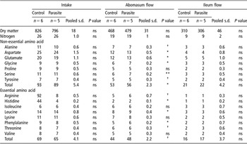
Feed intake was similar during the course of the experiment (826 parasite v. 796 control ± 18.2 g DM/day; Table 1). The parasite infected lambs were lighter (P < 0.01) than the control lambs before infection (34 parasite v. 36 control ± 1.4 kg). Therefore, liveweight gain during the course of the experiment was adjusted for initial liveweight. There were, however, no differences in liveweight gain due to the presence of an established parasite infection (19 parasite v. 24 control ± 10.4 g/day; Bermingham et al., Reference Bermingham, McNabb, Sutherland, Sinclair, Treloar and Roy2007a).
Dry matter, nitrogen and amino acid intake and flows (g/day) through the abomasum and ileum in lambs fed fresh Lucerne (Medicago sativa) and infected with Trichostrongylus colubriformis or kept as parasite free controls

ns = not significantly different.
†P < 0.10.
Faecal egg production peaked at 1800 eggs/g wet faeces (back-transformed values) on day 26 after infection, before displaying a consistent decline to 600 eggs/g wet faeces by day 48 (Bermingham et al., Reference Bermingham, McNabb, Sutherland, Sinclair, Treloar and Roy2007a). Back-transformed worm burdens in the small intestine at the end of the experiment were 22 000 in the parasitised lambs compared to 440 in the control lambs (P < 0.001; s.d. = 1100 adult worms).
The presence of parasites in the small intestine did not affect DM or N fluxes (Table 1). Similarly, the digestibility of both DM (65 parasite v. 65 control ± 1.2%.; P > 0.05) and N (72 parasite v. 74 control ± 2.0%; P > 0.05) was also unaffected by parasitic infection. Parasitic infection had no effect on the excretion of N in urine (18 parasite v. 16 control ± 3.0 g N/day) and faeces (7 parasite v. 6 control ± 0.3 g N/day) and consequently N retention was also unaffected (1 parasite v. 4 control ± 2.9 g N/day).
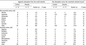
Concentrations of all measured AA in abomasal and ileal digesta were unaffected by treatments (data not shown) and AA intakes were similar between the control and parasite-infected lambs (Table 1). AA flux through the abomasum of infected lambs was increased (P < 0.05) by parasitic infection (Table 1) except for proline, isoleucine, lysine and valine, which were unaffected. Despite these higher fluxes, the fluxes of AA at the ileum were similar (Table 2). The apparent absorption of AA in the small intestine was unaffected by the presence of parasites (Table 2) as was the apparent digestibility of AA in the small intestine (63 parasite v. 62 control ± 8.3%).
Apparent amino acid absorption from the small intestine and net amino acid absorption across the mesenteric drained viscera (g/day) in lambs fed fresh Lucerne (Medicago sativa) and infected with Trichostrongylus colubriformis or kept as parasite free controls

1 Negative values represent a net release by the mesenteric drained viscera and positive values represent a net uptake.
ns = not significantly different.
†P < 0.10.
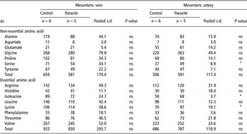
Plasma flow across the MDV was unaffected by the presence of parasites (772 parasite v. 545 control ± 222.1 ml/min) as was the concentration of AA in the mesenteric vein (Table 3). In the mesenteric artery, there was an increase in isoleucine (P < 0.05) in parasitised animals (Table 3). Serine and lysine tended to increase (P < 0.10) during parasite infection (Table 3). With the exception of glutamic acid (which was taken up by the small intestine for both parasite and control animals) there was a net release of AA by the small intestine. The arterial supply of total (1191 parasite v. 804 control ± 391.2 μmol/min), non-essential (694 parasite v. 467 control ± 207.9 μmol/min) and essential AA (692 parasite v. 462 control ± 239.2 μmol/min) to the MDV was unaffected by the presence of parasites. Similarly, the net flux of AA across the MDV was also unaffected by the presence of an established parasite infection (Table 2).
Plasma amino acid concentration (μmol/l) in the mesenteric vein and mesenteric artery in lambs fed fresh Lucerne (Medicago sativa) and infected with Trichostrongylus colubriformis or kept as parasite free controls

ns = not significantly different.
†P < 0.10.
Discussion
This study reports for the first time the apparent and net intestinal absorption of AA in lambs infected with parasites. The presence of an established T. colubriformis infection in growing lambs fed fresh Lucerne did not affect the apparent or net absorption of AA from the small intestine, nor did the arterial supply of AA to the MDV, suggesting that in this study, established adult parasites do not affect AA absorption across the small intestine.
Although the fluxes of DM and N through the small intestine were not affected by the presence of an established parasitic infection, the flow of AA through the abomasum increased in the current study. The DM AA flow values presented here are comparable to those obtained using dual markers, with abomasal flow approximately 60% of intake and duodenal flow approximately 40% of intake (Waghorn et al., Reference Waghorn, Ulyatt, John and Fisher1987 and Reference Waghorn, Shelton, McNabb and McCutcheon1994; Bermingham et al., Reference Bermingham, Hutchinson, Revell, Brookes and McNabb2001). The cause of the DM AA flow increase is unclear, as feed intake was similar between treatments. T. colubriformis inhabits the proximal third of the small intestine. Previous authors have noted that parasitic infections in the small intestine alter the morphology and digestive capabilities of sections of the digestive tract, both proximal (Barker and Titchen, Reference Barker and Titchen1982) and distal (Scofield, Reference Scofield1980; Cheema and Scofield, Reference Cheema and Scofield1982) to where the parasites live. This may explain the increase in AA flow at the abomasum observed in the current study or it is possible that this result is an artefact due to the difficulties in measuring digesta flow in vivo.
Despite increased AA flux in the abomasum, the ileal flux of AA was similar in control and parasitised lambs in the current study. This suggests an increase in AA disappearance from the small intestine of the infected lambs, although the apparent absorption from the small intestine and net flux of AA across the MDV were similar between treatments. The net flux of AA across the MDV in the current study (1 to 5 g/day) is similar to the 4 to 6 g/day reported by MacRae et al. (Reference MacRae, Bruce, Brown, Farningham and Franklin1997b) at similar intakes, but the similar net fluxes of AA across the MDV between control and parasitised lambs contrast with the observations for leucine by Yu et al. (Reference Yu, Bruce, Calder, Milne, Coop, Jackson, Horgan and MacRae2000). While mesenteric plasma flows are within the ranges in the literature for sheep at a similar age and DM intake (700 to 1000 ml/min; Janes et al., Reference Janes, Weekes and Armstrong1985; MacRae et al., Reference MacRae, Bruce, Brown, Farningham and Franklin1997b; Yu et al., Reference Yu, Bruce, Calder, Milne, Coop, Jackson, Horgan and MacRae2000; Bermingham et al., Reference Bermingham, McNabb, Sutherland, Sinclair, Treloar and Roy2007b), they are highly variable (CV of 28% to 40% in the control and parasitised lambs, respectively), and this variability may have masked any effects of infection on MDV AA fluxes. High variability (>25%) in blood flow across the MDV has been observed previously (MacRae et al., Reference MacRae, Bruce, Brown, Farningham and Franklin1997b; Yu et al., Reference Yu, Bruce, Calder, Milne, Coop, Jackson, Horgan and MacRae2000), and might be attributed to either incomplete mixing of the dye in those blood vessels or movement of the catheter due to peristaltic intestinal contractions (MacRae et al., Reference MacRae, Bruce, Brown, Farningham and Franklin1997b).
Possible reasons for the discrepancies between the current study and previous studies with respect to DM flow at the ileum (Gregory et al., Reference Gregory, Wenham, Poppi, Coop, MacRae and Miller1985), N flow (Poppi et al., Reference Poppi, MacRae, Brewer and Coop1986; Kimambo et al., Reference Kimambo, MacRae, Walker, Watt and Coop1988) and intestinal absorption (Yu et al., Reference Yu, Bruce, Calder, Milne, Coop, Jackson, Horgan and MacRae2000) may be due to the infection procedure used, namely the short-term dose in the current study and the trickle dose methods used by these authors. It is likely that the damage caused by the larvae could affect the ability of the intestine to transport AA. In contrast, the short-term dose method used here would initially increase the immune response (as the infective larvae inhabit the small intestine), and this response would decrease as the parasite’s reproductive phase begins. The choice of a short-term dose method of infection has been discussed in greater detail in Bermingham et al. (Reference Bermingham, McNabb, Sutherland, Waghorn, Sinclair, Treloar and Roy2006); however, it has the advantage of measuring the effects of an established adult population of parasites on the absorption of AA from the small intestine without the confounding effects of new larval challenge that multiple dosing brings.
Why the adult parasite population did not affect growth, or the apparent intestinal AA absorption and net AA flux, is puzzling. In the current study, the lambs were dosed with approximately 36 000 L3 T. colubriformis larvae over 6 consecutive days, with worm burdens of around 22 000 at slaughter. Faecal egg count rapidly declined from the peak of 1800 eggs/g wet faeces on day 26 after infection to only 600 eggs/g wet faeces at slaughter (Bermingham et al., Reference Bermingham, McNabb, Sutherland, Sinclair, Treloar and Roy2007a). This rapid decline is consistent with the results observed by Roseby (Reference Roseby1973) and Barnes and Dobson (Reference Barnes and Dobson1990) using a similar infection protocol and is presumably due to the onset of immunity in the lamb. Although a few T. colubriformis were recovered from the small intestine of the control lambs, any effects would have been negligible given their very low numbers. Given that continuous larval challenge decreased intake and liveweight gain in previous studies, (Sykes and Coop, Reference Sykes and Coop1976; Sykes et al., Reference Sykes, Poppi and Elliot1988; van Houtert et al., Reference van Houtert, Barger, Steel, Windon and Emery1995), the results of the current study suggest that it may be the continuous larval challenge itself which has these effects, and not the presence of adult worms. The negative impacts of continuous larval challenge may be due to the damage caused to the intestinal wall as the parasite burrows into the epithelial layer (Familton and McAnulty, Reference Familton and McAnulty1997).
Measurements of AA flux across the small intestine show large animal to animal variation in parasitised lambs (CV 69% for TAA), as do measurements of blood flow (CV 37%). These, together with highly variable individual responses to parasitic challenge (Barger, Reference Barger1985), suggest that measurement of changes in net fluxes of AA may be more appropriate at earlier periods, perhaps around peak egg production (up to day 26 post infection in this study; Bermingham et al., Reference Bermingham, McNabb, Sutherland, Sinclair, Treloar and Roy2007b), where larger differences for these parameters are likely to be found between parasitised and control lambs.
Differences in diets may also explain some of the discrepancies in the intestinal and MDV fluxes of DM and AA between the current study and those observed previously for the control lambs. For example, there was a net release of total AA by the MDV in the control lambs (about 32 g/day), but this was lower than the apparent absorption from the small intestine (about 60 g/day), suggesting that there was a net use of dietary AA by the small intestine of around 50%. Previous estimates (MacRae et al., Reference MacRae, Bruce, Brown and Calder1997a; Yu et al., Reference Yu, Bruce, Calder, Milne, Coop, Jackson, Horgan and MacRae2000) have suggested that around 20% of dietary AA is used by the small intestine. This difference may be due to feeding fresh (current study) rather than conserved forages (Poppi et al., Reference Poppi, MacRae, Brewer and Coop1986; MacRae et al., Reference MacRae, Bruce, Brown and Calder1997a; Yu et al., Reference Yu, Bruce, Calder, Milne, Coop, Jackson, Horgan and MacRae2000), as fresh forages tend to have a higher post-absorption AA availability than conserved forages (Ulyatt, Reference Ulyatt1981).
From the current experiment, it seems apparent that measuring digesta flow and mesenteric blood flow during the presence of an established parasite infection may not be appropriate, due to the potential effects that parasite infections will have directly on these parameters. Additionally, measuring the effects of parasite infections 48 days post infection may also not be appropriate as the faecal egg production is no longer at its peak, presumably reflecting a low immunological challenge to the lambs. There may have been more of an effect of an established parasite infection on day 26 post infection, which coincides with maximal faecal egg production (Bermingham et al., Reference Bermingham, McNabb, Sutherland, Sinclair, Treloar and Roy2007a).
In conclusion, these data suggest that the presence of an established adult parasite population in the small intestine does not affect the digestibility and absorption of nutrients (such as N and AA) in the small intestine and their availability to tissues in lambs fed fresh Lucerne. This result is reinforced by the similar N retention between the control and parasitised lambs. An established T. colubriformis population in the small intestine of lambs appears to impose little nutritional cost on the host 48 days after infection. It is likely that any parasite effects occurred during the earlier stages of infection and the data highlight a need to identify which stage(s) of intestinal parasitism need to be targeted in metabolic studies that attempt to elucidate the effects of parasite infection on nutrient partitioning.
Acknowledgements
This study was funded by the C. Alma Baker Trust, New Zealand Foundation for Research, Science and Technology and Meat Wool New Zealand. Emma Bermingham thanks Meat Wool New Zealand for funding her PhD studies. Many thanks to Gordon Reynolds, Brett Guthrie (Massey University, Palmerston North, New Zealand) for assistance with the surgical procedure and Matthew Deighton, and Bryan Treloar (AgResearch Limited) for analytical support.