Woven coronary artery disease is an extremely rare abnormality with unknown aetiology. In this entity there are branching thin channels and distal reanastomosis. It was reported in a few adult patients.Reference Gregorini, Perondi, Pomidossi, Saino, Bossi and Zanchetti1–Reference Kursaklioglu, Iyisoy and Celik3 There is completely normal blood flow after the distal segment of the abnormal coronary artery.Reference Kursaklioglu, Iyisoy and Celik3 Kawasaki disease is an acute, self-limiting, systemic vasculitis of unknown aetiology. However, clinical and epidemiological features strongly suggest infections. The inflammatory process preferentially involves the coronary arteries, potentially resulting in coronary arteritis, aneurismal or ectasial lesions, arterial thrombotic occlusion and it may lead to myocardial infarction, sudden death, or ischaemic cardiac disease.Reference Newburger, Takahashi, Gerber, Gewitz, Tani and Burns4 This is the first report of woven coronary artery in an infant associated with Kawasaki disease. We report a 9-month-infant with Kawasaki disease showing woven right and left coronary artery aneurysm.
A case report
A 9-month-old infant presented to a paediatric clinic with the complaints of high fever more than 5 days, redness in eyes and lips as well as swollen masses in the neck. The physical examination revealed bilateral non-exudative conjunctivitis, erythema of the lips and oral mucosa, rash, and cervical lymphadenopathy. In his family history, there was no sudden death or cardiac bypass operation.
Elevation of acute phase reactants, such as erythrocyte sedimentation rate and C-reactive protein, white blood cell count was assessed and the results showed 12,000 per cubic millimetres, platelet counts of 670,000 per cubic millimetres, erythrocyte sedimentation rate of 85 millimetres per hour and C-reactive protein of 58 international unit.
The echocardiographic assessment revealed left coronary artery dilatation and internal vessel diametres were measured as right coronary artery of 0.23 centimetre and left coronary artery of 0.45 centimetre. In electrocardiography, prolonged PR wave interval was marked, whereas ST and T wave changes were not observed.
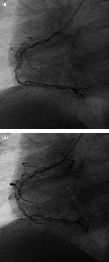
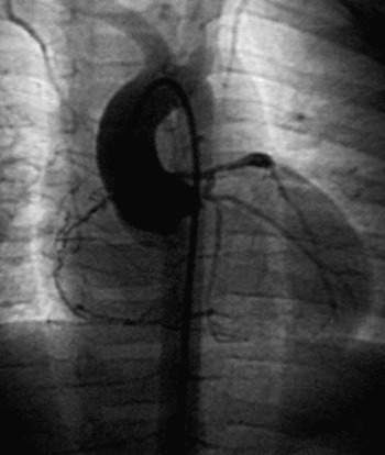
Intravenous infusion of two gram per kilogram immunoglobulin was started on the first day of the hospital administration. About 75 milligrams per kilogram of aspirin was added in four doses a day and was reduced to three milligram per kilogram after 2 weeks which was planned to continue until the coronary artery aneurysm regressed. In serial echocardiographic assessments, no regional wall motion abnormalities indicating myocardial ischaemia were seen. Patient was clinically asymptomatic for cardiac problems but coronary angiography was planned to assess the coronary arteries 1 year later and a woven form in right coronary artery and an aneurysm of left anterior descending coronary artery with an internal diameter of 0.44 centimetre was showed. This diameter is considered as an aneurysm based on American Heart Association’s classifications.Reference Newburger, Takahashi, Gerber, Gewitz, Tani and Burns4 Right coronary artery was subdivided into thin channels. These thin channels showed intertwining formation along their course and woven channels fused again soon after (Figs 1a, 1b and 2). Serial echocardiographic monitoring showed no regional wall motion abnormalities indicating myocardial ischaemia. After 1 year, a coronary angiogram was repeated and no changes were recorded. In the follow-up period for 4 years the patient was free of symptoms as well as cardiac events with aspirin.
Selective right coronary angiogram. Proximal thin channels in a woven coronary artery continue to become a normal conduit.

Aortic root angiography demonstrating woven right coronary artery and aneurysm of the left anterior descending artery.

Discussion
Woven coronary artery is an extremely rare coronary malformation and it was first described by Sane et alReference Sane and Vidaillet5 in 1988. There are only a few case reports in adult patients and none in children.Reference Kursaklioglu, Iyisoy and Celik3–Reference Sane and Vidaillet5 Woven structures were showed in right and left coronary arteries. Woven structure may mimic intracoronary thrombus. This condition may be caused by recanalisation of a thrombus. The coronary artery dissections may occur spontaneously or may present related to Kawasaki arteritis. Twisting of thin channels can cause intracoronary thrombus according to the distance of the anomalous segment. Up to now, this entity have not clearly defined and aetiolgy of woven coronary artery was not fully elluciated.Reference Gregorini, Perondi, Pomidossi, Saino, Bossi and Zanchetti1, Reference Martuscelli, Romeo, Giovannini and Nigri2, Reference Sane and Vidaillet5 We need more information about the characteristics and development of such a malformation.
Usually a woven coronary artery is accepted as a benign condition. In the cases reported by Martuscelli et alReference Martuscelli, Romeo, Giovannini and Nigri2 and Kusaklıoglu et alReference Kursaklioglu, Iyisoy and Celik3 no adverse coronary events occurred during the 4–5 years follow-up period. In our patient, who was diagnosed as Kawasaki disease, an evaluation of coronary arteries revealed aneurysm of left coronary artery in the echocardiograpic study and this required an angiographic study. A woven right coronary artery was detected coincidentally. The patient was followed-up for 4 years without any occurrence of cardiac event.
A woven coronary artery abnormality was not reported in a child or infant previously. We would like to present this case to discuss a woven coronary artery can be confused with other conditions such as a thrombus, dissection or a stenosis that can threaten life in a child with Kawasaki disease. Angiographic images of our patient presents suspicion for dissection. However, as the patient’s general condition was stable and the clinical and laboratory findings are not relevant with the findings of dissection, a diagnosis of dissection was not considered. A thrombus or a stenosis may require further medical or surgical intervention. In woven coronary arteries, filling is from proximal to distal without any problem and this is an important differential finding from other conditions such as thrombus or stenosis. Especially in Kawasaki disease there is a tendency for the development of thrombus and this condition should not be confused with a woven coronary artery. Therefore, the paediatric cardiologist should be aware that a woven coronary artery can be observed in Kawasaki disease. Advanced studies are needed on this unknown entity to show the pathological causes of this rare event.