Introduction
Fotemustine is increasingly being used in the treatment of disseminated malignant melanomas and primary brain tumours, with favourable oncological results.Reference Fischel, Barbe, Berlion, Formento, Berrile and Bizzari1, Reference Lokiec, Beerblock, Deloffre, Lucas and Bizzari2 It has the ability to cross the blood–brain barrier and achieve therapeutic levels in the central nervous system (CNS). However, it may also cause toxicity in the CNS. There is no published research on the effects of fotemustine on the inner ear.
Some chemotherapeutic agents can cause hearing loss via inner-ear toxicity. Proposed mechanisms of toxicity have been attributed to alteration in the arrangement of outer hair cell stereocilia.Reference Hatzopoulos, Di Stefano, Campbell, Falgione, Ricci and Rosignoli3 During the course of drug administration, threshold shifts and significant depression of transient evoked otoacoustic emission and distortion product otoacoustic emission amplitudes can appear.Reference Sockalingam, Freeman, Cherny and Sohmer4 In experimental studies using systemic chemotherapy, inner-ear damage has been reported.Reference Hatzopoulos, Petruccelli, Laurell, Avan, Finesso and Martini5 Currently, no chemotherapy strategies aiming to protect the cochlea have been adopted. Although a number of agents with the potential for protection from neuropathy have been developed, none has yet been used widely.Reference McMahon and Priestley6
The goal of this study was to elucidate the effects of fotemustine on the cochlea in a Sprague Dawley rat model, in terms of distortion product otoacoustic emission and transmission electron microscopy findings.
Materials and methods
The main experimental outcome in this study was the comparison of pre- and post-fotemustine distortion product otoacoustic emissions. Electron microscopy was used only in three rats in order to demonstrate morphological effects on the cochlear basal turn (distortion product otoacoustic emission measurements did not cover this high frequency zone, for technical reasons).
Initially, 10 three-month-old, male, Sprague Dawley rats (weight 255–280 g) were obtained. The rats were handled in accordance with the regulations of the Gazi University Medical School Ethical Board of Animal Research Studies. The background noise level in the colony room was about 40 dB and the room temperature was controlled to 23°C. The room was lit from 6:30 a.m. to 6:30 p.m. Food and water were available at all times.
Intraperitoneal ketamine (33 mg/kg) with xylazine (7 mg/kg) was used for anaesthesia. Under anaesthesia, a total of 20 ears of 10 rats were subjected to distortion product otoacoustic emission measurement, and followed by intraperitoneal administration of fotemustine (100 mg/m2 or 7.4 mg/kg).Reference Guaitani, Corada, Lucas, Lemoine, Garattini and Bartosek7 Four of the rats died over the next few weeks. Therefore, six rats and 12 ears were subjected to distortion product otoacoustic emission evaluation at the eighth week, to assess the effect of fotemustine. Then, three of the rats were sacrificed and their temporal bones assessed by transmission electron microscopy.
Distortion product otoacoustic emmission recording
Distortion product otoacoustic emissions were recorded in a sound-treated room, using the computer-based ILO 92DP otoacoustic emission version of Echoport Plus equipment (Otodynamics, London, United Kingdom). The frequency bandwidth of the distortion product otoacoustic emission responses was set at 1067 Hz (referenced to f 1) to 9512 Hz (referenced to f 2), and eight points were sampled per octave. However, due both to irregular recordings (as a result of noise) and to the fact that rat external ear canals are narrow, frequencies lower than 3056 Hz (referenced to 2f 1 − f 2) were not included in the study. Distortion product otoacoustic emission measurement was realised through construction of a DP-gram (i.e., graphics of average values of the amplitudes of DPOAE measurements at eight points between 1067 Hz (referenced to f 1) and 9512 Hz (referenced to f 2). The primary tone ratio f 2/f 1 was set to 1.22. Each record was the average of 24 responses with a noise tolerance of 10 dB SPL. The responses were evoked by a distortion product otoacoustic emission protocol, using equal primary tone stimulus intensities, i.e. L 1 = L 2. The protocol used was L 1 = L 2 = 70 dB. The test period was established as a minimum of 100 seconds. Signals were analysed by fast Fourier transform (a technique on which the parameter estimation of the Distortion product otoacoustic emissions (DPOAE's) has been based. DPOAE's are acoustic signals generated from the cochlea when stimulated by dual tone stimulus. The measurement of the DPOAE's is useful in objectively assessing the hearing functionality of humans).
The distortion product otoacoustic emission 2f 1 − f 2 recordings were analysed as signal-to-noise ratios. This decision was made because the distortion product otoacoustic emission amplitudes and the noise floor level values varied between the pre- and post-recording sessions. It was decided to use the noise level during each recording as a reference point, so that the resulting signal-to-noise ratio could reflect in a more efficient way the induced alterations in the distortion product otoacoustic emission response. A distortion product otoacoustic emission response was considered to be present when the distortion product otoacoustic emission amplitude was at least 3 dB SPL above the noise level.
In order to record a distortion product otoacoustic emission response from the anaesthetised rat, a funnel-shaped external ear canal probe designed for rats (total length 22 mm; large inner diameter 4.5 mm; small inner diameter 2.5 mm) was held at the entrance of the rat's external auditory meatus. The probe's calibration was done in a 1 cm3 acoustic calibration cavity.
Morphological analysis
After the electrophysiological measurements, three randomly selected animals were sacrificed and then perfused, via a transcardiac route, with a solution of phosphate-buffered, 2.5 per cent glutharaldehyde and 2 per cent paraformaldehyde. An incision was made over the pinna to expose the ear canal, and the middle ear was entered at the lateral rim of the bony canal. The ossicles were removed to gain access to the cochlear promontory. Cochleae were removed immediately by exposing the otic capsule. Both the round and oval windows were opened and the apex of the cochlea was drilled open to facilitate perfusion.
Post-fixation was achieved using a phosphate-buffered, 2 per cent solution of osmium tetroxide for one hour. The specimens were then dehydrated in a series of increasing grades of ethanol. No signs of outer- or middle-ear pathology were seen during temporal bone dissection. Later, these specimens were blocked and 1–2 μm, semi-thin sections obtained, using an LKB Nova ultratome (LKB Nova, Bromma, Sweden), from the basal cochlear turns. These sections were stained with toluidine blue and viewed with a Nikon Ophtipot light microscope (Nikon, Tokyo, Japan). The same ultratome was used to obtain 60–90 nm thin sections, which were contrasted with uranyl acetate and lead citrate and viewed with a Jeol Jem 1200 transmission electron microscope (JEOL, Tokyo, Japan).
Since the distortion product otoacoustic emission equipment tested only the mid-range to high frequencies (2f 1 − f 2 = 2549 − 6076 Hz), in terms of the rat hearing acuity range,Reference Muller8 morphological observations were used in order to assess the higher frequency zone of the cochlea.
Statistical methods
The distortion product otoacoustic emission responses in the right and left ears prior to chemotherapeutic administration were compared with each other using the Wilcoxon signed ranks test; no statistically significant difference was found at any frequency (p > 0.05). The responses from both sites were accrued in the same group for each frequency; thus, 12 ears of six surviving rats were evaluated. The data sets for pre- and post-fotemustine distortion product otoacoustic emissions were compared, using the Kruskal–Wallis variance analysis test. When a difference was found, the Mann–Whitney U test with Bonferroni correction was used in order to find the frequency involved. Statistical analysis was carried out using the Statistical Package for the Social Sciences version 10.0 software (SPSS Inc, Chicago, Illinois, USA). The criterion for statistical significance for measures on the two groups was p < 0.05.
Results and analysis
Distortion product otoacoustic emission findings
Six of the 10 fotemustine-administered rats survived for two months and thus were included in the analysis. Therefore, analysis was performed on 12 ears, eight weeks after fotemustine administration.
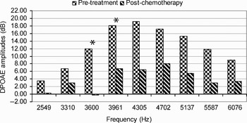
Before fotemustine administration, in 20 ears, the average distortion product otoacoustic emission amplitudes and standard deviations (referenced to 2f 1 − f 2) at 2549, 3310, 3600, 3961, 4305, 4702, 5137, 5587 and 6076 Hz frequencies were 3.45 ± 7.47, 6.66 ± 7.61, 11.95 ± 7.52, 18.09 ± 7.49, 19.25 ± 6.6, 17.19 ± 5.85, 15.24 ± 5.95, 11.81 ± 6.86 and 8.96 ± 6.67 dB, respectively. In the eighth week after fotemustine administration, average values for the same parameters and frequencies were 0.22 ± 9.99, 3.00 ± 8.38, −0.26 ± 9.45, 6.74 ± 12.11, 6.38 ± 13.64, 8.06 ± 13.26, 5.53 ± 16.73, 2.98 ± 19.54 and 3.43 ± 15.69 dB, respectively (see Table I).
Mean DPOAE measurements before and eight weeks after fotemustine administration, by frequency

* Referenced to 2f 1−f 2. †n = 20; ‡n = 12. **p < 0.05. DPOAE = distortion product otoacoustic emission; SD = standard deviation
Regarding distortion product otoacoustic emission amplitudes prior to and following fotemustine administration, there was statistical significance at 3600 and 3961 Hz frequencies (referenced to 2f 1 − f 2) (chi-square = 14.104, p = 0.003 and chi-square = 8.955, p = 0.03, respectively) (Figure 1). Hearing changes at other frequencies were found to be non-significant. These measurements demonstrated that, following fotemustine administration, only a limited frequency range showed significant alteration.
Mean signal-to-noise histogram of DP-gram responses, showing average distortion product otoacoustic emission amplitudes by frequency (range 2.55–6.08 kHz, referenced to 2f 1 − f 2), before and after fotemustine administration. The responses were evoked by a protocol in which L 1 = L 2 = 70 dB. *p < 0.05, comparing pre- and post-fotemustine data.

Transmission electron microscopy findings
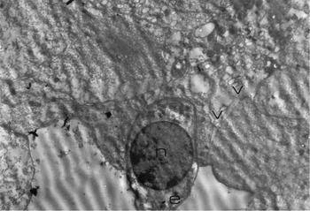
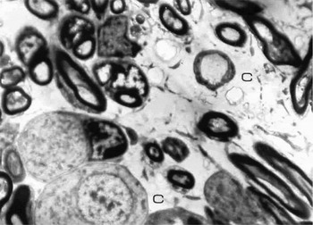
As transmission electron microscopy involved no parametric data, we did not use any controls for statistical analysis of results. Ultrastructural changes at the outer hair cells, stria vascularis and cochlear ganglion in the basal turn of the cochlea were assessed. The most notable findings were oedematous intracytoplasmic spaces and margination of the chromatin in the nuclei of the outer hair cells. Following intraperitoneal fotemustine administration, cytoplasmic changes in the supporting cells included formation of numerous vacuoles (Figure 2). The stria vascularis also showed some changes. Eight weeks after intraperitoneal fotemustine administration, perivascular oedema and intracytoplasmic vacuoles were observed. There were large, oedematous areas between the cochlear nerve axons. No changes occurred in the ganglion cell perikarya (Figure 3).
Transmission electron micrograph of rat cochlear basal turn and supporting cell, showing oedematous intracytoplasmic spaces and chromatin margination of the nuclei, plus numerous cytoplasmic vacuoles. e = oedematous intracytoplasmic spaces, n = nucleus, v = vacuoles in supporting cells (×4000)

Transmission electron micrograph of rat cochlear basal turn and cochlear nerve axon, showing large, oedematous areas between the cochlear nerve axons. The ganglion cell perikarya are intact. c = oedema between cochlear axons (×4000)

Discussion
It is very well documented that various chemotherapeutic agents can cause sensorineural hearing loss at therapeutic levels. Ototoxic dosage and reversibility can vary based on individual susceptibility. Knowledge of the adverse effects of these drugs and of the signs of ototoxicity can prevent permanent inner-ear damage.
Fotemustine belongs to the nitrosourea group of drugs. These are chemotherapeutic agents and have been most widely used in the treatment of metastatic melanoma.Reference Lotze, Dallal, Kirkwood, Flickinger, De Vita, Hellman and Rosenberg9 Since this group of drugs can cross the blood–brain barrier, they have also been used in the treatment of primary and metastatic brain tumours, as well as recurrences. (In fact, the primary treatment of brain metastases is irradiation. The temporal bone and cochlea may be included in the treatment portals during such irradiation of the brain. Thus, irradiation could potentially have a negative effect on the cochlea.)Reference Akmansu, Eryilmaz, Korkmaz, Sennaroglu, Akmansu and Gocer10 Our study aimed to assess whether fotemustine itself had a negative effect.
To assess cochlear damage in an animal model, it is common to use electrophysiological techniques such as electrocochleography and auditory brainstem responses. These techniques record global responses from the cochlea and from the afferent auditory nerve fibres, but do not allow direct evaluation of subtle alterations to the functional status of the outer hair cells, which represents the most likely pathway of ototoxicity following systemic chemotherapy. It is considered that distortion product otoacoustic emissions might provide a useful tool for detecting any induced functional changes of the cochlear structures. Although a number of studies have employed distortion product otoacoustic emission protocols to investigate chemotherapeutic ototoxicity, we have not found any such report for fotemustine.Reference Hatzopoulos, Di Stefano, Campbell, Falgione, Ricci and Rosignoli3, Reference Henley, Owings, Stagner, Martin and Lonsbury-Martin11
When the cochlea is stimulated simultaneously with two pure tones at frequencies f 1 and f 2 (with f 2 > f 1), it generates additional frequency components, termed intermodulation distortion tones, which are not present in the original stimuli and are viewed as products of two-tone interactions caused by the non-linearity of the cochlea. The frequencies of these distortion tones or products are termed primaries f 1 and f 2. For two given primaries, numerous families of distortion product otoacoustic emissions exist; however, in the mammalian ear, the cubic distortion product 2f 1 − f 2 is the most prominent one. There is evidence that the 2f 1 − f 2 product is generated by the outer hair cells, and that it is sensitive even to small cochlear alterations.Reference Henley, Owings, Stagner, Martin and Lonsbury-Martin11
Fotemustine is a relatively new chemotherapeutic agent among the nitrosoureas; however, it is becoming widely popular for treatment of the aformentioned diseases. Over the past decade, a few articles have reported its adverse effects, but we have not encountered any research on potential cochlear side effects. It was initially expected that alteration of the signal-to-noise ratios of the distortion product otoacoustic emission responses would correspond to the higher frequencies, as occurs with chemotherapeutics, aminoglycosides, noise and ageing.Reference Lautermann, Crann, McLaren and Schacht12, Reference Ravi, Somani and Rybak13 In our study, we determined that significant hearing alterations occurred at the mid-range to high frequencies (i.e. 3600 and 3961 Hz) (Figure 1). However, it should be remembered that our experimental equipment covered the frequency range of 2549 to 6076 Hz (referenced to 2f 1 − f 2). Therefore, it was not possible to detect changes outside this range.
In the present study, fotemustine-induced ototoxicity was also assessed by morphological analysis of the rat cochleas. This analysis was performed after the distortion product otoacoustic emission measurements in the eighth week post-fotemustine. As the normal rat cochlea has a hearing range of 1200–54 000 Hz, our sections through the basal turn reflected a higher frequency zone than that assessed by distortion product otoacoustic emission measurements.Reference Crofton, Janssen, Prazma, Pulver and Barone14
• Fotemustine is a chemotherapeutic agent which crosses the blood–brain barrier and is used in the treatment of primary and metastatic brain tumours and metastatic melanoma
• This study examined the potential otoxicity of fotemustine, using the rat as an animal model
• Fotemustine was determined to have potential for ototoxicity at 3600 and 3961 Hz. Electron microscopic morphological analysis revealed morphological effects in the cochlear basal turn
• Further experimental and clinical studies are required in order to determine if the changes seen in this pilot study are reversible, and also to analyse the effect in humans
On electron microscopy, the normal outer hair cell is cylindrical, and the cell surface contains the cuticular plate with stiff stereocilia. The nucleus is round and inferiorly placed in the cell. Mitochondria are collected in the subcuticular and infranuclear regions. Well developed layers of subsurface cisterns line the plasma membrane in the supranuclear part. Hensen bodies are often found, which are concentrically arranged endoplasmic reticula and mitochondria.Reference Kim and Shin15
In our study, damage could be observed mainly in the outer hair cells, stria vascularis and cochlear ganglion at the basal turns of the cochlea. The most notable findings at the stria vascularis were perivascular oedema and intracytoplasmic vacuoles. Although there were large, oedematous areas between the cochlear nerve axons, the ganglion cell perikarya were well preserved. The oedema was considered to represent a change in vessel permeability.Reference Kim and Shin15 Hair cell damage was studied qualitatively. The most characteristic findings in outer hair cells were margination of the chromatin and the presence of oeematous intracytoplasmic spaces. Euchromatin is indicative of active chromatin, whereas heterochromatin is regarded as inactive chromatin. Generally, metabolically active cells possess large amounts of euchromatin. Thus, extension of the heterochromatin indicates a decline in the metabolic activity of the hair cells. This phenomenon has been observed in hair cells after conventional radiation and conversely in radioresistant in nerve cells that have large amounts of euchromatin after irradiation.Reference Masurovsky, Bunge and Bunge16, Reference Winther17 Distortion in the chromatin content in the form of margination leading to heterochromatin formation, was observed in our transmission electron microscopy findings. Changes in the cytoplasm of the supporting cells included the presence of numerous cytoplasmic vacuoles. The ultimate result of the autolytic process is observed in the vacuolated cells.
Our study demonstrated the early effects of fotemustine on the rat cochlea, both morphologically and electrophysiologically. Hearing loss was restricted to the 3.6–4 kHz range; there was no observed damage at other frequencies. Once hair cell loss occurs, nerve fibre degeneration begins and hearing loss becomes irreversible. Recovery of temporary threshold shifts within the first few days may partly reflect synaptic repair. However, it is not possible to predict if the limited effect observed in our study was reversible. With these study results, further, long term, follow-up studies can be designed prospectively in cancer patients receiving fotemustine. Oncologists who prescribe this agent to their patients should be aware of our findings, and should ensure that patients undergo audiography before commencing treatment. Patients at increased risk, such as those with existing cochlear hearing loss (for whatever reason), should be handled meticulously. Following research both in animal models and in vitro, it has been suggested that the ototoxic effects of chemotherapeutic agents might be reduced by using agents (such as fosfomycin, D-methionine, sodium thiosulphate, derived neurotrophic factors and melanocortins) with the potential to confer protection from neuropathy.Reference Campbell, Rybak, Meech and Hughes18–Reference Schweitzer, Rarey, Dolan, Abrams, Litterst and Sheridan26
Conclusion
Fotemustine was found to have the potential for ototoxicity at 3600 and 3961 Hz. Morphological analysis, using electron microscopy, revealed morphological effects in the cochlear basal turn. Oedematous intracytoplasmic spaces and perivascular areas of the stria vascularis were detected, as well as distorted chromatin content, thereby suggesting the potential ototoxic effects of this agent. Further experimental and clinical studies are required in order to determine whether the effect observed in this pilot study is reversible, and also to analyse the effect of fotemustine in humans.
Acknowledgements
We thank Beyza Akdag MS, of the Hacettepe University Department of Biostatistics, Ankara, Turkey, for performing statistical analysis of the data, Suleyman Boynuegri MD, of the Ankara Numune Hospital Department of Otorhinolaryngology, for figure preparation, and Ayse Iriz MD, of the Ankara Numune Hospital Department of Otorhinolaryngology, for proofreading the manuscript.