Introduction
Megaspora (Clauzade & Cl. Roux) Hafellner & V. Wirth was first introduced as a subgenus of Aspicilia A. Massal. (Clauzade & Roux Reference Clauzade and Roux1984), but was later raised to generic level (Wirth Reference Wirth1987). It was distinguished from Aspicilia by its large, up to c. 60 µm long, thick-walled ascospores and by having apothecia immersed in thalline verrucae. Megaspora has so far generally been regarded as a monotypic genus with the mainly terricolous M. verrucosa (Ach.) Hafellner & V. Wirth. Corticolous and lignicolous specimens have often been referred to Aspicilia mutabilis (Ach.) Körb., which has been reduced to synonymy with M. verrucosa (e.g. Hafellner & Türk Reference Hafellner and Türk2001) or given subspecific rank (M. verrucosa ssp. mutabilis Nimis & Cl. Roux). Megaspora agrees with Aspicilia in several important characters: ascus-type (asci with a nonamyloid to pale amyloid tholus), presence of the pigment Caesiocinerea-green (Meyer & Printzen Reference Meyer and Printzen2000) in the epihymenium, simple and hyaline ascospores, and branched and anastomosing paraphysoids.
Megaspora is currently placed in Megasporaceae (Pertusariales), together with Aspicilia and Lobothallia (Clauzade & Cl. Roux) Hafellner (Lumbsch & Huhndorf Reference Lumbsch and Huhndorf2007). The position in Pertusariales has been confirmed by molecular studies (e.g. Wedin et al. Reference Wedin, Wiklund, Crewe, Döring, Ekman, Nyberg, Schmitt and Lumbsch2005; Miądlikowska et al. Reference Miądlikowska, Kauff, Hofstetter, Fraker, Grube, Hafellner, Reeb, Hodkinson, Kukwa and Lücking2006; Schmitt et al. Reference Schmitt, Yamamoto and Lumbsch2006), even if the status of Megasporaceae was questioned by Miądlikowska et al. (Reference Miądlikowska, Kauff, Hofstetter, Fraker, Grube, Hafellner, Reeb, Hodkinson, Kukwa and Lücking2006) who placed its members in Pertusariaceae. Nonetheless they form a well supported group, as shown by Nordin et al. (Reference Nordin, Savić and Tibell2010) in a study based on nuLSU and mtSSU focused on Megasporaceae. In this study a subdivision of Aspicilia was proposed and Lobothallia was recircumscribed. Megaspora was found to be closely related to the species transferred to Circinaria Link.
During field work performed by TV between 2001 and 2010, an unidentified sorediate species was frequently observed growing on the bark of various trees, and material from several localities was collected. Subsequently terricolous and saxicolous specimens were also discovered. Wheneventually apothecia were found, it became clear that it belonged either to Aspicilia or to Megaspora. The large ascospores rather suggested the latter, which was finally also confirmed by comparison of ITS sequences. Hence a second Megaspora species, M. rimisorediata, is here described as new to science.
Material and Methods
The material was collected by TV between 2001 and 2010. Morphology was studied under a stereo microscope. Anatomical characters were studied by light microscopy on hand-cut sections mounted in water or 10% KOH. Measurements of ascospores are recorded as (minimum–) x-σx –[x] – x+σx (–maximum) followed by the number of measurements (n). The amyloid reactions in the hymenium were tested using Lugol's solution with and without pre-application of KOH. Sulphuric and nitric acids were used for the detection of calcium oxalate and the Caesiocinerea-green pigment respectively. TLC was performed according to standard methods (Orange et al. Reference Orange, James and White2001).
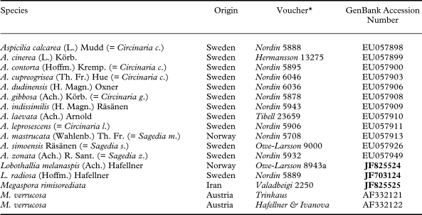
Nuclear rDNA ITS1-5.8S-ITS2 sequences from a number of specimens representing the five genera of Megasporaceae accepted by Nordin et al. (Reference Nordin, Savić and Tibell2010) (Table 1) were assembled and processed using the phylogenetic network program SplitsTree4 (Huson & Bryant Reference Huson and Bryant2006). Three new sequences were produced. Extractions of DNA and amplifications were carried out according to methods described in Nordin et al. (Reference Nordin, Tibell and Owe-Larsson2007). The sequences were aligned using ClustalW as implemented by the Bioedit software packet (http://www.mbio.ncsu.edu/RNaseP/info/programs/BIOEDIT/bioedit.html). No variable sequence portions were removed before analysis. In the program SplitsTree4, the neighbour-net option (Bryant & Moulton Reference Bryant and Moulton2004) with default settings was used for network construction.
Voucher specimens and GenBank accession numbers of the ITS sequences used in the phylogenetic network analysis. New sequences in bold
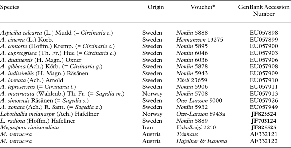
* All voucher specimens are deposited in UPS except Megaspora rimisorediata (TARI) and M. verrucosa (GZU).
Results
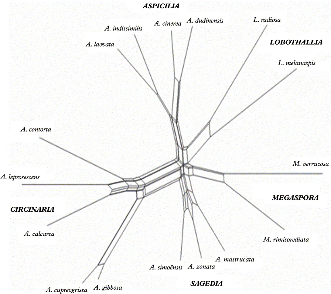
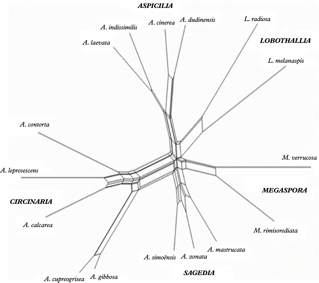
The total length of the alignment was 210 sites and the estimated proportion of invariant sites 0·39579415. In the graph resulting from the neighbour-net analysis (Fig. 1) the splits are displayed as parallel edges separating the fractions, where the length of the edges is proportional to the weight of the splits. Megaspora rimisorediata and M. verrucosa appear as more closely related to each other than to other members of Megasporaceae, although their sequences differ in more than 50 bases. The groups formed by the other species agree with those found in Nordin et al. (Reference Nordin, Tibell and Owe-Larsson2010), where the genera Circinaria and Sagedia Ach. were reintroduced and a number of new combinations proposed (cf. Table 1). Here, however, the names used in GenBank have been used both in Fig. 1 and Table 1.
Graphic representation of the relationships between Megaspora rimisorediata and other members of the Megasporaceae, resulting from a neighbour-net analysis of ITS sequences implemented in the program SplitsTree4. The splits are displayed as parallel edges. The length of the edges is proportional to the weight of the splits.
The Species
Megaspora rimisorediata Valadbeigi & A. Nordin sp. nov
MycoBank No.: MB 518433
Thallus rimosus, pallide ochraceus vel caesiocinereus, ad latera rimarum sorediatus. Apothecia rara, immersa in thallo plano vel in areoleis paulo elevatis. Ascosporae ellipsoideae, (32·8–) 35·3–[38·8]–42·2 (–45·2) ×(21·5–) 22·9–[24·9]–26·9 (–28·3) µm. Conidia brevia, c. 7 µm longa. Materiae chimicae secundariae non detectae.
Typus: Iran, Ilam, Tonele Reno, 33°42′N, 46°24′E, on bark of Quercus brantii, alt. c. 2000 m, 12 July 2009, T. Valadbeigi 10250 (TARI—holotypus; B, BR, hb. Valadbeigi—isotypi).
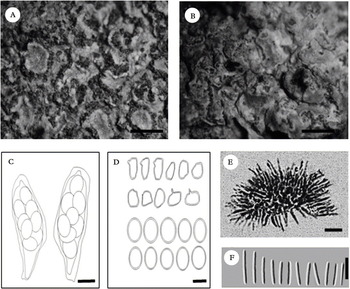
(Fig. 2A–F)
Aspicilia rimisorediata (holotype). A, sorediate thallus; B, thallus with apothecia; C, ascus; D, mature and immature ascospores; E, conidiophores and conidiogenous cells with conidia; F, conidia. Scales: A & B = 1 mm; C & D = 25 µm; E & F = 5 µm.

Thallus rimose, up to 4 cm diam., pale ochraceous to bluish grey, sorediate. Soredia dark blue-greenish, globules of 50–70 µm diam., produced on the sides of elongate, c. 0·2 mm wide cracks, often forming a dense net over the thallus. Cortex c. 30 µm thick, paraplectenchymatous; cells c. 5–9 µm diam.; upper part brownish and inspersed with calcium oxalate crystals; lower part hyaline or partly slightly greenish, K+ brown, N+ intensifying green (Caesiocinerea-green). Photobiont chlorococcoid, 12–17 um diam.; algal layer not developed below the apothecia. Medulla densely packed with calcium oxalate crystals.
Ascomata apothecia, rare, to 1·5 mm diam., first immersed, later forming separate, slightly raised areoles surrounded by cracks and bordered by a thalline margin, up to 0·4 mm wide; disc dark brown to black, ± concave, very irregular in shape. Proper exciple distinct, greenish brown, c. 45 µm wide in upper part. Hypothecium colourless to slightly yellowish brown. Hymenium to 150 µm tall, I+ blue. Epithecium olivaceous green, K+ brown, N+ intensifying green (Caesiocinerea-green). Paraphyses branched and anastomosing, not moniliform at the tips. Asci of Aspicilia-type, I−, 145 × 46 µm, 4–8-spored. Ascospores (32·8–) 35·3–[38·8]–42·2 (–45·2) × (21·5–) 22·9–[24·9]–26·9 (–28·3) (n = 20); wall c. 2–3 µm thick.
Pycnidia not observed under the dissecting microscope, but conidia and conidiogenous cells from entirely immersed pycnidia were seen in cross sections of the thallus. Conidiogenous cells c. 4–8 × 1–2 µm, irregularly thickened and tapering towards the end. Conidia bacilliform, straight, c. 5·5–8 × 1 µm.
Chemistry. K−, C−, Pd−. No substances detected by the standardized TLC method.
Etymology. The species epithet refers to the sorediate cracks.
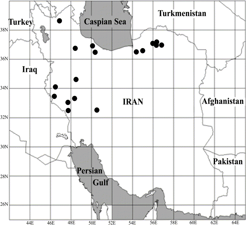
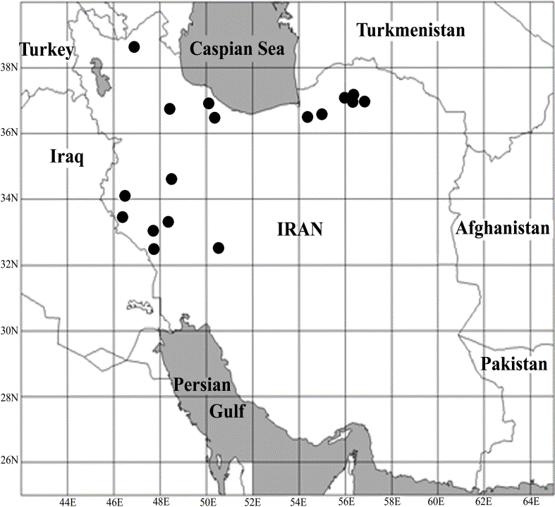
Habitat and distribution. Corticolous on bark of scattered Juniperus sp., Quercus brantii and Q. castaneifolia in steppe vegetation on dry mountain slopes; rarely muscicolous and saxicolous on calciferous rocks. The specimens collected during the winter were larger than those collected during the summer. Furthermore, the soredia were more abundant in the specimens collected during the winter. In other words, in the same locality (eg. Ilam, Tonele Reno), the new species covered most of branches of Q. brantii in the winter. It seems that the amount of water is important for extension of the thallus. To date the species is known only from the northern half of Iran (Fig. 3).
Distribution map of Aspicilia rimisorediata (●).
Remarks. Megaspora rimisorediata was usually encountered as a sterile, sorediate crust with characteristic soralia. The dark bluish green soredia contrast with the pale ochraceous thallus and are produced on the sides of cracks which form a dense net with meshes of about 0·5 mm over the thallus. The production of the soredia causes the cracks to become wider and finally the whole thallus surface becomes eroded.
Megaspora verrucosa is also known from Iran, but lacks soralia and usually has abundant apothecia immersed in thalline verrucae. Further, it differs from M. rimisorediata by having a taller hymenium (200–250 µm), larger asci (200–230 × 45–50 µm), and larger ascospores (30–60 × 21–42 µm) (James & Fletcher Reference James, Fletcher, Smith, Aptroot, Coppins, Fletcher, Gilbert, James and Wolseley2009).
Compared to sorediate Aspicilia species (often originally referred to Lecanora), Megaspora rimisorediata has larger ascospores. However, sterile specimens also differ from Aspicilia. In no sorediate Aspicilia species do the soralia develop along cracks and form mesh-like patterns. In A. caesiocinerea var. isidiata (H. Magn.) Oxner they develop from isidiate verruciform areoles. In A. capituligera (Poelt) Poelt, A. grisea Arnold, A. insolata (H. Magn.) Hav., A. mashiginensis (Zahlbr.) Oxner and A. simoënsis Räsänen they usually form discrete, rounded patches, which in A. capituligera and A. mashiginensis sometimes become distinctly raised and convex. In A. corallophora the initially formed ± convex soralia soon develop into coralloid isidia. Aspicilia excrescens (J. Steiner) Szatala and A. leprosescens (Sandstede) Hav., on the other hand, are mainly scurfy granular or subisidiate, and are only occasionally irregularly sorediate.
Contrary to M. rimisorediata, the sorediate Aspicilia species usually contain secondary metabolites (all tested by TLC except A. excrescens, for which no information is available). Aspicilia casiocinerea and A. leprosescens contain aspicilin; A. grisea, A. insolata and A. simoënsis norstictic acid; A. corallophora and A. simoënsis stictic acid; and A. capituligera and A. mashiginensis substictic acid.
Additional specimens examined. Iran: Ilam: Abdanan, Kabir Kouh, 2000 m, 33°04′ N, 47°71′E, on the bark of Quercus brantii, TV 1879 [TARI, hb. Valadbeigi]; Dinar Kouh, 2200 m, 32°57′N, 47°72′E, Q. brantii, TV 1803 [TARI, hb. Valadbeigi]; Sarabe Bagh, 32°53′N, 47°34′E, 1200 m, Q. brantii, TV 11137 [TARI, hb. Valadbeigi]; Kolm, mountain between Dareh Shahr and Kolm, 32°53′N, 47°34′E, 1100 m, Q. brantii, TV 11140 [TARI, hb. Valadbeigi]; Sarabe Kolm, the mountains near fish culture pool, 33°20′N, 46°54′E, 1300 m, Q. brantii, TV 11149 [TARI, hb. Valadbeigi]; Tonele Reno, 33°42′N, 46°24′E, 1300 m, Q. brantii, TV 10139 [TARI, hb. Valadbeigi]; Tonele Reno, 33°42′N, 46°24′E, 2000 m, mosses, TV 10178 [TARI, hb. Valadbeigi]; rocks, TV 11130 [TARI, hb. Valadbeigi]; Shishkalan mountains, 33°42′N, 46°23′E, 1100 m, Q. brantii, TV 11150 [TARI, hb. Valadbeigi]. Gorgan: Tuskestan forest, 1400 m, 36°52′N, 54°50′E, bark, TV 1601 [TARI, hb. Valadbeigi]; Golestan National Park, Almeh, 1700 m, 37°27′N, 56°20′E, Juniperus, TV 1160 [TARI, hb. Valadbeigi]; Almeh, 8 km from Mirza-Baylu Lodge, 1500 m, 37°21′N, 56°11′E, bark, TV 9100 [TARI, hb. Valadbeigi]; Sharleg, 1100 m, 37°00′N, 56°04′E, TV 234 [TARI, hb. Valadbeigi]; Tuskestan forest, 20 km SE of Gorgan along minor road to Shahrud, 1500 m, 36°41′N, 54°34′E, Q. castaneifolia, TV 1010 [TARI, hb. Valadbeigi]. East Azarbayejan: Arasbaran protected area, top mountain to Mazgar, 2200 m, 38°40′N, 47°00′E, calciferous silicate rocks, TV 10115 [TARI, hb. Valadbeigi]. Gilan: on the road to Javaherdasht, 800 m, 37°00′N, 50°18′E, Q. castaneifolia, TV 1222 [TARI, hb. Valadbeigi]. Khorasan: between Ashkhaneh and Roberte Gharabil, 1100 m, 37°25′N, 56°40′E, bark, TV 2444 [TARI, hb. Valadbeigi]. Kordestan: 15 km after Eslam Abad, 1500 m, 34°03′N, 46°29′E, Quercus, TV 307 [TARI, hb. Valadbeigi]. Mazandaran: Kelardasht to Abbas Abad, 1000 m, 36°46′N, 50°33′E, bark, TV 10110 [TARI, hb. Valadbeigi]. Lorestan: S of Qal'eh Mozaffari, Kakareza, 2000 m, 33°43′N, 48°16′E, bark, TV 1659 [TARI, hb. Valadbeigi]. Hamedan: Kivarestan area, 1000 m, 34°51′N, 48°25′E, bark, TV 1555 [TARI, hb. Valadbeigi]. Zanjan: Tarom, 2200 m, 36°43′N, 48°47′E, bark, TV 10787 [TARI, hb. Valadbeigi]. Chaharmahal & Bakhtiyari: S of Shahrekord, between Tanghanak and Shalamzar, 1700 m, 32°29′N, 50°54′E, bark, TV 1778 [TARI, hb. Valadbeigi].
TV is grateful to Dagmar Triebel (M), Andreas Beck (M) and Harrie Sipman (B) for their hospitality and support in connection with herbarium visits, and to Siahgis Zolfaghari (Ilam, Iran) for good companionship during the field work. The work was financially supported by a grant from Shahid Beheshti University, Tehran, awarded to Dr H. Riahi.