Pulmonary atresia with intact ventricular septum is a rare disease characterised by the absence of communication between the right ventricle and either the pulmonary trunk or the left ventricle. The lesion is morphologically heterogeneous, with varying degrees of right ventricular and tricuspid valvar hypoplasia. The size and morphology of tricuspid valve, the size of right ventricular cavity, the presence of fistulous communications between the right ventricle and the coronary arteries, and the presence of a right ventricular dependent coronary arterial circulation are the key determinants of prognosis. Unlike the situation when pulmonary atresia is found in the setting of tetralogy of Fallot, it is usual to find confluent pulmonary arteries, which are fed through a persistently patent arterial duct. It is exceedingly rare to encounter non-confluent pulmonary arteries supplied through either systemic-to-pulmonary collateral arteries or bilateral arterial ducts.Reference Daubeney, Delany and Anderson 1
Velocardiofacial syndrome, DiGeorge syndrome, and a variety of other clinical syndromes have a hemizygous deletion of chromosome 22q11.2 as their cause. This syndrome is extremely common, with nearly one in 3000 children being affected.Reference Sullivan 2 The most frequent anomalies involve palatal function, facial features, and congenital cardiac defects. In addition, learning disabilities and psychiatric issues are common.Reference Hay 3 The most commonly associated cardiac lesions involve the ventricular outflow tracts, usually tetralogy of Fallot with pulmonary stenosis or atresia, or common arterial trunk. In patients with tetralogy with pulmonary atresia, it is common to find the pulmonary arteries supplied by systemic-to-pulmonary collateral arteries. To our knowledge, however, there is but a single case reported of supply through collateral arteries in association with deletion of 22q11.2 when pulmonary atresia is found in the setting of an intact ventricular septum.Reference Li, Chudley, Soni and Divekar 4 We describe here a child with pulmonary atresia and 22q11.2 deletion syndrome with confluent branch pulmonary arteries, emphasising the significance of considering the possible association of 22q11.2 deletion syndrome in patients with pulmonary atresia with intact ventricular septum.
Case
Our female patient was born at 36 weeks to non-consanguineous parents at an outside hospital and diagnosed to have pulmonary atresia with an intact ventricular septum. She had been placed on prostaglandin with the intention of maintaining ductal patency for pulmonary blood flow, and had undergone balloon atrial septostomy on the first day of life. She was intubated and mechanically ventilated pending any surgical procedure before transfer at our institute owing to urgent hurricane evacuation. At presentation, she was 45 days old. She had a small jaw, but no other dysmorphic feature. A generalised maculopapular rash was recognised to be due to fungal infection. Saturations of oxygen were measured at 75–85% on mechanical ventilation with 70% FiO2. She had a normal first heart sound and single second heart sound, with a grade II/VI continuous systolic murmur audible at the upper right sternal border. Her liver was enlarged, and peripheral pulses were bounding.
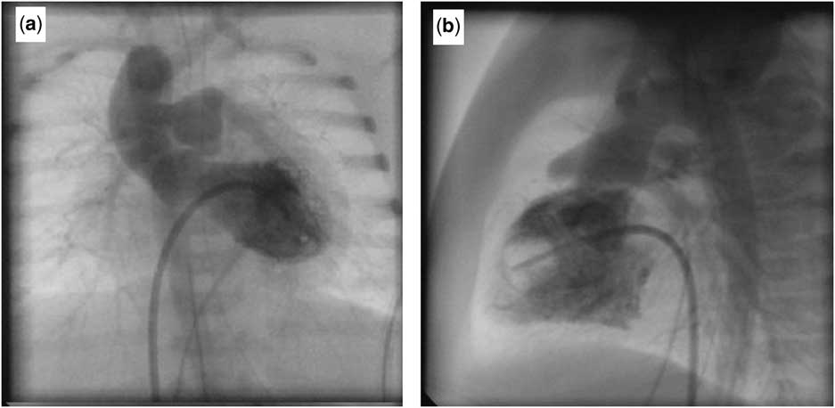
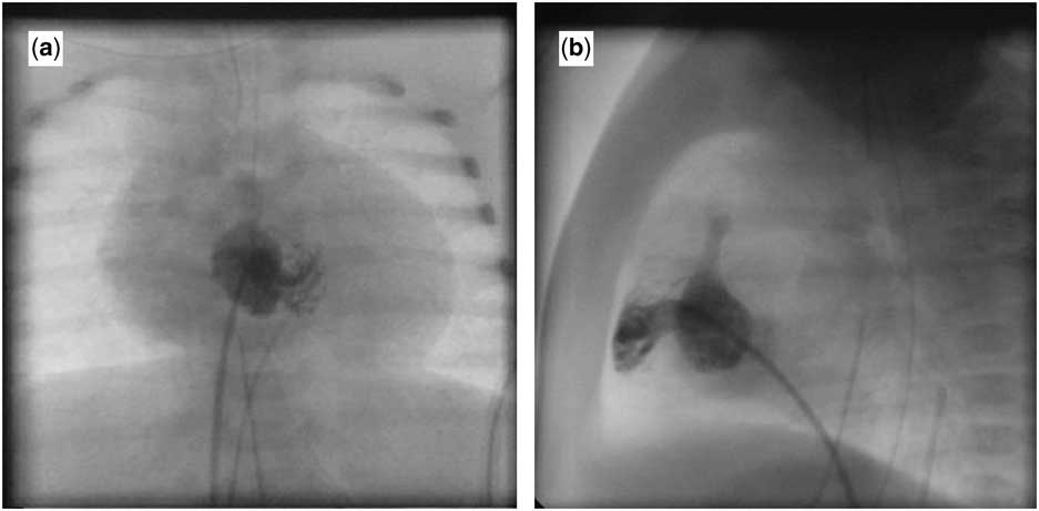
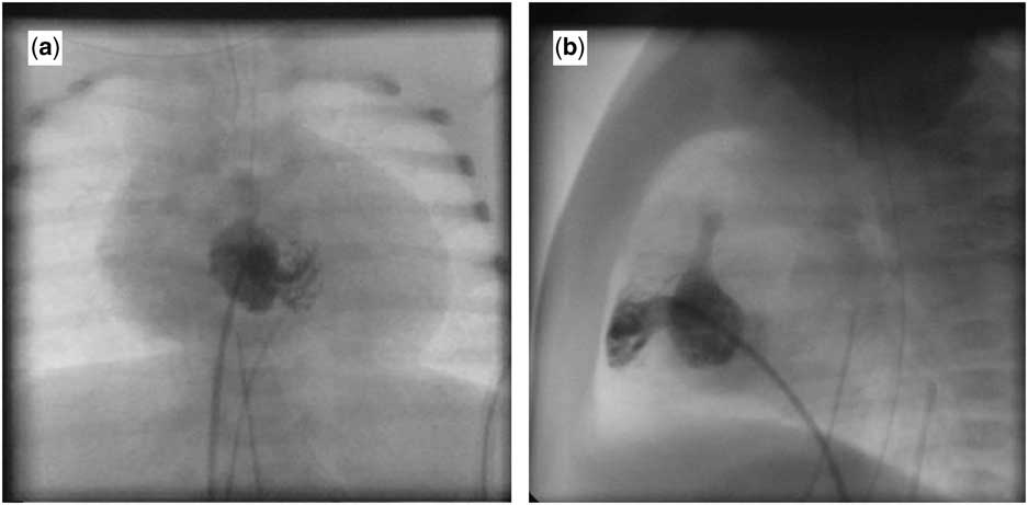
Transthoracic echocardiogram confirmed the diagnosis of pulmonary atresia with an intact ventricular septum, with a severely hypoplastic but tripartite right ventricle. The diameter of the tricuspid valve was measured at 9 mm (z score −1.6), with limited excursion of its leaflets. The atrial septal defect was small. The arterial duct was patent, with shunting of blood noted from the aorta to pulmonary arteries. Her laboratory evaluation revealed significant hypocalcaemia and lymphopaenia. Her cardiac catheterisation (Figs 1 and 2) confirmed the diagnosis, and failed to reveal the presence of any fistulous communications with the coronary arteries. The right ventricular pressure was supra systemic, with a right ventricular to systemic systolic pressure ratio of 1.4:1.
Antero-posterior ( a ) and lateral ( b ) projections demonstrating the right aortic arch and confluent pulmonary arteries supplied by an arterial duct. Note that contrast is injected in the left ventricle with the catheter advanced through the atrial septal defect into the left ventricle via the left atrium.

Right ventricular angiogram in anterio-posterior and lateral projections, demonstrating the hypoplastic right ventricle with an imperforate pulmonary valve at the ventriculo-arterial junction.
She was stabilised and taken to the operating room for a palliative surgical procedure. Intra-operatively, the diagnosis was confirmed, with an imperforate pulmonary valve noted at the right ventriculo-arterial junction. There was no thymic tissue. The right ventricle was hypoplastic and deemed unsuitable for a complete biventricular repair. She underwent atrial septectomy, pulmonary valvotomy, resection of right ventricular infundibular myocardium, and construction of a 3.5mm modified left Blalock–Taussig shunt (Goretex®, W.L. Gore and Associates, Inc., Flagstaff, AZ). Her arterial duct was ligated. Her left ventricular function was mildly depressed in the postoperative period, necessitating the use of milrinone, from which she was subsequently successfully weaned. Further growth of the right ventricle in follow-up will dictate the feasibility of a complete biventricular repair in future.
The presence of hypocalcaemia and lymphopaenia and the absence of thymus prompted further evaluation. Chromosome analysis on peripheral blood demonstrated 46,XX.ish del(22) (q11.2q11.2) (TUPLE1-). T-cell study demonstrated normal T-cell percentage but severe lymphopaenia. Mitogen study was normal, indicating normal T-cell function. She was transferred back after about a month of her surgery to the parent institute for further management and care closer to her home.
Discussion
The 22q11.2 deletion syndromeReference Momma 5 is seen in one in 3900 to one in 9700 children.Reference Goodship, Cross, LiLing and Wren 6 Cardiovascular anomalies are present in up to four-fifths of these neonates. In addition, most patients suffer developmental delay, facial dysmorphism, palatal dysfunction, and difficulties with feeding. At least three genes have been identified on chromosome 22q11.2, namely TBX1, CRKL and ERK2, whose haploinsufficiency causes dysfunction of the neural crest cell and anterior heart field, thus producing the typical anomalies.Reference McDonald-McGinn, Sullivan and Marino 7 The most common cardiac lesions involve the ventricular outflow tracts and include tetralogy of Fallot with either pulmonary stenosis or atresia, common arterial trunk associated with interruption of the aortic arch, double-outlet right ventricle, and discordant ventriculo-arterial connections.Reference Ryan, Goodship and Wilson 8 In a review of reported cardiovascular anomalies in chromosome 22q11.2 deletion syndrome, however,Reference Momma 9 pulmonary atresia in the setting of an intact ventricular septum had not been encountered. Indeed, our review of the English literature yielded but a solitary case reportReference Li, Chudley, Soni and Divekar 4 describing 22q11.2 deletion in a patient with pulmonary atresia in the setting of intact ventricular septum.
Pulmonary atresia with an intact ventricular septum, although anatomically simple, is complex in terms of the meticulous surgical and medical management required for optimal treatment. Despite the substantial improvement of the overall prognosis of patients with CHDs, recent epidemiological studies have outlined that about 30% of them present a genetic syndrome with one or more extra-cardiac anomalies. This association is particularly significant for those presenting as neonates.Reference Harris, Francannet and Pradat 10 According to the current literature, the presence of 22q11.2 deletion syndrome in patients with tetralogy of Fallot with pulmonary atresia does worsen the surgical prognosis.Reference Carotti, Digilio and Piacentini 11 In the first reported case of pulmonary atresia with intact ventricular septum and 22q11.2 deletion,Reference Hay 3 pulmonary arterial supply was derived from systemic-to-pulmonary collateral arteries. The patient suffered septicaemia, and in light of a worsening neurological state the parents decided to discontinue life support.
Our experience indicates that chromosome 22q11 deletion should not be excluded as a potential factor in patients having pulmonary atresia in the setting of an intact ventricular septum when there are other clinical or laboratory pointers, such as hypocalcaemia, absence of the thymus, or lymphopaenia. Recognition of the deletion would help in identifying these children, who might need specialised care for other co-morbidities associated with the genetic malformation. In addition, as we likely identify more patients having pulmonary atresia with intact ventricular septum and chromosome 22q11 deletion, future prognostic information will be stouter.
Acknowledgements
None.
Financial Support
This research received no specific grant from any funding agency, commercial or not-for-profit sectors.
Conflicts of Interests
None.