Implication
In this study, we show that intramammary treatment with lipopolysaccharide induced nitric oxide burst that was associated with nitrosative stress that reflected the formation of hazardous compounds in milk: nitrotyrosines, carbonyls and lipid peroxides. These changes were associated with impairment in milk quality. Currently, very little is known about the presence of oxidative substances in commercial milk and their industrial and health implications. As mastitis is a prevalent problem in the dairy industry, this study stresses the importance of gaining further information on the effect of different forms of mastitis on milk composition.
Introduction
Milk plays a fundamental role in modern western diets. It is an easily accessible matrix rich in a wide variety of essential nutrients such as minerals, vitamins and easily digestible proteins with balanced amino acid profiles, which are important in supporting most body functions (Drewnowski and Fulgoni, Reference Drewnowski and Fulgoni2008; Silanikove et al., Reference Silanikove, Leitner, Merin and Prosser2010). High-quality raw milk constituents are required to obtain high yields and good-quality dairy products such as yogurt and cheese (Oliveira et al., Reference Oliveira, Fernandes, Neto, Fonseca, Silva and Balian2002; Leitner et al., Reference Leitner, Silanikove, Jacobi, Weisblit, Bernstein and Merin2008). Nowadays, dairy plants grade milk upon reception according to its hygienic quality based on acceptable standards. In most countries, these standards comprise limits on maximal transporting temperature, maximal bacterial count, antibiotic residues and somatic cell count (SCC) (McLaughlin, Reference McLaughlin2006; PMO, 2007).
Activation of indigenous enzymatic systems in milk plays an important role in promoting these changes. The simultaneous reduction in milk quality and milk secretion in infected glands is related to the activation of plasminogen activator (PA), plasminogen (PG) and plasmin (PL)-derived active peptides from milk β-casein (Silanikove et al., Reference Silanikove, Shamay, Shinder and Moran2000, Reference Silanikove, Shapiro, Shamay and Leitner2005, Reference Silanikove, Merin and Leitner2006 and Reference Silanikove, Shapiro and Shinder2009a; Shamay et al., Reference Shamay, Shapiro, Mabjees and Silanikove2002 and Reference Shamay, Shapiro, Leitner and Silanikove2003). Enzymes linked to the metabolism of nitric oxide (NO) have also been shown recently to considerably affect the milk composition of inflamed mammary glands (Silanikove et al., Reference Silanikove, Shapiro, Shamay and Leitner2005, Reference Silanikove, Merin and Leitner2006, Reference Silanikove, Shapiro and Shinder2009a and Reference Silanikove, Shapiro, Silanikove, Merin and Leitner2009b). Xanthine oxidase (XO), lactoperoxidase (LPO) and their respective substrates xanthine/hypoxanthine and NO are components of the mammary innate immune system. These components interactively function to create an effective bactericidal environment towards major mammary gland pathogens. Accordingly, H2O2 and NO are being constantly surged from the surrounding epithelial cells and milk leukocytes. NO cycles in milk through its auto-oxidation to nitrite and the conversion of nitrite into NO2• by H2O2-dependent LPO activity (Silanikove et al., Reference Silanikove, Shapiro, Shamay and Leitner2005). In turn, NO2• contributes towards the formation of a bacteriocidic environment in milk and interacts with thiol-bearing groups on proteins to form thiyl radicals (Silanikove et al., Reference Silanikove, Shapiro, Shamay and Leitner2005) and with tyrosine on proteins to form nitrotyrosine (Ntyr; Silanikove et al., Reference Silanikove, Shapiro, Silanikove, Merin and Leitner2009b). The interaction of NO with thiyl radicals forms nitrosothiols, which serve as a pool that constantly delivers NO into the system (Silanikove et al., Reference Silanikove, Shapiro, Shamay and Leitner2005 and Reference Silanikove, Shapiro, Silanikove, Merin and Leitner2009b). The above-described NO cycle includes two important regulatory modes that modulate and restrain NO2• activity. The first is catalase, which converts nitrite, the substrate of LPO, into the more stable metabolite nitrate, and the second is the formation of nitrosothiols, as an alternative to the formation of much more reactive Ntyr (Silanikove et al., Reference Silanikove, Shapiro, Shamay and Leitner2005 and Reference Silanikove, Shapiro, Silanikove, Merin and Leitner2009b). A few recent studies have shown that under certain conditions, such as extended storage and mastitis, milk nitrite could be in the low μM range, which is above the permitted level in foods (Silanikove et al., Reference Silanikove, Shapiro, Shamay and Leitner2005; Titov et al., Reference Titov, Kosenko, Fisinin and Klimov2010).
Mastitis is one of the most common diseases in dairy cows, inflicting 25% to 50% of the udders in modern dairy herds (Rainard and Riollet, Reference Rainard and Riollet2006). This indicates that mastitis has a major effect on milk quality and potentially may also affect its safety for human consumption; therefore, it is imperative that its effect on milk composition be methodically understood.
Lipopolysaccharides (LPS) are glycolipids from the outer membrane of Gram-negative bacteria. LPS stimulates immune system cells, resulting in acute increased production of pro-inflammatory cytokines, overexpression of cell adhesion molecules and matrix-degrading enzymes (Heumann and Roger, Reference Heumann and Roger2002). Exposure of a tissue to LPS induces rapid inflammation, which resembles, to a large extent, the inflammatory response induced by Escherichia coli infection. Therefore, LPS challenge is a frequent model to study the inflammatory response induced by E. coli and its endotoxins in the mammary gland (e.g. Bruckmaier, Reference Bruckmaier2005).
The general aim of this study was to explore the effects of an intramammary LPS challenge on milk composition and the effect of these changes on milk quality and safety. The specific aims were to determine (i) how LPS affects the NO cycle in milk and milk composition, (ii) how LPS affects the PA–PG–PL-casein-derived peptides (proteose peptones) and how these changes affect milk yield (MY) and composition and (iii) to show that catalase plays a role in restraining the nitrosative stress induced by LPS challenge.
Material and methods
All protocols were approved by the Institutional Animal Care Committee of the Agricultural Research Organization, which is the legitimate body for such authorizations in Israel.
Study layout
The layout details of the study are described in Silanikove et al. (Reference Silanikove, Rauch-Cohen, Shapiro, Blum, Arieli and Leitner2011). Briefly, 12 Israeli Holstein heifers with low SCC (27 000 ± 5000 cells/ml) and no bacterial detection that produced 33.2 ± 3.1 milk/day were divided into two groups of six cows each. In the control cows, two mammary quarters, one front and one rear were infused with 10 ml of a sterile non-pyrogenic saline solution (Teva Pharmaceutical Industries Ltd, Tel Aviv, Israel) (r-Cont). The other two counter quarters (c-Cont) served as controls to the procedure. In the experimental cows, two mammary quarters, one front and one rear were infused with LPS; 10 μg of LPS (E. coli, O55B5; Sigma Chemical Co., St Louis, MO, USA) dissolved in 10 ml of sterile non-pyrogenic saline (e-LPS), while the two counter quarters served as controls to the treatment (c-LPS). Intramammary infusion was injected using a special applicator following careful sterile cleaning of the teat. During the 4 days of the study (−24, 0, +24, +48 and +72 h, where 0 h refers to the day of infusion), every quarter of each cow was separately milked into individual containers and milk samples were taken after recording MY. Milk was discarded 7 days following the infusion.
Analytical procedures
For the determination of fat, protein, lactose, urea and SCC, one sub-set of samples was sent to a central laboratory. The amount of milk components secreted and the concentration of particular components at each time point were calculated from the MY. The quality of milk for cheese production was tested by measuring curd firmness (CF) and rennet clotting time (RCT) using an Optigraph (Ysebaret, Frepillon, France) (Merin et al., Reference Merin, Fleminger, Komanovsky, Silanikove, Bernstein and Leitner2008). Additional sub-sets of milk samples were defatted under cold conditions (Silanikove and Shapiro, Reference Silanikove and Shapiro2007) and analyzed according to previously described procedures: concentrations of casein, whey proteins, proteose peptones, lactoferrin (enzyme-linked immunosorbent assay – ELISA), albumin (ELISA), Na+ and K+, activities of PA, PLG and PL (Silanikove et al., Reference Silanikove, Shamay, Shinder and Moran2000 and Reference Silanikove, Shapiro, Silanikove, Merin and Leitner2009b), activities of XO, LPO and catalase and the concentrations of nitrite (fluorometrically by the DAN reagent) and nitrate (colorimetrically by the Griess reaction). The formation of potential harmful products in milk was assessed by analyzing the concentration of Ntyr in whey proteins by ELISA, the concentration of carbonyls in whey protein and the formation of lipid peroxides (Lpx) ion milk lipids by colorimetric reactions (Silanikove et al., Reference Silanikove, Shapiro, Shamay and Leitner2005).
Statistical analysis
The results of this study were analyzed using repeated-measures analysis modeling correlated residuals within cow (SAS Institute, 1990) as described before (Shamay et al., Reference Shamay, Shapiro, Leitner and Silanikove2003). Briefly, the analysis considered the effects of treatment, day and treatment–day interactions. The effect of days in milk was not significant (P > 0.25) and, therefore, was not included in the analyses presented here.
The model used was

where Yijkm is the variable within cow, treatment, quarter and day, Ci is the cow class effect, Tj is the treatment class effect, Dk is the day class effect, TjDk is the treatment–day interaction effect, Qm (TjCI) is the quarter within cow treatment error term for treatment effect and eijkm is the residual error.
Results
None of the measured metabolites were affected in the control cows, whether the glands were treated with saline or not, which indicates that the responses were specific to the LPS treatment.
MY, SCC and lactose
This data have been reported elsewhere (Silanikove et al., Reference Silanikove, Rauch-Cohen, Shapiro, Blum, Arieli and Leitner2011). Briefly, the dose of LPS applied in this experiment induced acute inflammation that was reflected by an increase in SCC (up to 5 to 6×10⁶ cells/ml), rectal temperature, plasma cortisol concentration and NAGase activity. These changes were associated with a maximum decrease of ∼30% in MY at 24 h post-treatment in the experimental glands. MY recovery occurred after 4 to 5 days. MY in c-LPS glands decreased by ∼20%, indicating that LPS induced a negative effect on MY in the non-treated glands, although the effect was significantly milder in comparison with the effect on treated glands and was apparent only 24 h post-treatment. LPS challenge induced a significant transient decrease in lactose concentration (∼20% at 24 h post-treatment) and lactose secretion (∼40% at 24 h post-treatment) in the e-LPS glands. A decrease of ∼9% and ∼6% in lactose concentration at 24 and 48 h post-treatment and of ∼14% and ∼11% in lactose secretion was recorded in treatment c-LPS, respectively.
Casein, whey proteins and proteose peptones
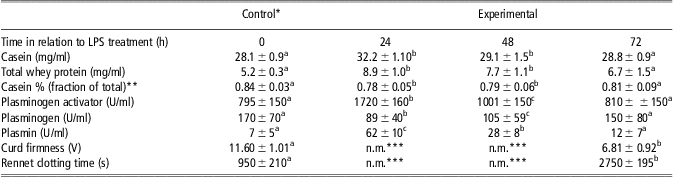
LPS challenge in the e-LPS glands induced a significant transient increase in casein, whey proteins and proteose peptone concentrations. Casein concentration increased ∼1.2-fold at 24 h post-treatment (Table 1); however, despite the increase in its concentration, casein secretion decreased by ∼21% at 24 h post-treatment. Furthermore, the proportion of casein/total protein in the LPS-challenged glands was lower than that in the control treatments (0.78 v. 0.84; Table 1). Whey proteins concentration increased ∼1.6-fold at 24 h post-treatment (Table 1). Thus, despite the decrease in MY, the secretion of whey proteins actually increased by 1.21-fold at 24 h post-treatment in the LPS-challenged glands (P < 0.05). No changes were recorded in the concentration of whey proteins in the control treatments (Table 1). Thus, the ∼20% decrease in whey protein secretion in treatment c-LPS was proportional to the decrease in MY.
Effect of LPS on the concentrations of casein, total whey protein and casein %, and the activities of plasminogen activator, plasminogen and plasmin (mean ± s.d.)

LPS = lipopolysaccharide.
*Pooled data of control treatments: these levels did not change in quarters treated with non-pyrogenic saline (r-Cont, c-LPS) and non-treated control quarters: c-Cont.
**Casein % = casein/(casein + total whey protein).
***n.m. – not measurable.
Parameters within a row with no common superscript differ significantly (P < 0.001) or are lower.
LPS challenge in the e-LPS glands induced a marked 4.4-fold transient increase in the concentration of proteose peptones at 24 h post-treatment (Table 1). Thus, despite the decrease in MY, the yield of proteose peptones actually increased ∼2.7-fold at 24 h post-treatment in the LPS-challenged glands (P < 0.01). No changes in the proteose peptone concentration were recorded in the control treatments (Table 1). Thus, the ∼20% decrease in the proteose peptone content in treatment c-LPS was proportional to the decrease in MY.
PA–PG–PL system and milk clotting
LPS challenge in the e-LPS glands induced a transient ∼2.1-fold increase in PA activity at 24 h post-treatment and a corresponding ∼1.7-fold decrease in PG activity, an increase of ∼11-fold in PL activity (Table 1), which was reflected by a decrease in the PG/PL ratio from ∼27 to ∼1.5 at 24 h post-treatment. Whereas milk-coagulating parameters were in the normal range in the control samples (CF 11.60 ± 1.01 V; RCT 950 ± 210 s), it was not possible to curdle the milk in samples collected 24 and 48 h after LPS challenge (Table 1). At 72 h post-treatment, milk clotting parameters were significantly different from those in the control (CF 6.81 ± 0.92 V; RCT 2750 ± 195 s), indicating impairment in milk quality (Table 1).
Soluble components of the innate immune system: lactoferrin and albumin
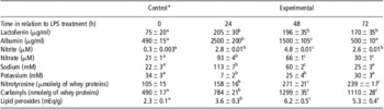
LPS challenge induced a marked transient increase in lactoferrin (2.7-fold) and albumin (4.7-fold) concentrations at 24 h post-treatment in the e-LPS glands (Table 2). These increases in lactoferrin and albumin concentrations were greater than the decline in MY, indicating that a net increase in their secretion occurred. The response was specific to LPS treatment as no changes in lactoferrin and albumin concentrations were detected in c-LPS.
Effect of LPS on the concentrations of albumin, nitrite, nitrate, sodium, potassium, nitrotyrosine, carbonyls and lipid peroxides and milk clotting parameters, curd firmness and rennet clotting time in milk components (mean ± s.d.)

LPS = lipopolysaccharide.
*Pooled data of control treatments: these levels did not change in quarters treated with non-pyrogenic saline (r-Cont, c-LPS) and non-treated control quarters: c-Cont.
Parameters within a row with no common superscript differ significantly (P < 0.001) or are lower.
Enzymes of the innate immune system along with components of their precursors and products
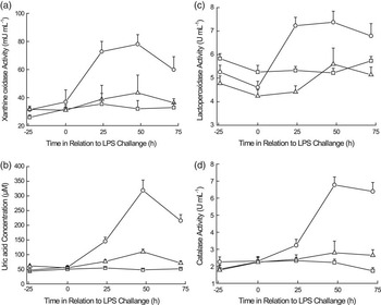
LPS challenge in the e-LPS glands induced a transient 2.3-fold increase in XO activity at 24 h post-treatment and a 2.5-fold increase at 48 h post-treatment. The corresponding figures for c-LPS increased by 1.4-fold at 24 h post-treatment and by 2.0-fold at 48 h post-treatment (Figure 1a). These increases in XO activity were greater than the decline in MY, indicating that a net increase in its secretion occurred.
Effect of lipopolysaccharide (LPS) on xanthine oxidase activity (mU/ml; panel a), uric acid concentration (μM; panel b), lactoperoxidase activity (U/ml; panel c) concentrations and catalase activity (U/ml; panel d) in treated (circles) control-LPS (triangles) and control-control (squares) glands. The results are presented as mean ± s.d. Results at 24, 48 and 72 h in the experimental treatment are significantly higher (P < 0.001) than those in the controls.

Uric acid in milk is a product of XO activity in milk (Silanikove et al., Reference Silanikove, Shapiro and Leitner2007). LPS challenge in the e-LPS glands induced a transient increase in uric acid concentration, which was parallel to the changes in XO activity: a 3.1-fold increase at 24 h post-treatment and a 6.7-fold increase at 48 h post-treatment. The corresponding figures for c-LPS were increase of 2.3-fold at 24 h post-treatment and 4.8-fold increase at 48 h post-treatment (Figure 1b). These increases in uric acid concentrations were greater than the decline in MY, indicating that a net increase in the secretion of its precursors, xanthine and hypoxanthine, occurred (Silanikove et al., Reference Silanikove, Shapiro and Leitner2007).
LPO is a major whey protein (Silanikove et al., Reference Silanikove, Shapiro, Shamay and Leitner2005), explaining its high initial activity (∼5 U/ml). LPS challenge in the e-LPS glands induced a transient 1.4-fold increase in LPO activity at 24 and 48 h post-treatment (Figure 1c). The corresponding figure for c-LPS was increase of 1.1 LPO activity. The increase in LPO in e-LPS glands and c-LPS glands was roughly equal in the case of treated glands or less than the decline in MY in c-LPS, indicating that unlike XO, no net increase in the secretion of LPO occurred.
LPS challenge in the e-LPS glands induced a marked transient increase in the nitrite and the nitrate concentration of 9- and 4. 4-folds, respectively, at 24 h post-treatment, and 16- and 3.1-folds, respectively, at 48 h post-treatment. These responses were sharp and, at 72 h post-treatment, nitrite and nitrate concentrations decreased to the pre-treated values (Table 2).
Fresh raw milk catalase activity was ∼2 U/ml (Figure 1d). LPS challenge in the e-LPS glands induced a transient 3 to 3.5-fold increase in catalase activity to ∼7 U/ml at 48 h post-treatment and ∼6.2 U/ml at 72 h post-treatment (Figure 1d). Thus, the increase in catalase activity lagged behind the increase in XO and LPO activities and remained high for a longer period. The increase in catalase activity was significantly higher than the reduction in MY, indicating a net increase in catalase secretion. No increase in catalase activity was recorded in c-LPS glands.
Oxidized substances
Fresh raw milk contained ∼100 nmole/g of Ntyr in whey proteins (Table 2). LPS challenge in the e-LPS glands induced a transient increase in Ntyr concentration, which was parallel to the changes in LPO activity: an increase of 5.3-fold at 24 h post-treatment and 5.7-fold at 48 h post-treatment. The response was specific to LPS treatment as no changes in Ntyr concentration were detected in c-LPS glands.
The concentration of carbonyls and Lpx followed similar kinetics to that of Ntyr. A summary of the control and the values of Ntyr, carbonyls and Lpx at 24, 48 and 72 h are presented in Table 2. The data show a peak in their concentration at 48 h post-treatment, which was ∼3-fold higher than that in the controls. Although the concentrations of Ntyr, carbonyls and Lpx started to decrease at 72 h post-treatment, it was still significantly higher (∼2-fold) than that in the controls (Table 2).
Changes in Na+ and K+ concentrations
LPS challenge in the e-LPS glands induced a transient ∼4-fold increase in Na+ concentration (from ∼22 to ∼113 mM). LPS challenge in the e-LPS glands induced a transient ∼4-fold decrease in potassium concentration (from ∼33 to ∼8 mM) at 24 h post-treatment and the kinetics was inversely proportional to that of sodium concentration (Table 2). These responses were sharp and at 48 h post-treatment, Na+ and K+ concentrations resumed to approximately half-way of the pre-treatment level and reverted to the pre-treatment values at 72 h post-treatment. No significant changes in Na+ and K+ concentrations were recorded in c-LPS glands.
Discussion
Changes in milk composition with respect to milk quality and safety
The present study provides significant evidence that supports the generally accepted rule in the dairy industry that milk from clinical mastitic udders should not be used for human consumption. Considerable amounts of milk are typically used in many western countries for the production of cheese. Typically, nowadays, the SCC content in the bulk milk tank (BMT) in most European Union countries is around 250 000/ml, whereas the upper permitted level is 600 000/ml (EEC Council Directive 94/71/EC, 1994). Mastitis usually infects a single gland and typically has SCC 1 × 10⁶/ml and above (Rainard and Riollet, Reference Rainard and Riollet2006). Thus, according to the current SCC-based hygienic criterion, such milk in small quantities but with high SCC may perhaps enter the BMT, and the milk will still meet the above-described criteria. The amount of low-quality milk that can enter the BMT without violating these criteria is inversely related to the SCC level. Analysis of the quality of bulk milk in 11 tanks of Israeli dairy farms has shown huge variability in milk quality for curdling that was not related to SCC (Leitner et al., Reference Leitner, Silanikove, Jacobi, Weisblit, Bernstein and Merin2008). It was suggested that this variability was related to mixing milk from post-clinical infection when the milk appears normal and from sub-clinically infected udders with the general milk. However, mixing of milk from infected udders with milk from non-infected ones could not be detected by measures such as determination of SCC, proteose peptone content and % of casein, which worked well at the individual cow level as predictors of udder inflammation (Leitner et al., Reference Leitner, Silanikove, Jacobi, Weisblit, Bernstein and Merin2008). Thus, the results of this and former studies (Silanikove et al., Reference Silanikove, Shapiro and Leitner2007; Leitner et al., Reference Leitner, Silanikove, Jacobi, Weisblit, Bernstein and Merin2008; Forsback et al., Reference Forsback, Lindmark-Mansson, Andren, Akerstedt and Svennersten-Sjaunja2009 and Reference Forsback, Lindmark-Mansson, Andren and Svennersten-Sjaunja2010) indicate that it is important for the dairy industry to develop analytical tools that will allow to prevent combining low-quality milk, such as milk rich in somatic cells and nitrite, with high-quality milk in order to ensure optimal yield and quality of curd from milk designated for cheese production.
The concentration of nitrite in milk infected by bacteria may exceed the permitted safety levels of ∼1 μM in milk (present study; Silanikove et al., Reference Silanikove, Shapiro, Silanikove, Merin and Leitner2009b; Titov et al., Reference Titov, Kosenko, Fisinin and Klimov2010). Whether nitrites and nitrates in foods and drinking water affect human health adversely is a controversial issue (Milkowski et al., Reference Milkowski, Garg, Coughlin and Bryan2010). However, as discussed below, high levels of Ntyr, Lpx and carbonyls in mastitic milk are associated with a high level of nitrite and nitrate in that milk. Ntyr, Lpx and carbonyls are considered as causative agents and hallmarks of cancer, atherosclerosis and other inflammatory diseases (Ohshima et al., Reference Ohshima, Tatemichi and Sawa2003). Thus, obviously, their presence in food is undesirable, although, as recently mentioned, there is lack of information regarding the significance of their occurrence in foods (Silanikove et al., Reference Silanikove, Shapiro, Silanikove, Merin and Leitner2009b).
A critical question that arises from this study is: does the current milk quality hygienic criteria are sufficient to ensure the highest quality that the present stage of knowledge permits? A single report from Brazil, based on a study carried out in 1998, has shown that all samples of bulk milk collected from 36 farms exceeded the Brazilian legislation level for nitrite, and that 14.3% of commercial pasteurized milk samples were above that level (Seraphim et al., Reference Seraphim, Bastos De Siqueira, Galego and de Fernicola1988). Because the present experimental setup induced a very strong inflammatory response, it is still premature to draw conclusions pertaining to more common situations that prevailed in dairy farms. We also could not find more information regarding the effect of mastitis on milk safety in the scientific literature. Thus, more research at the basic cow/gland level and at the BMT level in different sites, both within and between countries, is required to clarify this issue.
LPS challenge is reflected by accumulation of inflammatory indices in milk
The present results are consistent with previous findings that have shown that the inflammatory response induced by LPS disrupts tight junction integrity as reflected by changes in Na+ and K+ concentrations in milk (Shamay et al., Reference Shamay, Shapiro, Mabjees and Silanikove2002 and Reference Shamay, Shapiro, Leitner and Silanikove2003) and increases the secretion of soluble constituents of the innate immune system (Silanikove et al., Reference Silanikove, Merin and Leitner2006; Ibeagha-Awemu et al., Reference Ibeagha-Awemu, Ibeagha, Messier and Zhao2010), including the secretion of albumin from mammary epithelial cells (Shamay et al., Reference Shamay, Homans, Fuerman, Levin, Barash, Silanikove and Mabjeesh2005).
Our results are also consistent with previous studies showing that LPS challenge induces significant casein degradation and identifies milk PL as a major cause for this (Moussaoui et al., Reference Moussaoui, Michelutti, Le Roux and Laurent2002; Leitner et al., Reference Leitner, Chaffer, Shamay, Shapiro, Merin, Ezra, Saran and Silanikove2004a, Reference Leitner, Merin and Silanikove2004b and Reference Leitner, Merin, Silanikove, Ezra, Chaffer, Gollop, Winkler, Glikman and Saran2004c; Silanikove et al., Reference Silanikove, Merin and Leitner2006; Rossi et al., Reference Rossi, Galante, Fusi, Luini, Dell'Orto and Baldi2009). The biological implications of these findings have been discussed before (Leitner et al., Reference Leitner, Krifucks, Merin, Lavi and Silanikove2006 and Reference Leitner, Merin and Silanikove2011; Silanikove et al., Reference Silanikove, Merin and Leitner2006) and, therefore, these aspects are not discussed here in detail.
LPS-induced acute nitrosative response and accumulation of oxidative substances in milk
It is known that LPS challenge and inflammatory mediators (cytokines) induce the formation of NO burst from various cells, including mammary gland cells (Boulanger et al., Reference Boulanger, Bouchard, Zhao and Lacasse2001; Silanikove et al., Reference Silanikove, Shapiro, Shamay and Leitner2005; Zheng et al., Reference Zheng, Watson and Kerr2006; Ara et al., Reference Ara, Iijima, Asanuma, Yoshitake, Ohara, Shimosegawa and Yoshimura2008; Ito et al., Reference Ito, Iijima, Ara, Asanuma, Endo, Asano, Koike, Abe, Imatani and Shimosegawa2010). Here, we show for the first time that such NO burst is reflected by accumulation of NO-derived metabolites that in turn impaired the oxidative stability of proteins and lipids in bovine milk. In a previous study carried out during mammary gland involution, it was suggested that the main bactericidal effect of NO in milk may be related to the conversion of nitrite into NO2• in a hydrogen peroxide-dependent manner by LPO according to reactions (1) to (3) (Silanikove et al., Reference Silanikove, Shapiro, Shamay and Leitner2005 and Reference Silanikove, Shapiro, Silanikove, Merin and Leitner2009b):


This proposition is further supported in the present study by the increased LPO activity and huge increase in the Ntyr content in whey proteins. Ntyr cannot be produced directly by NO, but it can be formed by interaction with NO2• (Johnston and DeMaster, Reference Johnston and DeMaster2003; Sala et al., Reference Sala, Nicolis, Roncone, Casella and Monzani2004).
The present results are also consistent with the previous proposition that increased NO2• production is also responsible for the accumulation of carbonyls and oxidized fat in milk (Silanikove et al., Reference Silanikove, Shapiro, Shamay and Leitner2005 and Reference Silanikove, Shapiro, Silanikove, Merin and Leitner2009b), as also found in other tissues and cells (Jung et al., Reference Jung, Engels, Klotz, Kroncke and Grune2007).
Nitrosative stress can be defined as a condition that occurs when the production of highly reactive nitrogen-containing chemicals, such as NO2•, exceeds the ability of biologically regulated systems such as milk to neutralize and eliminate it and, consequently, can lead to reactions that alter normal protein and other organic components structure in that system. The rather drastic increase in the contents of Nytr, carbonyls and oxidized fat in milk indicates that acute inflammation, such as that induced by LPS challenge, induces nitrosative stress. This suggests that the formation of NO2• at a peak inflammatory response exceeds the capacity of the two regulatory systems that function to modulate NO2• to eliminate it. These are formation of nitrosothiols instead of more reactive NO2• (reactions (5) and (6)) and conversion of nitrite into nitrate (discussed below):


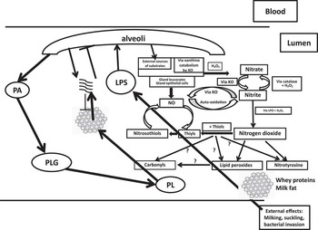
The association between thiols and NO (RSNO) is relatively weak, resulting in a slow dissociation of NO from nitrosothiols (Silanikove et al., Reference Silanikove, Shapiro, Shamay and Leitner2005). Thus, RSNO are probably the main reason for the constant cycling and accumulation of NO-derived species in milk (Silanikove et al., Reference Silanikove, Shapiro, Shamay and Leitner2005). As the rate of NO2• formation is second order with regard to NO-nitrite-mediated oxidation (reactions (1) to (3)), nitrosation reactions are limited by the availability of nitrite, which is a product of NO auto-oxidation. Thus, by maintaining a constant NO cycle, the ability to respond rapidly to a bacterial infection is preserved as rather small increase in NO surge will exceed the capacity of RSNO formation and nitrite oxidation by catalase to restrain the formation of NO2•. All in all, the continuous accumulation of nitrate following LPS treatment supports this model as summarized in Figure 2.
Schematic model that describes the simultaneous activation of the PA–PLG–PL system and the nitric oxide (NO)-derived cycle following challenge of the mammary gland with lipopolysaccharide (LPS), and the interaction between them. LPS challenge activates the PL system, and the increased PL activity releases peptides from the casein micelle, which in turn downregulate milk secretion and casein micelle clotting; see Leitner et al. (Reference Leitner, Merin and Silanikove2011) for further details. In parallel, LPS upregulates NO-cycle metabolism, which results in increased formation of nitrotyrosine, carbonyls and lipid peroxide; see Silanikove et al. (Reference Silanikove, Shapiro, Shamay and Leitner2005 and Reference Silanikove, Shapiro, Silanikove, Merin and Leitner2009b) for further details. The increased content of oxidized components in milk most likely increased their susceptibility to proteolysis Silanikove et al. (Reference Silanikove, Merin and Leitner2006). Casein-derived active peptides:  , casein micelle:
 , PA = plasminogen activator; PG = plasminogen; PL = plasmin; XO = xanthine oxidase; LPO = lactoperoxidase.

NO products accumulate in milk mainly as nitrate: the role of catalase
Catalases are enzymes ubiquitously found in all kinds of living organisms and are best known for catalyzing the decomposition of hydrogen peroxide to water and oxygen. However, catalase contains porphyrin heme (iron) groups in its center and can thus oxidize various acceptors, including nitrite, by functioning as peroxidase according to classical 3-step reactions of peroxidases (reactions (7) to (9)):


We have previously shown that conversion of nitrite into nitrate by catalase is the main function of milk catalase that serves as a basic mechanism for the prevention of excessive nitrosative stress in milk (Silanikove et al., Reference Silanikove, Shapiro, Shamay and Leitner2005 and Reference Silanikove, Shapiro, Silanikove, Merin and Leitner2009b). The increase in nitrate to levels of ∼80 μM in the treated glands suggests that the need to convert nitrite into nitrate under LPS challenge increased substantially (Silanikove et al., Reference Silanikove, Shapiro, Silanikove, Merin and Leitner2009b). The increase in uric acid (the product of xanthine or hypoxanthine oxidation in milk) concentration to ∼300 μM implies a similar increase in the secretion of XO substrates, xanthine and/or hypoxanthine, that led to a proportional increase in hydrogen peroxide production (Silanikove et al., Reference Silanikove, Shapiro and Leitner2007). The levels of uric acid measured in the present experiment suggest that V max of catalase exceeds the normal catalase activity in milk (Silanikove et al., Reference Silanikove, Shapiro, Silanikove, Merin and Leitner2009b). Thus, the drastic increase in catalase activity in the present results suggests that it plays an important role in protecting the gland from nitrosative stress and is consistent with a recent finding that the presence of intact catalase in milk is essential for preventing the accumulation of basal Ntyr, Lpx and carbonyls (Silanikove et al., Reference Silanikove, Shapiro, Silanikove, Merin and Leitner2009b). Thus, the present results reemphasize our previous conclusion that in light of its essential contribution to maintenance of milk quality and because of the importance of cow's milk in human nutrition, it is important to ensure that this enzyme is fully functional in each cow acquired by a commercial dairy farm.
LPS induced an increase in XO activity in milk
Hydrogen peroxide formation is essential for the conversion of nitrite into NO2• by LPO and oxidation of nitrite to nitrate by catalase. XO was identified as the major source of hydrogen peroxide in bovine milk (Silanikove et al., Reference Silanikove, Shapiro and Leitner2007 and Reference Silanikove, Shapiro, Silanikove, Merin and Leitner2009b). Under subclinical infection with E. coli, which induced much milder nitrosative stress than in this study, XO activity was upregulated by increasing the secretion of xanthine + hypoxanthine, which are the main physiological substrates of XO in milk. However, under acute inflammation, as in the present work and under involution, the activity of XO was upregulated both by an increase in its net secretion into the milk and by an increase in the secretion of its substrates, xanthine + hypoxanthine. This most likely reflects the large increase in the need for H2O2 and is reflected by the large increase in the uric acid concentration, consistent with previous reports (Silanikove et al., Reference Silanikove, Shapiro and Leitner2007 and Reference Silanikove, Shapiro, Silanikove, Merin and Leitner2009b).
The conversion of xanthine + hypoxanthine into uric acid is stoichiometrically linked to superoxide and H2O2 formation (reactions (10) to (13); Silanikove et al., Reference Silanikove, Shapiro and Leitner2007):


Whether superoxide or hydrogen peroxide would be coupled to xanthine + hypoxanthine oxidation at the molybdenum active site of XO depends on the number of electrons produced during the reaction, which depends on the reaction conditions. Fridovich (Reference Fridovich1970) has originally shown that under physiological conditions, ∼80% H2O2 and ∼20% O2•− are produced, whereas the production of 100% O2•− requires an environment of 100% O2 at pH 10, which is quite unphysiological. In accordance, it was shown that superoxide is not produced by milk XO (Silanikove et al., Reference Silanikove, Shapiro, Shamay and Leitner2005) and is scarcely produced by XO attached to the apical surface of bovine aortic endothelial cells (Kelley et al., Reference Kelley, Khoo, Hundley, Malik, Freeman and Tarpey2010), whereas hydrogen peroxide is the main product of XO oxidation under both conditions. The lack of evidence for O2•− production in milk is consistent with the fact that milk is much more hypoxic in comparison with blood.
In systemic fluid, XO is considered frequently as a source for the formation of harmful radicals, such as peroxynitrite and superoxide. The situation in milk is quite complicated. On the one hand, it provides H2O2 for the conversion of NO into NO2•, which is essential for the glandular defense, but impairs milk composition. On the other, it provides H2O2 for the conversion of nitrite into nitrate by catalase, which is essential for the resolution of inflammatory response and in maintaining milk quality during its storage in the udder and under commercial farming conditions (Silanikove et al., Reference Silanikove, Shapiro, Silanikove, Merin and Leitner2009b).
Conclusions
This study supports the concept that LPS induced NO burst from the surrounding mammary epithelial cells. The consequential nitrosative stress was most likely induced by the formation of NO2• from nitrite by LPO. This response was associated with an ∼3-fold increased formation of hazardous compounds, Ntyr, carbonyls and Lpx, and is associated with impairment in milk quality. Currently, very little is known about the presence of oxidative substances in commercial milk and about the health implications of their presence. Thus, our study stresses the importance of gaining further information in this direction.
Acknowledgment
This study was partially supported by the Israel Dairy Board.