Introduction
The candidate criteria for cochlear implantation have changed significantly since the first multichannel device was implanted in the late 1970s. In general, the pool of candidates has gradually expanded and now includes patients with residual hearingReference Fraysse, Macias, Sterkers, Burdo, Ramsden and Deguine 1 and cochlear malformations,Reference Buchman, Copeland, Yu, Brown, Carrasco and Pillsbury 2 and children younger than 12 months old.Reference Colletti 3 With improved techniques and experience, surgeons are more willing to perform cochlear implantations in challenging cases, especially in cases with severe cochlear malformations. Here, we report a case of cochlear implantation in a patient with severe cochlear hypoplasia.
Case report
We report the case of a female child, who, at the age of two years, was identified as having profound bilateral hearing loss. At three years of age, she received bilateral hearing aids and aural rehabilitation. Cochlear implantation was recommended at the age of four years because of poor hearing and speech ability.
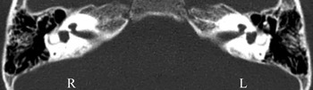
Auditory testing revealed profound, bilateral hearing loss. Computed tomography (CT) scans showed severe, bilateral cochlear hypoplasia with a rudimentary cochlear bud (Figure 1). The height of the bud was less than 5 mm. Both cochleae had formed as small, round cavities; the internal architecture of the cochlea, including the modiolus, was not visible.
Axial computed tomography image of patient's inner ears, demonstrating severe, bilateral cochlear hypoplasia. R = right; L = left

After thorough family counselling about the potential complications of a cochlear implantation, such as a cerebrospinal fluid (CSF) ‘gusher’, the insertion of the electrode array into the internal auditory canal and the likelihood of a less favourable outcome, it was agreed that the surgeon would perform a cochlear implantation in the patient's right ear.
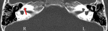
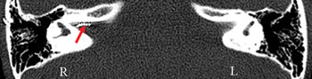
A suprameatal approach was used for the cochlear implantation. There were no complications during the operation with respect to facial nerves and middle-ear structures. As this patient did not have a normal cochlear structure, cochleostomy was performed on the right cochlear bud, parallel to where the cochlear axis should have been (Figure 2). During the cochleostomy, a CSF gusher occurred, which gradually subsided after approximately 10 minutes. Full insertion of the implant was not possible because of poor cochlear development. Thus, only seven electrodes of the Med-El Combi40+ implant (MED-EL, Innsbruck, Austria) were inserted through the newly created tunnel, and part of the electrode array was inserted into the internal auditory canal (Figure 3). Intra-operative monitoring demonstrated that the impedances of the seven inserted electrodes were appropriate; all other electrodes showed abnormal impedance measurements. A small piece of temporal muscle was placed around the electrode array to seal the cochleostomy.
Axial computed tomography image showing the cochleostomy site (arrow) on the right cochlear bud parallel to the imaginary cochlear axis. R = right; L = left

Axial computed tomography image of the right inner ear following cochlear implantation; the arrow indicates the part of the electrode array inserted into the internal auditory canal. R = right; L = left

Post-operative recovery was fairly stable, and the patient did not experience any facial nerve dysfunction or vertigo attacks.
One month after surgery, the cochlear implantation device was turned on. Neural responses could be evoked from the seven electrodes, all of which demonstrated appropriate impedance. Stimulation through two of the electrodes was found to cause facial twitching. Thus, only five of the electrodes were activated in the electrode mapping, using a continuous, interleaved sampling strategy.
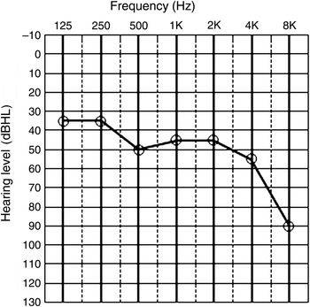
The patient received no formal speech training in the first year post-surgery due to financial constraints; during this period, her auditory and speech abilities did not improve. The patient began receiving speech training in the second year after the implantation, and the effectiveness of the implant improved thereafter. Furthermore, the two non-active electrodes were put into use a year after implantation because at this stage they no longer caused facial nerve twitching. Two years after implantation, the patient's audiogram thresholds ranged from 30 to 55 dBHL, with a flat hearing loss below 4 kHz and a severe hearing loss of 90 dBHL at 8 kHz (Figure 4). Table I shows the patient's mapping parameters with the seven activated electrodes at the last programming of the implant. At that time, the stimulation values were all within the normal range.
Pure tone audiogram of right ear with cochlear implant activated, two years after implantation.

Fitting parameters at last programming*

* For the Med-El Combi 40+ implant. Pulse rate/channel = 977.2 pulses per second. Strategy: continuous, interleaved sampling. MCL = Most comfortable level; CUL = current level; THR = threshold; DYN = dynamic value; PD = pulse duration
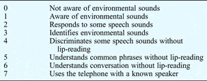
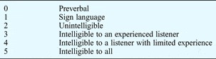
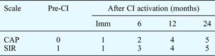
Improvements in auditory and speech functions were clearly demonstrated using the categories of auditory performance scale (CAP) categoriesReference Ahmad, Saeid, Paryaneh, Hessamaddin and Mohammad 4 (Table II) and the speech intelligibility rating (SIR) scaleReference Ahmad, Saeid, Paryaneh, Hessamaddin and Mohammad 4 (Table III) devised by Nikolopoulos and colleagues. The results are summarised in Table IV. Before cochlear implantation, the auditory performance scale and speech intelligibility ratings were 0 and 1, respectively; 24 months after cochlear implantation, they had both increased to 5. Although the patient's speech intelligibility was still poor compared with that of children of the same age with normal hearing, her family was satisfied with the outcome as she could go to a normal school and communicate with others. The patient's speech therapists were also satisfied and reported continual improvements.
Categories of auditory performance scale

Reproduced with permissionReference Ahmad, Saeid, Paryaneh, Hessamaddin and Mohammad 4
Speech intelligibility rating scale

Reproduced with permissionReference Ahmad, Saeid, Paryaneh, Hessamaddin and Mohammad 4 .
Auditory and speech results

CI = cochlear implant; Imm = immediately; CAP = categories of auditory performance scale; SIR = speech intelligibility rating scale
Discussion
Although cochlear implantation is a widely accepted procedure for hearing rehabilitation in patients with profound hearing loss, cochlear implantation surgery in ears with congenital malformation can be troublesome for the surgeon. Major concerns include CSF leakage, the insertion of the electrode array into the internal auditory canal, unpredictable distribution of neural tissue in patients with inner-ear anomalies, abnormal facial nerve activation, post-operative meningitis and uncertainty regarding the post-operative outcome.Reference Luntz, Balkany, Hodges and Telischi 5 – Reference Weber, Dillo, Dietrich, Maneke, Bertram and Lenarz 7 The risks increase with the degree of cochlear malformation.
In order to classify the various malformations of the inner ear, and to determine the surgical issues and rehabilitation outcomes associated with certain types of malformations, most reports use the classifications based on embryonic life devised by Jackler and colleagues.Reference Jackler, Luxford and House 8 The stage at which embryonic development of the cochlea is arrested can affect the severity of the resulting malformation. Thus, a malformation of the cochlea may consist of: total aplasia; severe cochlear hypoplasia; mild cochlear hypoplasia (basal turn only); a common cavity; severe, incomplete partition; mild, incomplete partition; or a subnormal cochlea that does not reach the full 2.5 turns.
Among these varied degrees of malformation, total aplasia is an absolute contraindication to cochlear implantation because such a cochlea has no spiral ganglion cells for the cochlear implant to stimulate.Reference Jackler, Luxford and House 9 However, cochlear implantation is recommended for patients with mild incomplete partition, a subnormal cochlea that does not reach the full 2.5 turns, mild cochlear hypoplasia (basal turn only) or severe incomplete partition. These patients have similar rehabilitation results as those with a normal inner ear structure,Reference Eisenman, Ashbaugh, Zwolan, Arts and Telian 10 despite the fact that implantation in the former patients often involves only partial insertion, even when a compressed electrode array is used.Reference Mylanus, Rotteveel and Leeuw 11 Patients with severe inner ear malformations, such as a common cavity or severe hypoplasia, are expected to have a less favourable outcome than patients with a normal cochlea, because they are likely to have fewer ganglion cells. They also present more complex surgical challenges, even to the most experienced otoneurologist.Reference Eisenman, Ashbaugh, Zwolan, Arts and Telian 10
Thus, patients with severe hypoplasia or a common cavity are controversial candidates for cochlear implantation, and, until recently, many cochlear implantation centres deferred implantation in children with these malformations. The patient presented in this report initially showed poor auditory and speech skills following implantation, although her delayed speech training may have partly contributed to this outcome. However, the (eventual) resulting benefits of cochlear implantation in this patient and others are encouraging, indicating that severe cochlear hypoplasia may not be a contraindication for multichannel cochlear implantation. The quality of life of such patients can be significantly improved with cochlear implantation.
When cochlear implantation is performed in a patient with severe cochlear hypoplasia, full insertion of all the stimulating electrodes is often not possible. Electrode insertion can also be complicated by misplacement of the electrode array through the deficient modiolus into the internal auditory canal or carotid canal. This may result from deeper insertion of the electrode(s) into a severely hypoplastic cochlea with insufficient available space. Surgeons often refuse to insert the electrode array into the internal auditory canal because an electrode in this position may stimulate the facial nerve.Reference Sennaroglu, Sarac and Ergin 12 However, the success of the case presented here indicates that such deep insertion of the cochlear implantation electrodes into the internal auditory canal is feasible and beneficial, despite the difficulty in the intra-operative electrode insertion and the possible occurrence of unwanted facial nerve stimulation after implant programming.
-
• Severe cochlear hypoplasia need not be a contraindication for cochlear implantation
-
• Patients with severe cochlear hypoplasia could benefit from an implant
-
• If full insertion is impossible, the electrode array can be partly inserted into the internal auditory canal
-
• If facial nerve stimulation occurs, the relevant electrodes can be turned off and tried later
In fact, even in cochlear implantation cases with normally structured cochleae, unwanted facial nerve stimulation may occur through the electrodes closest to the labyrinthine part of the nerve. Nevertheless, this is not an insurmountable problem in multichannel cochlear implants. The problem can usually be resolved by turning off the electrodes that cause facial nerve stimulation. In our case, only five of seven inserted electrodes were initially activated because the other two electrodes caused unwanted facial nerve stimulation. The patient's relatively poor auditory and speech skills immediately after implantation, compared with other implant patients, may have been partly attributable to the small number of activated electrodes. However, the two non-activated electrodes were turned on a year after implantation, without causing facial nerve activation, and thereafter the patient was able to benefit from these two extra electrodes.
Conclusion
The outcome of this case indicates that severe cochlear hypoplasia need not be a contraindication for cochlear implantation. If severe cochlear hypoplasia limits the available space, this may be compensated for by electrode insertion into the internal auditory canal. The problem of facial nerve stimulation can be resolved by switching off the affected electrodes; these electrodes may be able to be activated at a later point if they no longer cause facial nerve stimulation at that stage. Although the treatment outcome in patients with cochlear hypoplasia is less favourable than in those with a normal cochlear structure, the benefits can outweigh the risks and difficulties.
Acknowledgements
This study was supported by grants from The National Natural Science Foundation of China (30271410/C030310 and 81070778/H1303).