Case report
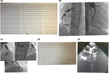
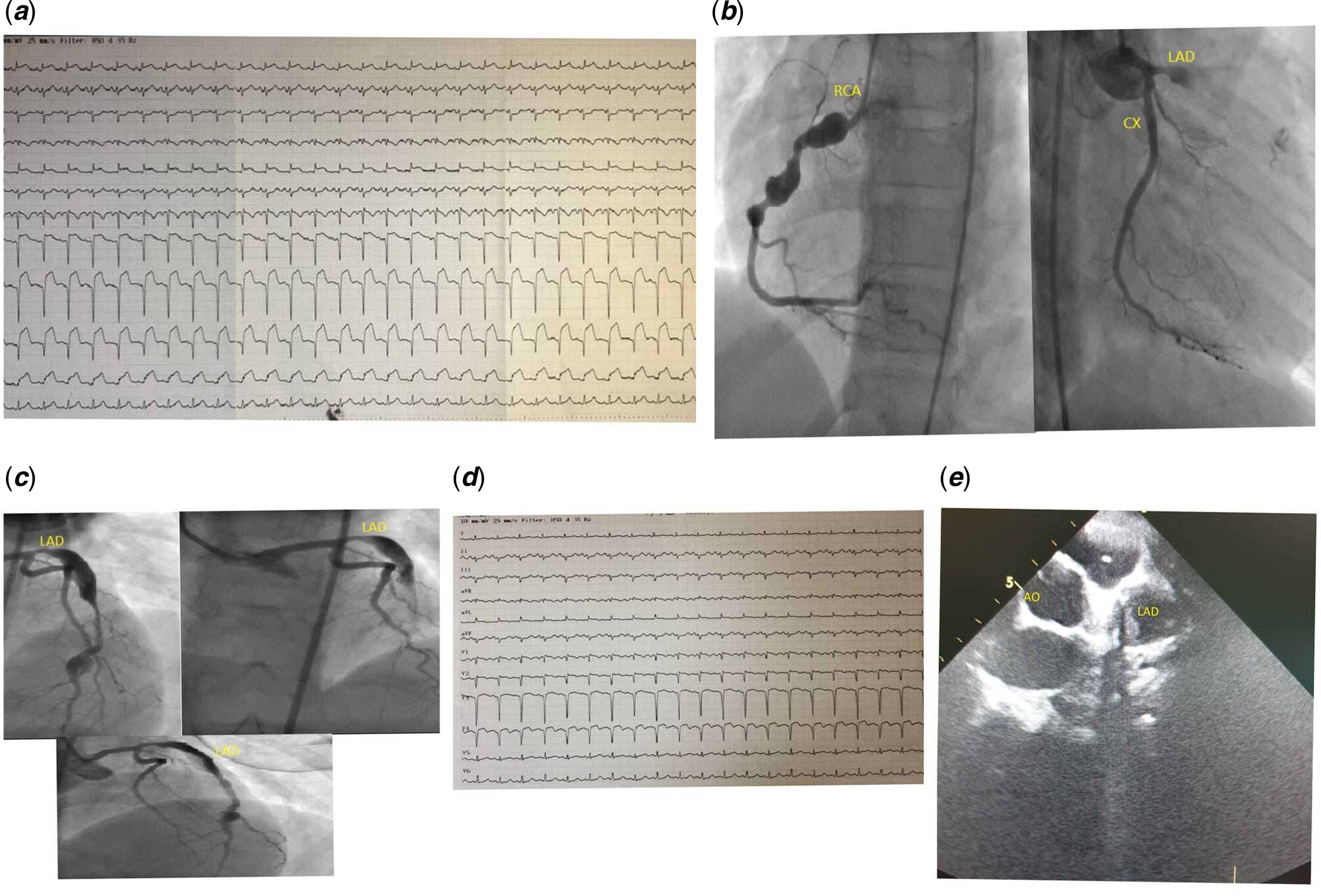
A 10-year-old girl patient diagnosed with Kawasaki disease 2 years ago and followed up in an external centre due to coronary aneurysm admitted to our emergency department with the complaints of chest pain, weakness, and palpitations. There were segment elevation (ST) elevations in V2, V3, V4, and V5 in the electrocardiogram (Fig 1a). CK-MB (mass): >300.000 ng/mL, hs Tropon I: >26.548 ng/L, and emergency selective coronary angiography were performed due to acute anterior myocardial infarction. Two giant aneurysms were seen in the right coronary artery in coronary angiography. Circumflex coronary artery was found normal. The left anterior descending artery was 100% totally occluded from the proximal (Fig 1b). Although left anterior descending artery was opened through proximally by coronary balloon angioplasty, a significant stenosis was observed in the middle segment. Giant aneurysm was observed in two separate locations both proximal and distal of the stenosis of the left anterior descending artery. Then, a coronary stent was implanted in the area of stenosis. The stenosis was fully opened in the left anterior descending artery (Fig 1c). Unfractionated heparin infusion was given only 1 day. Then unfractionated heparin was stopped and low-molecular-weight heparin (enoxaparin sodium) was started. After percutaneous coronary intervention, it was observed that ST elevations were significantly improved on the electrocardiogram (Fig 1d). There was a significant decrease in control cardiac enzymes. Enoxaparin sodium was discontinued after the INR level reached between 2 and 3 sn following a few days. Acetylsalicylic acid and warfarin sodium treatment was continued. On echocardiography, a giant aneurysm with a diameter of 16 mm in the left anterior descending artery was detected (Fig 1e). Ejection fraction was normal. The patient who had regressed myocardial infarction findings on electrocardiogram and had an INR around 2–3 was discharged with beta-blocker, angiotensin converting inhibitor, acetylsalicylic acid, and warfarin sodium treatment.
( a ) ST elevations in V2, V3, V4, and V5 in the electrocardiogram (ECG). ( b ) Two giant aneurysms were seen in the right coronary artery (RCA) in coronary angiography. Circumflex coronary artery (CX) was found normal. The LAD was 100% totally occluded from the proximal. ( c ) Although left anterior descending artery was opened through proximally by coronary balloon angioplasty, a significant stenosis was observed in the middle segment. Giant aneurysm was observed in two separate locations both proximal and distal of the stenosis of the left anterior descending artery. Then, a coronary stent was implanted in the area of stenosis. The stenosis was fully opened in the left anterior descending artery. ( d ) ST elevations were significantly improved on the ECG. ( e ) On echocardiography (ECHO), a giant aneurysm with a diameter of 16 mm in the LAD was detected. AO = aort; LAD = left anterior descending artery.
Discussion
Acute myocardial infarction is the most important cause of morbidity and mortality in patients with Kawasaki.Reference Kato, Sugimura and Akagi1,Reference McCrindle, Rowley and Newburger2 Therefore, anticoagulant therapy is recommended along with antiplatelet therapy in patients with Kawasaki who have giant aneurysm.Reference Tsuda, Hamaoka and Suzuki3 The blood flow that slows down as a result of the enlargement of the coronary artery may cause ischaemia and myocardial infarction by causing turbulent flow and thrombosis.Reference Markis, Joffe, Cohn, Feen, Herman and Gorlin4 Kawasaki patients with coronary artery aneurysm should receive regular acetylsalicylic acid and warfarin sodium therapy and INR levels should be kept between 2 and 3 to prevent myocardial infarction.Reference Newburger, Takahashi and Gerber5 Although our patient received acetylsalicylic acid and warfarin treatment, we found that the INR level was constantly between 1.2 and 1.8. Acute myocardial infarction may be due to this.
Successful results have been obtained with coronary bypass surgery in some Kawasaki patients with giant aneurysms.Reference Muta and Ishii6 Although percutaneous coronary intervention is the standard treatment procedure in adults, percutaneous coronary intervention applicability is limited in children due to the small size of the patient, the inability to find an appropriate size catheter, and lack of technical expertise. Therefore, the paediatric cardiologist should make a percutaneous coronary intervention decision considering both the patient and the current conditions.Reference Mason and Takahashi7
Percutaneous coronary intervention and coronary bypass are the procedures that should be performed in Kawasaki patients with coronary pathology. However, there is no consensus on whichever should be done first. In a study, it was thought that percutaneous coronary intervention was less effective than coronary bypass in Kawasaki patients.Reference Muta and Ishii6
If the lesion in the coronary arteries is localised and short, transcatheter intervention is recommended. However, if the patient has severe left ventricular dysfunction, the best first intervention is cardiac bypass surgery.Reference Ishii, Ueno and Akagi8
In another study, as in our patient, a patient diagnosed Kawasaki with giant coronary aneurysm admitted with anterior myocardial infarction and the occluded vessel was recanalised by immediately percutaneous coronary intervention and it was determined that the patient’s ejection fraction was preserved. In the control of this patient after 2 years, it was found that ventricular contraction was preserved. In this study, it was advocated that percutaneous coronary intervention is an effective and a safe treatment method in the long term in Kawasaki patients with giant coronary aneurysm who developed acute myocardial infarction.Reference Maurizio Mongiovì, Annalisa Alaimo and Federica Vernuccio9
Although myocardial infarction in childhood is rare, myocardial infarction should be considered in children with a history of Kawasaki disease. It should be questioned whether there may be a coronary aneurysm. Also, it should be known that patients with aneurysm should take acetylsalicylic acid and warfarin and the INR level should be kept between 2 and 3 in the Kawasaki patients. Although our patient was a child, we successfully performed percutaneous coronary intervention and stabilised our patient. However, more studies are needed to determine whether percutaneous coronary intervention or coronary bypass is the most appropriate procedure for these patients.
Acknowledgements
None.
Financial support
This research received no specific grant from any funding agency, commercial, or not-for-profit sectors.
Conflicts of interest
None.
Ethical standards
Not applicable.
Authors’ Contributions
MT and AA: Concept, patient management, radiosurgery, manuscript writing, review, critical evaluation, submission; FE and AAO: Patient management, critical evaluation of manuscript; MT: Patient management, critical evaluation of manuscript, endocrinological management; AA and MT: patient management; AA and SS: Radiology support, patient management; MT and FE: Patient management, literature review; AAO: Literature review and administration support. MT: will act as guarantor for this paper.