The presence of coarctation of the aorta in patients with tetralogy of Fallot or pulmonary atresia with ventricular septal defect is thought to be an exceedingly rare condition, with only 11 cases described in the medical literature.Reference Yip, Deekollu and Arnold 1 – Reference Momma 9 In fact, the coexistence of these conditions seemingly contradicts many of the foetal haemodynamic theories regarding the development of coarctation.Reference Rudolph 10 This combination of defects can be quite challenging to diagnose and has resulted in early post-operative mortality in a patient following tetralogy repair.Reference Gunthard, Murdison and Wagner 2 We present three patients with variants of tetralogy of Fallot who were subsequently found to have coarctation of the aorta.
Case presentations
Case 1
Patient 1 was a 31-week estimated gestational age female infant with a birth weight of 1.33 kg with tetralogy of Fallot with ductal-dependent pulmonary blood flow. She was maintained on prostaglandin until 2 months of life when she underwent transannular patch-type repair. She was discharged home but subsequently presented at 3 months of life with vomiting. Echocardiography revealed a 50-mmHg peak gradient in her descending aorta. Owing to this surprising finding, a cardiac magnetic resonance imaging was obtained, demonstrating coarctation of the aorta with a minimum diameter of 1.7 mm. She underwent patch augmentation of her transverse and proximal descending aorta. At 6.5 months of life, she underwent balloon angioplasty of recurrent coarctation of the aorta. Her ventricular function remains normal.
Case 2
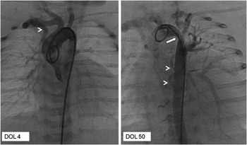
Patient 2 was a 35 6/7-week, 2.54-kilogram male with a prenatal diagnosis of pulmonary atresia. Newborn cardiac catheterisation demonstrated pulmonary atresia/ventricular septal defect with absent native branch pulmonary arteries and the presence of major aortopulmonary collateral arteries arising from the right subclavian artery and the descending aorta. There was no evidence of coarctation of the aorta. The patient tolerated discontinuation of prostaglandin infusion and was discharged to outpatient care. After 6 weeks, he presented acutely ill with feeding intolerance and cyanosis, with echocardiography demonstrating severely decreased left ventricular function and coarctation of the aorta with a peak gradient of 70 mmHg. He underwent cardiac catheterisation, confirming coarctation of the aorta with a peak-to-peak gradient of 42 mmHg (Fig 1) and a left ventricular end-diastolic pressure of 18 mmHg. He underwent balloon angioplasty and stent placement, allowing time for recovery of ventricular function before patch aortoplasty and unifocalisation of left-sided collaterals. He later developed recurrent coarctation of the aorta and required balloon angioplasty. At present, he is awaiting further surgical palliation.
Angiography of patient 2 on day of life (DOL) 4 while on continuous prostaglandin infusion demonstrating a widely patent aortic arch with multiple aortopulmonary collateral arteries (MAPCAs) including a prominent vessel off of the right subclavian artery supplying the right lung (arrow head). The second image of the same patient on DOL 50 demonstrates discrete coarctation of the aorta at the site of takeoff of the left subclavian artery. In addition, two large aortopulmonary collateral arteries are seen arising from the descending aorta (arrow heads).

Case 3
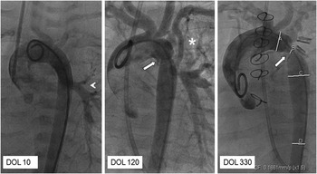
Patient 3 was a 39 1/7-week, 2.37 kg female with oesophageal atresia/tracheoesophageal fistula and tetralogy of Fallot with discontinuous left pulmonary artery. She was maintained on prostaglandin. Cardiac catheterisation confirmed that the discontinuous left pulmonary artery was supplied by the persistent ductus arteriosus; there were no additional collaterals. A left modified Blalock–Taussig Shunt was placed on day of life 25. She underwent a routine cardiac catheterisation at 4 months of life, demonstrating no pressure gradient in the aorta by direct measurement. She then underwent tetralogy repair with reimplantation of the left pulmonary artery at 8 months of life. At follow-up, she was noted to have progressive ventricular dysfunction with an ejection fraction of 30%. This prompted a cardiac catheterisation that showed a discrete coarctation with a 22-mmHg peak-to-peak gradient and a left ventricular end-diastolic pressure of 17 mmHg (Fig 2). She underwent coarctation repair with extended end-to-end anastomosis at 11 months of life. At follow-up, she was noted to have gradual but incomplete recovery of her ventricular function.
Angiography of patient 3 on day of life (DOL) 10 while on continuous prostaglandin infusion demonstrating a widely patent aortic arch with a discontinuous left pulmonary artery supplied by the patent ductus arteriosus (arrow head). The second frame is of the same patient on DOL 120 demonstrating patency of a left modified Blalock–Tausig Shunt (*) supplying the left pulmonary artery and an area of narrowing of the aorta distal to the site takeoff of the left subclavian artery (arrow). This narrowing ultimately developed into a focal coarctation on DOL 330 measuring 3 mm.

Discussion
The presence of both left- and right-sided outflow tract obstruction has been described to be an exceedingly rare occurrence. We present three patients with right ventricular outflow tract obstruction who subsequently developed coarctation of the aorta. Importantly, the significance of the arch obstruction in each of these patients was initially overlooked. Owing to the fact that it is perceived to be such a rare entity in this setting, further imaging was pursued to clearly establish the diagnosis before intervention. Patient 1 had extensive imaging, largely because the diagnosis was in doubt. Timely surgical intervention occurred in this patient, before left ventricular function deteriorated. Patient 2 presented with acute decompensation, and prompt transcatheter intervention was critical to the recovery of ventricular function. Patient 3 had echocardiographic evidence of coarctation of the aorta, but intervention was delayed because the significance of the imaging findings was not appreciated until there was a significant clinical change. This patient has yet to fully recover left ventricular function despite 18 months of medical therapy. One might speculate as to the anatomic basis for the development of coarctation in our patients. The substrate for the coarctation for patient 1 may have been ductal tissue in the juxtaductal aorta, which constricted after discontinuation of chronic prostaglandin infusion, similar to isolated coarctation of the aorta. In patient two, it is possible that there was a large amount of flow through the right subclavian artery into the largest collateral vessel, thus reducing flow in the distal aorta. However, this physiology is common with major aortopulmonary collaterals and coarctation remains quite rare. In patient 3, the development of coarctation of the aorta is even more perplexing.
Conclusion
We present three patients with right ventricular outflow tract obstruction who subsequently developed coarctation of the aorta. These patients were identified within the last 6 years of our clinical practice, suggesting that the coexistence of coarctation of the aorta in patients with variants of tetralogy of Fallot may be significantly more common than previously suspected. Our experience with these patients underscores the critical importance of evaluating the aorta in patients with various types of right ventricular outflow tract obstruction during their entire course. Delayed diagnosis of this rare association can result in substantial morbidity as the resultant left ventricular dysfunction may not fully resolve.