Introduction
Endoscopic resection of skull base tumours can be very challenging due to difficulties with safe access. The risk of life-threatening bleeding secondary to injury of the internal carotid or internal maxillary artery often forces surgeons to opt for a safer, open surgical approach.
However, over the past few years new technological advances have allowed surgeons to operate in previously inaccessible areas. The ultrasonic dissector has been part of the armamentarium of neurosurgeons and general surgeons for many years; however, it is also a useful instrument for otorhinolaryngologists, for endoscopic resection of specific skull base lesions.
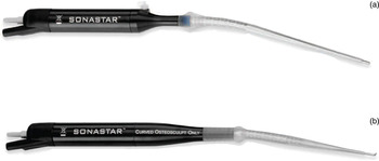
In this paper, we present five patients who underwent endoscopic skull base surgery using the Misonix SonaStar ultrasonic dissector (Misonix; Farmingdale, New York, USA) (Figure 1).
Misonix SonaStar ultrasonic (a) soft tissue and (b) bone dissector.

Soft tissue ultrasonic dissectors have been utilised since the 1980s. Their main use has been in laparoscopic, liver and thyroid surgery.Reference Cirocchi, D'Ajello, Trastulli, Santoro, Di Rocco and Vendettuoli1 Ultrasonic bone dissectors have been developed and utilised in spinal and dental surgery.
Electrical energy is generated by an electronic generator and then converted into mechanical vibrations through piezoelectric crystals located in the hand-piece of the instrument. Ultrasound is mechanical vibration at a frequency above human hearing; the ultrasonic dissector functions at more than 22.5 kHz.
Medical ultrasound works by expanding and compressing tissues thousands of times per second, causing cell walls to break and tissues to disrupt, together with coagulation of vessels, depending on the frequencies used. Tumours can therefore be resected with dissection onto vessels but without vessel injury. This is a major advantage, especially when tumours encroach on major arteries.
The endoscopic approach to the petrous apex necessitates the use of an instrument that can navigate through a very narrow corridor between the internal carotid artery (ICA) and the dura. Intranasal drills are classically used for this approach, but even with neuro-navigation the risk to the ICA is significant.Reference Chatrath, Nouraei, De Cordova, Patel and Saleh2 Injury to this vessel usually has catastrophic consequences, and any device that could potentially reduce this risk will be of value to the otolaryngologist and neurosurgeon.
Endoscopic surgeons who perform advanced skull base surgery in which instruments need to reach beyond the sphenoid sinus frequently experience specific problems. Instruments are often too short, and intranasal drills may not have built-in irrigation and suction; furthermore, visualisation may be reduced secondary to the smoke generated. When working on the ICA during advanced petrous apex or sphenoid sinus surgery, surgery cannot be performed safely unless a bloodless field is created. In this context, the ultrasonic dissector has the potential to reduce the risk of intra-operative bleeding and to facilitate dissection when lesions abut vital structures.
Case reports
Case one
A 58-year-old woman presented with a one-year history of unilateral sensorineural hearing loss and headaches.
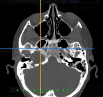
A computed tomography brain scan revealed a large petrous apex cholesterol granuloma (Figure 2).
Axial computed tomography scan of case one, showing a petrous apex cholesterol granuloma.

Endoscopic drainage of petrous apex cholesterol granulomas has definite advantages over the open surgical approach. A permanent drainage stent may be inserted, with drainage into the nasal cavity, and post-operative recovery is quicker when compared with a craniotomy.
This first patient's surgery was particularly difficult due to the narrow surgical corridor that had to be created superior to the internal carotid artery (ICA). The cystic lesion displaced the ICA inferiorly, and the margin for error was less than 2 mm.
Endoscopic access to the petrous apex is usually obtained using an extra-long intranasal drill. The danger with this instrument is the risk of lacerating the ICA when working in such a narrow surgical corridor, especially since the tip of the drill cannot be visualised during this stage. This is a major obstacle when operating in this area, since the navigation system will not be useful during this stage of the surgery. The smoke generated by the drill makes this surgical stage even more difficult.
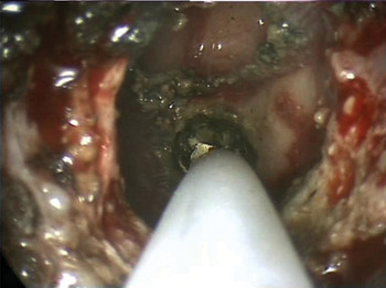
The ultrasonic dissector has many benefits during endoscopic trans-sphenoidal surgery to the petrous apex. Firstly, the instrument has a specialised hand-piece dissection probe that can reach quite comfortably the petrous apex or lesions at the posterior aspect of the anterior skull base. Secondly, the ultrasonic bone dissector is invaluable when creating a narrow surgical corridor, as the tip is small enough to be visualised throughout the dissection process (Figure 3). Thirdly, the dissector will only dissect bone and will not injure any soft tissue structures or the ICA.
Endoscopic view of the ultrasonic bone dissector proceeding through the posterior wall of the maxillary sinus, the pterygopalatine fossa and the posterolateral wall of the sphenoid sinus.

The only disadvantage of the ultrasonic dissector during endoscopic surgery is the mist created by the irrigation system, which forces one to keep the endoscope at a distance, reducing visualisation to some degree. Irrigation can be decreased to avoid this but the dissecting probe will heat up, with possible thermal damage to nearby structures. A study by Emam and Cuschieri showed that tissue temperatures rose to 140°C 1 cm from the tip of the ultrasonic dissector used in their study on an animal model.Reference Emam and Cuschieri3
Our patient developed a brief bradycardia as surgical dissection approached the ICA. This immediately resolved when irrigation was increased. The cystic lesion was easily identified and drained, and the patient was discharged the following day with no complications.
Case two
A 70-year-old man presented with a one-week history of severe, right-sided trigeminal pain with complete numbness in the distribution of the second branch of the trigeminal nerve.
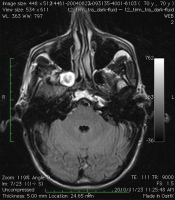
An MRI scan showed a brightly enhancing lesion in the pterygopalatine fossa (Figure 4). A diagnosis of schwannoma was suggested.
Axial magnetic resonance imaging scan of case two, showing a pterygopalatine fossa abscess.

Endoscopic resection required dissection on the internal maxillary artery. The ultrasonic dissector was used to facilitate dissection on the internal maxillary artery, without injuring the vessel.
Dissection within the pterygopalatine fossa is always complex due to the difficulty in differentiating between fat, tumour, blood vessels and nerves. The soft tissue ultrasonic dissector only dissects soft tissue and is thus unable to cut through bone or vascular tissue.
In this second patient, the sphenopalatine artery was identified and followed back to the internal maxillary artery, without injury to the latter. The mass was exposed and found to be an abscess of the pterygopalatine fossa.
Case three
A 14-year-old boy presented with a six-month history of right-sided nasal obstruction.
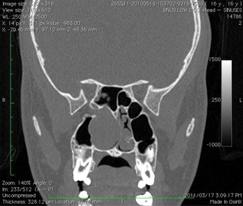
Imaging studies revealed a large angiofibroma involving the sinonasal cavity, pterygopalatine fossa and sphenoid sinus (Figure 5).
Coronal computed tomography scan of case three, showing an angiofibroma.

An endoscopic resection was planned. The patient underwent embolisation two days before surgery to minimise blood loss. The soft tissue ultrasonic dissector was used, in order to assess its feasibility for dissection of a previously embolised vascular tumour. Although the ultrasonic dissector facilitated dissection in the pterygopalatine fossa and on the lateral wall of the sphenoid sinus in close proximity to the ICA, it was of limited use unless optimal embolisation had been achieved. In areas where this was not the case, dissection was difficult as the ultrasonic dissector could not disrupt viable blood vessels.
As a result of this experience, we propose that vascular tumours should be embolised five to seven days before ultrasonic dissector surgery, to allow the tumour to become hard and fibrotic.
Case four
A 43-year-old man was diagnosed with a rare neuro-endocrine tumour involving the right fossa of Rosenmüller. Only a handful of previous cases have been documented in the literature.Reference Mohebbi, Daneshi and Emami4 The response to radiotherapy is unpredictable, and the patient was therefore referred for endoscopic resection before adjuvant treatment was considered. The major limitation of and risk to surgery in the fossa of Rosenmüller is the presence of the ICA. The ultrasonic dissector was therefore used, in conjunction with neuro-navigation, to safeguard the ICA.
In this case, the ultrasonic dissector was invaluable due to its length, bloodless dissection field, and ability to avoid injury to the ICA during close dissection.
Case five
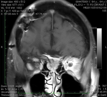
A 63-year-old woman was referred for endoscopic resection of a large sinonasal meningioma (Figure 6). The tumour was located centrally, over the cribriform plate, and extended to the posterior ethmoids bilaterally.
Coronal magnetic resonance imaging scan of case five, showing a sinonasal meningioma.

The lesion was resected using the ultrasonic dissector, enabling precise, bloodless dissection onto the skull base. The dura was easily identified, minimising the risk of a cerebrospinal fluid leak. As meningiomas are soft tumours, this patient's tumour was quickly and efficiently debulked using the ultrasonic dissector.
Discussion
There is no previously published English language literature on the use of the ultrasonic dissector in skull base surgery and otorhinolaryngology. A PubMed search using ‘ultrasonic dissector’ as the main search term revealed that only 27 articles have been published on the use of ultrasonic dissectors since 1986. None of these articles discussed the potential use of this instrument in skull base surgery or otorhinolaryngology.
Concern has previously been raised regarding the potential thermal risk to surrounding tissues posed by ultrasonic dissection.Reference Emam and Cuschieri3 However, new ultrasonic dissection systems have central irrigation channels in the titanium probe which continually cool the device.
An advantage of the Misonix SonaStar ultrasonic dissector used was the availability of both bone and soft tissue dissection attachments. The bone dissector attachment is especially useful when bone needs to be removed close to vital structures. A perfect example is the creation of a pathway to the petrous apex; using the bone dissector, one is able to create a narrow surgical pathway which avoids injury to the dura and the internal carotid artery (ICA).
The tip of the bone dissector is shaped as a small, flat hook (Figure 1b) approximately 2 mm in diameter. This shape enables the surgeon to use the instrument as a drill, by pushing down onto the flat surface of the hook. The real advantage of the design lies in the fact that the hook can be placed under a ledge of bone, its tip applied to the underside of the ledge, and then traction applied to pull the instrument towards the surgeon, thereby enlarging the surgical corridor and enabling the surgeon to work away from any vital structures.
The main difference between using an endonasal drill and an ultrasonic bone dissector is the fact that the tip of the bone dissector can always be visualised regardless of the size of the corridor being drilled. The ultrasonic bone dissector has a long, thin shaft, and this is an essential feature when using multiple instruments simultaneously in the nasal cavity.
Another major advantage of the ultrasonic bone dissector is the fact that the tip never becomes blunt, and there is thus no requirement for multiple drill bits during the one procedure.
Furthermore, the shape of the ultrasonic dissector allows one to perform different steps with the same instrument, obviating the need to change instruments to perform a simple task such as removing the face of the sphenoid sinus during trans-sphenoidal surgery. (This would usually entail using a sphenoid punch to enlarge the ostium, followed by a Kerrison punch or drill to remove the rostrum of the sphenoid.) When using a drill, special care needs to be taken to ensure the dura is not breached when removing bone over the pituitary fossa or planum sphenoidale. The ultrasonic bone dissector can be used as a drill by pushing on its tip, and as a Kerrison punch by hooking its tip around bony ledges, while at the same time avoiding injury to soft tissue and dura.
• The ultrasonic dissector is useful for endoscopic resection of skull base tumours
• It is invaluable for endoscopic trans-sphenoidal access to petrous apex lesions
• Its vessel-sparing properties are ideal for resection of pterygopalatine fossa lesions
The ultrasonic soft tissue dissector will cut only soft tissues, and will not injure any vessel with flow in it, nor cut through bony structures. The instrument has the feel of a nasal sucker. During dissection, tissue ablation is achieved by pushing the instrument against the relevant tissue. As pressure is released, the instrument acts as a sucker. The rate of soft tissue dissection depends on the pressure applied to the tissue; increased pressure leads to more rapid dissection.
In both the soft tissue and bone dissector attachments, irrigation runs constantly through the tip of the instrument, cooling it and avoiding thermal tissue damage.
Conclusion
Endoscopic skull base surgery has many challenges. New surgical technologies have enabled access to areas in which there is a significant risk of injury to the internal carotid artery, dura and other vital structures.
The five presented cases highlight potential uses of the ultrasonic dissector. The major advantages of this instrument are its long, tapered shaft, superior visual field (compared with an endonasal drill), protection of vital structures during dissection in difficult areas, and avoidance of the need for multiple drill bits during surgery.
Accessing the petrous apex via a trans-sphenoidal approach is especially difficult, and the ultrasonic dissector shows its greatest potential for this type of surgery.
Research is needed to determine whether ultrasonic dissection results in significant differences in blood loss and angiofibroma surgery outcomes, compared with conventional surgery.