Introduction
Epithelial–mesenchymal transition (EMT) is recognised as naturally occurring process during embryogenesis, including mesoderm formation and neural crest development, adult tissue repair and its maintenance. Loss of adherens junctions, desmosomes and tight junctions that connect epithelial cells laterally with the neighbouring cells as well as to the underlying basement to maintain apical–basal polarity is the characteristic property of EMT. As a result stationary cells exhibit plasticity, lose epithelial phenotype and gain mesenchymal phenotype, including migratory abilities and fibroblastoid morphology during developmental morphogenesis (Ref. Reference Boyer and Thiery1). EMT usually undergoes a reverse process known as mesenchymal–epithelial transition (MET), which is required during differentiation for fulfilling developmental tasks (Ref. Reference Lim and Thiery2). During the transition from epithelial (E) to mesenchymal (M) type, cells adopt partial EMT phenotype also called as hybrid E/M phenotype. Cells with mixed epithelial and mesenchymal properties gain maximal cellular plasticity and stemness during morphogenesis.
Significance of EMT program classified as type 1 EMT is well characterised in morphogenesis of different organs and embryonic development, whereas type 2 EMT contributes towards wound healing, organ fibrosis and tissue regeneration (Ref. Reference Garg3). Functions of type 3 EMT are well known in conferring tumour properties to epithelial cells, enhanced cellular plasticity, metastatic potential, cancer stem cell (CSC) phenotype and resistance to drug therapies at achievable doses, thereby result in tumour progression and recurrence in due course of time. EMT and its intermediate states dynamically transit between epithelial (E) and mesenchymal (M) phenotypes and identified as important drivers of organ fibrosis and tumour progression (Ref. Reference Nieto4). Tumour cells are identified to share number of molecular and morphological traits with neighbouring stromal fibroblasts. At the same time, constantly evolving tumour microenvironment bestows tumour cells to adopt epithelial/mesenchymal (E/M) hybrid phenotype and gain cellular plasticity. As a result, these tumour cells gain migratory potential, invade basement membrane, enter peripheral blood vessels and become circulating tumour cells (CTCs). These cells undergo MET process, either move to distant organs or self-seed back to original target organ, settle down, extravasate and proliferate to establish secondary metastasis (Ref. Reference Kim5).
Radiological examinations, pathological staging, grading and molecular markers are still the gold standard methods for diagnosing the clinical manifestation of tumour growth; however, unfortunately, they fail to provide accurate and reproducible information about the diagnosis and prognosis of ongoing metastasis. Presence of CTCs with E/M hybrid phenotype and cancer stemness property not only reflect the aggressiveness of tumour, but also set challenges for diagnosing malignancy, monitoring disease recurrence, detecting prognosis and achieving therapeutic success for increased survival of cancer patients. The aim of this review is to highlight and recommence our understanding on the significance of EMT program, various biological issues of CTCs with hybrid E/M or cancer cells with either E or M phenotype and their clinical relevance during every stage of cancer metastasis.
EMT
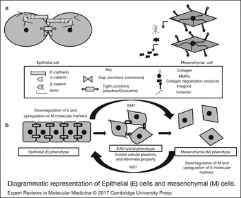
Developmental programs that control specification and differentiation are mechanistically governed by molecular/cellular changes that regulate EMT/MET program. EMT is characterised by the transcriptional repression of many epithelial junction proteins, including E-cadherin, α-catenin, γ-catenin/plakoglobin, desmosomes and cytokeratins. Promoter hypermethylation, protein phosphorylation followed by degradation of epithelial marker proteins are observed during EMT. Expression of fibronectin (FN), an extracellular protein, required for mesenchymal cell migration, is induced during EMT (Fig. 1). Enhanced activity of PDGF (platelet-derived growth factor)/PDGFR (platelet-derived growth factor receptor) autocrine loop to induce invadopodia-mediated degradation of extracellular matrix (ECM) is required for Twist1 to promote cell migration and invasion (Ref. Reference Eckert6). Upregulated expression of nonepithelial cadherins, N-cadherin, cell surface proteins, CD44 (cluster of differentiation 44) and integrin β6, switch from cytokeratin to vimentin, synthesis of ECM components and matrix metalloproteinases (MMPs), validate the occurrence of EMT and endow cancer cells with fibroblastoid phenotype.
(a) Diagrammatic representation of epithelial (E) cells and mesenchymal (M) cells. E cells express epithelial marker proteins, but loose cell–cell contacts, gain motility, spread to surrounding tissues and acquire M phenotype during cancer. (b) Epithelial–mesenchymal transition (EMT) is a multistep biological process where epithelial cells undergo morphological change to motile M phenotype. E and M cells undergo transition during EMT program and adopt a hybrid E/M phenotype. Mesenchymal–epithelial transition (MET) is a reverse process where mesenchymal cells gradually become tightly linked, acquire polarised E phenotype and mark the progression of cancer disease. Cancer cells with partial E/M hybrid phenotype co-express both E and M marker proteins and are associated with increased cellular plasticity and stemness. Partial EMT and not complete EMT allows cells to gain migratory and invasive traits during cancer metastasis.

Experimental studies over the past few years document the role of signalling pathways and classify the molecules as signature molecules that act as inducers and regulators of EMT/MET during oncogenic transformation.
EMT core regulators and inducers
Three groups of core EMT regulators play significant role in inducing EMT and are thus responsible for metastatic progression. Zeb family consists of well-conserved zinc finger/homeodomain proteins Zeb1 and Zeb2, which trigger EMT by binding to E-box in the promoter region to suppress its expression. Distantly related group of Snail zinc finger family comprises three members: Snail, Slug and Smuc. These members share zinc finger cluster at C-terminal region and SNAG domain at N-terminal region; bind to E-box of E-cadherin promoter and repress its expression. Basic helix-loop-helix (bHLH) is another family of EMT-transcription factors that consists of Twist1, Twist2 and E12/47 and regulates the expression of target genes, acts as co-repressor of E-cadherin, and activates programs required for tumour invasion (Ref. Reference Garg3).
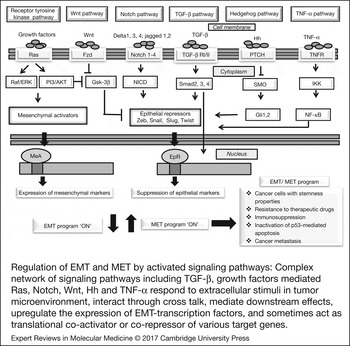
Signalling pathways, including transforming growth factor receptor-β (TGF-β), insulin growth factor (IGF), epidermal growth factor (EGF), hepatocyte growth factor (HGF), fibroblast growth factor (FGF), Notch, Wnt (wingless type), Hh (hedgehog) and tumour necrosis factor α (TNFα) interact through crosstalk, influence EMT regulatory network and thus are referred as motor of cellular plasticity. These pathways respond to extracellular stimuli in tumour microenvironment and couple EMT/MET program with other vital cellular properties such as metabolism, apoptosis, immune evasion and cell cycle. Aberrant activation of signalling pathways results in expression of specific downstream target genes. These targets when interact with EMT core regulators, initiate cancer growth and metastasis (Fig. 2). Serine–threonine kinase receptors, TβRI (TGFβ receptor I) and TβRII (TGFβ receptor II), when bind with TGF-β ligand, activate Smad 2 and Smad3. Smad2/3 interacts with Smad 4, get translocated to nucleus to activate the expression of target genes (Ref. Reference Attisano and Wrana7). This pathway is considered as primary inducer of EMT and is shown to promote EMT core transcription factors, including Zeb1, Zeb2/Sip1 (smad interacting protein 1), Snail1, Snail2/Slug and Twist1. This pathway collaborates with receptor tyrosine kinase (RTK) pathway where growth factors, HGF, EGF and FGF activate RTKs/Ras followed by the activation of downstream effectors, mitogen-activated protein kinases (MAPK) or PI3 K (phosphoinositide 3-kinase) (Ref. Reference Grünert, Jechlinger and Beug8). Activation of Wnt signalling requires the binding of Wnt ligands to (Fz) Frizzled and lipoprotein receptor, followed by inhibition of destruction complex consisting of APC (adenomatous polyposis coli), Axin, GSK 3β (glycogen synthase kinase 3β) and casein kinase-1α. This stabilises and translocates β-catenin to the nucleus where it interacts with LEF/TCF (lymphoid enhancer factor/T cell factor) and acts as coactivator for the transcription of target genes. Intracellular protein Axin 2 stabilises Snail and acts as EMT inducer (Ref. Reference Gordon and Nusse9). EMT induction also depends upon the cooperative activity of β-catenin/LEF and TGF-β signalling pathways. Binding of Notch receptors, including Jagged1, 2, Delta like 1, 3 and 4 initiate notch signalling by facilitating intramembrane cleavage of notch receptor by γ-secretase followed by release of notch intracellular domain and its translocation to nucleus to activate target genes’ expression (Ref. Reference Wang and Zhou10). However, this signalling fails to induce adequate EMT, but when coordinates with TGF-β through smad3, notch activity gets increased which upregulates the expression of Jagged1, HEY1 (hairy/enhancer of split related repressor) and Slug. Hedgehog signalling is initiated by binding of Patched (PTCH) receptor with Hh ligands, including Sonic, Desert and Indian which results in de-repression of SMO (smoothened), activation of GLI-1, -2 and -3 zinc-finger transcription factors and induction of EMT program by increasing the expression of Snail1 (Refs Reference Feldmann11, Reference Varnat12). Colorectal xenografts are observed with increased expression of EMT marker, including GLI-1, epithelial morphology and metastatic potential (Ref. Reference Varnat12).
Regulation of epithelial–mesenchymal transition (EMT) and mesenchymal–epithelial transition (MET) by activated signalling pathways: Complex network of signalling pathways including TGF-β, growth factors mediated Ras, Notch, Wnt, Hh and TNF-α respond to extracellular stimuli in tumour microenvironment, interact through cross talk, mediate downstream effects, upregulate the expression of EMT-transcription factors, and sometimes act as translational co-activator or co-repressor of various target genes. Aberrant activation of signalling pathways elicit EMT/MET regulatory network, initiate cancer growth and metastasis. TGF-β, transforming growth factor receptor-β; Wnt, Wingless type; Hh, hedgehog; TNF-α, tumour necrosis factor-α.

Proinflammatory cytokine, TNFα, produced in tumour microenvironment behaves as inducer of EMT program and facilitates tumour development. TNFα regulates Twist1 expression via inhibitor of nuclear factor κB kinase (IKK-β) and nuclear factor-κB (NF-κB) p65 activation, while it stabilises Snail1 via NF-κB activation (Refs Reference Wu13, Reference Li14). Activation of Stat3 (signal transducer and activator of transcription 3) via JAK (Janus Kinase) kinases and thereby induction of Twist1 is identified as another function of cytokines. Twist1 and Snail1 are shown to be induced by HIF-1 (hypoxia inducible factor-1), mediated by hypoxia conditions in tumour environment (Ref. Reference Mak15). EMT has been identified to be involved in cancer pathogenesis as well as immune evasion of cancer. Immunohistochemical expression of epithelial markers (E-cadherin), mesenchymal markers (Zeb1, Snail, Slug, vimentin), PD-L1, CD8 and PD-1 has been examined in 477 cases of pulmonary adenocarcinoma (pADC). PD-L1 score has been observed significantly higher in patients with mesenchymal and epithelial–mesenchymal (E/M) phenotypes compared with those with epithelial and unspecified phenotypes. This study demonstrates the significant association between EMT phenotype and PD-L1 overexpression in pADC cells and potentially explores PD-1/PD-L1-targeted immunotherapy as a promising therapeutic strategy for pulmonary adenocarcinoma (Ref. Reference Kim16).
Signalling pathways and many molecules cooperate to trigger epithelial plasticity in tumour cells. The diverse array of EMT effectors and core transcription regulators are widely used as EMT molecular markers in studying human cancer.
E, M and hybrid E/M phenotypes
Repeated cycles of transitions between E and M phenotypes during EMT and its reverse process, MET, are the hallmarks of cancer metastasis. Motile cancer cells when invade 3D environment, exhibit at least two distinct mesenchymal (M) and amoeboid (A) patterns. These cells display different adhesive and proteolytical interactions with ECM surrounding them. Spindle-shaped cells, which express mesenchymal markers, are observed to have filopodia/lamellopodia on their leading edge. These cells adhere strongly to ECM, secrete MMPs and act as path generators. Motile cells as a result of complete EMT are also observed to appear round-shaped. These amoeboid cells often show blebby structures, adhere weakly to ECM, squeeze themselves through the gaps in ECM owing to plastic nature and act as path finders. Mixed population of cells with hybrid A/M morphologies, having both lamellopodia and blebs can be seen. Response to external signals, genetic alterations and plasticity allow cancer cells to undergo transition between A and M types. Mutually repressive feedback loop between Rho A GTPase and Rac1 GTPase regulate cytoskeleton-dependent processes during cell migration. Signalling of Rho family of small GTPases governs the transition between A and M morphologies and plays important role in converting apico-basal polarity typical of epithelial cells to front back polarity required for motility during EMT. Activation of Rho A brings about actomyosin contractions, increases membrane blebbing and facilitates amoeboid phenotype. On the other hand, Rac 1 activation is important for actin polymerisation, focal adhesions, formation of lamellopodia and thus maintains the mesenchymal state of spindle-shaped cells (Ref. Reference Panková17).
EMT regulatory network consists of Zeb and Snail transcription factors and members of microRNA-200 (miR-200) and microRNA-34 family. MicroRNAs (miRs) are small noncoding RNA molecules that control gene expression post-transcriptionally as well as translationally and get aberrantly activated in cancer to influence tumour development. Cells with E phenotype express higher amounts of miR-200 and miR-34, whereas cells with M phenotype exhibit higher expression of Zeb and Snail. These factors and miRs repressively interact with each other and form mutually inhibitory feedback loops to regulate EMT (Ref. Reference Sánchez-Tilló18). Mutually exclusive inhibitory loop brings about bistable switch between two distinct E (high miR-200 and miR-34; low Zeb and snail expression) and M (low miR-200 and miR-34; high Zeb and snail expression) cell-fates. One of the variations observed is the co-expression of both the factors in a loop at an intermediate level, characteristic property of hybrid E/M phenotype (Ref. Reference Tian, Zhang and Xing19). Nevertheless, this assumption ignores experimentally identified parameters, which quantitatively determine the manifestation of partial EMT state in different types of cancer cells. Differences in number of binding sites of miR-34 on Snail mRNA and miR-200 on Zeb mRNA; transcriptional regulation versus miR-mediated translational regulation; and self-inhibition of Snail versus self-activation of Zeb are important in determining the fold change differences in the expression of epithelial/mesenchymal or both the protein types in a particular cancer cell population (Ref. Reference Jolly20).
Cancer cells with partial or hybrid E/M phenotype have both epithelial (cell–cell adhesion) and mesenchymal (migration) traits for a transient period during EMT. Malignant CTCs with hybrid E/M phenotype individually or collectively migrate in ECM through bloodstream. These CTCs as multicellular aggregates gain metastatic dissemination to promote cancer progression in breast, lung and prostate cancer patients. This process of tumour budding is a histological phenomenon and has been associated with poor clinical outcomes (Ref. Reference Grigore21). CTCs acquire greater metastatic potential and are more resistant to apoptosis as compared with cells with either E or M phenotype. Recent studies highlight the association of CTCs having hybrid E/M phenotype with increased cellular plasticity, tumour-initiating potential, and drug resistance. CTCs form finger-like projections, express P-cadherin marker protein and maintain partial EMT (Ref. Reference Ribeiro and Paredes22).
One of the research studies through a mechanism-based theoretical model demonstrates the downstream effect of Jagged-dominated Notch signalling in regulating EMT by allowing formation of clusters containing hybrid E/M cells (Ref. Reference Boareto23). Knockdown of GRHL2 and OVOL in H1975 lung cancer cells impairs the hybrid E/M state of cells and their collective migration. GRHL2 and OVOL classified as phenotypic stability factors (PSFs) couple to core EMT decision-making circuit (miR200/Zeb) and stabilise the hybrid E/M state. Higher levels of PSFs are identified to be associated with high tumour-initiating potential and can predict poor patient outcome (Ref. Reference Jolly20).
Understanding and investigating the key factors/mechanisms that control the formation and maintenance of population of CTCs would be helpful in designing antimetastatic strategies.
EMT/MET and cancer stemness
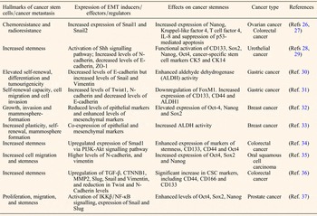
Processes of EMT/MET are orchestrated with the concurrent expression of specific subset of genes, required in the acquisition of stemness, thus bestow the cancer cells with the self-renewal program. The stemness characteristics of the cancer cells ensure the generation and survival of tumour mass required for cancer progression and metastasis and these cells may stay dormant for long period of time. EMT and its reverse process provide a mechanism for escaping CSCs to a new less adverse niche, confer resistance to them towards radiotherapy and or chemotherapy, inactivate p53-mediated apoptosis; thereby result in tumour cell invasion and metastasis in distant organs (Refs Reference Garg24, Reference Diepenbruck and Christofori25). Link between EMT and stemness has been first observed for immortalised human mammary epithelial cells. Similar findings are reported in many other cancers (Ref. Reference Garg3). Table 1 describes the findings of some of the recent studies that define the functions of EMT/MET in conferring cancer cells with stemness properties and cellular plasticity required for aggressive cancer metastasis in various cancer types. Relationship between stemness and tumour cells with E or M phenotype has been well studied. However, many recent research studies contradict this finding and strongly link stemness with hybrid E/M phenotype of tumour cells (Fig. 1). According to EMT gradient model, activation of EMT program in epithelial cells is bimodal in nature. EMT is coupled to stemness during early stage of tumour development but it becomes incompatible to stemness when cells acquire the mesenchymal status (Ref. Reference Ombrato and Malanchi38). Recent literature provides evidence on the connection between partial rather than complete EMT and tumour proliferation, quiescence, and stemness at tumour bud level. Tumour buds, which display collective cell migration, co-express epithelial and mesenchymal markers in multiple cancer types and are associated with poor cancer outcomes (Ref. Reference Grigore21).
Epithelial–mesenchymal transition (EMT) and its reverse process mesenchymal–epithelial transition (MET) allow tumour cells to gain stemness along with enhanced survival benefits required for cancer metastasis in various cancers

P53, tumour protein 53; Shh, Sonic hedgehog; ZO-1, Zona-Occludens-1; CD133, cluster of differentiation 133 (also known as prominin-1); Oct4, octamer-binding transcription factor 4; Ck5, cytokeratin5; CK14, cytokeratin14; Sox2, sex-determining region Y-box containing gene 2; CD44, Cluster of differentiation 44; FoxM1, forkhead box M1; TGF-β, transforming growth factor-β; CTNNB1, Catenin β1; MMP2, Matrix metalloproteinase2; IKKβ/NF-κB, Inhibitor of nuclear factor κB kinase/nuclear factor-κB; CD166, cluster of differentiation 166; PI3K-Akt, phosphoinositide3-kinase-protein kinase B; ALDH, aldehyde dehydrogenase; CSC, cancer stem cell.
Breast CSCs co-expressing both epithelial and mesenchymal marker proteins are examined to display increased plasticity, self-renewability, mammosphere formation and ALDH1+ (aldehyde dehydrogenase 1+) activity as compared with highly differentiated M cells (exhibit less plasticity) and E cells (show less self-renewability). Independent of cellular origin, increased stemness because E–M cooperation offers biological rationale for robust association between hybrid E/M state of cancer cells and poor prognosis. Further results suggest that elimination of hybrid E/M cells and cooperation between E and M cell-types by targeting E/M heterogeneity could improve breast cancer patient survival (Ref. Reference Grosse-Wilde33). Growing evidence suggests that partial reactivation of EMT program allows breast cancer cells to gain plasticity and pluripotency which leads to a stemness-like phenotype. Further, a complete EMT would be a dead end program and would not allow successful colonisation of distal target tissues (Ref. Reference Moyret-Lalle, Ruiz and Puisieux39). Hybrid E/M state of cells and phenotypic plasticity have been observed to be associated with the transformation of epithelial ovarian cells into cancer cells and in vivo xenograft tumour growth. Subset of ovarian cancer cells in a transitory E/M hybrid stage simultaneously express epithelial and mesenchymal markers. These highly enriched tumour -forming cells display features typical of CSCs and are characterised for membrane E-cadherin (low)/cytoplasmic E-cadherin (high)/CD133 (high), CD44 (high) and Tie2 (low) expression. Experimental studies on trans-differentiation of hybrid E/M cells into either mesenchymal or epithelial cells report loss of stem cell markers and tumourigenicity (Ref. Reference Strauss40). Isolation and characterisation of mesenchymal-like and EMT tumour cells in an autochthonous murine model of prostate cancer are examined to harbour both epithelial and mesenchymal markers. These cells exist with plasticity, display enhanced stemness and invasive character and contribute to multiple stages of the metastatic cascade (Ref. Reference Ruscetti41). Human breast cancer cells metastasised to axillary lymph nodes in a severe combined immunodeficiency (SCID) mouse model are examined to co-express vimentins and keratins. Experimental data suggest that E and M marker proteins expressed in breast cancer cells together confer them a potential selective advantage in their interpretation of signalling cues from the ECM. These cells are characterised for increased proliferative capacity, clonogenic potential, tumourigenicity and metastatic phenotype (Ref. Reference Hendrix42).
Another study by Jolly et al. used a theoretical framework to explain the positioning of ‘stemness window’ on the ‘EMT axis’. PSFs such as OVOL couples with miR200/ZEB/LIN28/let-7 circuit, increases the stemness of the hybrid E/M phenotype, finely tunes the EMT-stemness interplay and thereby pulls the ‘stemness window’ away from the M end of ‘EMT axis’ (Ref. Reference Jolly43). At genetic level, stemness gets regulated by LIN28/let-7 and EMT/MET is controlled by miR200/ZEB. Inhibition of LIN28 by miR-200 couples the two modules. NF-κB, but not c-MYC has been identified to control LIN28/let-7 regulation and elevates the likelihood of E/M phenotype to gain stemness (Ref. Reference Jolly44).
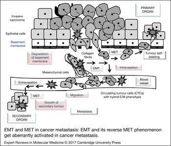
Simultaneous expression of EMT/MET master regulators, including epithelial and mesenchymal marker proteins at the genetic level represent valuable prognostic markers for increased plasticity and stemness. Partial EMT and not the complete EMT program render tumour cells to fully metastasise to distant organs (Ref. Reference Grigore21). Tumour metastasis, the cause of more than 90% of cancer cell mortality, is a multistep process. Tumour cells survive series of events known as metastatic cascade and are described in subsequent section along with their clinical significance (Fig. 3).
Epithelial–mesenchymal transition (EMT) and mesenchymal–epithelial transition (MET) in cancer metastasis: EMT and its reverse MET phenomenon get aberrantly activated in cancer metastasis. Cancer metastasis consists of series of events, including local invasion, intravastion, transport of tumour cells, extravasation followed by colonisation. As a result of genetic alteration, epithelial cells undergo EMT, lose cell–cell contact, acquire mesenchymal and invasive traits. Locally invaded tumour cells either seed back into primary organ and establish metastasis or enter into circulation and behave as circulating tumour cells (CTCs). CTC either moves individually or collectively in clusters, travel through blood vessels, extravasate, undergo MET, proliferate, seed into secondary organs and establish metastasis. Solid arrows indicate the direction of movement of tumour cells.

EMT and metastasis: clinical evidence
Local invasion, intravasation and clinical evidence
Epithelial cancer cells growing in a stromal microenvironment acquire stable genetic and epigenetic changes and undergo EMT. As a result these cells gain morphological changes, undergo transition between E and M phenotypes and exhibit survival benefits. Degradation of underlying basement membrane and ECM allows epithelial cancer cells to move and invade the membrane. Changes in the expression of EMT marker genes with distinct cellular functions alter cell phenotype and induce tumour invasion (Refs Reference Pasquier45, Reference Ombrato and Malanchi38). Proteomic profiling of serum from colorectal cancer patients with different stages identifies 917 total proteins. Out of 917, 93 were observed to be differentially expressed among normal and stages I–III cancer patients. These proteins have been predominantly recognised as those involved in cell adhesion, immune response, coagulation process and metabolism. Expression of collagen I has been reported to be dynamically changed from stages I to IV, with maximum expression in stage II. Serum levels of collagen I degradation telopeptide (CTx) correlates with the tumour staging and poor survival rate and supports the fact that its altered expression might be an early event during tumourigenesis of colorectal cancer (Ref. Reference Zou46). High levels of FN have been detected in the stroma of breast tumours. During tumourigenesis, epithelial cell adhesion switches from cell–cell contacts to mainly cell–ECM interactions, confirms its active role and establishes FN as a marker for EMT (Ref. Reference Jung, Fattet and Yang47). Further, FN-induced EMT mediated by Src kinase and extracellular signal-regulated kinase/mitogen-activated protein (ERK/MAP) kinase signalling, upregulation of Snail, N-cadherin, vimentin, MMP2, α-SMA (α-smooth muscle actin), phospho-Smad2 and acquisition of cell migratory behaviour facilitate breast tumour development (Ref. Reference Park and Schwarzbauer48).
Aberrant activation of EMT in gastric epithelial cells endows the cells with increased mesenchymal characteristics and less epithelial features. Repression of cellular adhesion molecules especially E-cadherin and enhanced stemness promote cancer initiation. Owing to this, tumour cells disseminate and spread throughout the body (Ref. Reference Huang, Wu and Xu49). Immunohistochemistry was carried out in 364 centrally confirmed triple negative breast cancers (TNBCs) to check the expression levels of β-catenin, Myc, E-cadherin, P-cadherin (cell-adhesion), EGFR and cytokeratin 5 (CK5) (identification of basal-like tumours). Low expression of β-catenin in the cell membrane, co-expression of E-cadherin and P-cadherin predict patient outcome in an EGFR-dependent manner (Ref. Reference Lakis50). Increased TGFβ/Smad signalling and enhanced expression of Notch ligand Jagged1 and Notch target gene HES1 (hairy and enhancer of split 1) in EOC (epithelial ovarian cancer) cells form a reciprocal positive regulatory loop, cooperatively regulate EMT and promote cancer cell motility and migration (Ref. Reference Zhou51). Gene expression analysis, including CHIP-seq (chromatin immunoprecipitation-sequencing) and functional tests identify the regulatory effect of transcription factor ∆Np63α in promoting basal-like breast cancer (BLBC) motility by inducing the expression of atypical cadherin FAT2 and mesenchymal genes, Slug and Axl. Induced expression of these genes confers hybrid E/M state to the cells, influences E-cadherin localisation to cell–cell contacts, restricts spontaneous cell movement and extends cellular protrusion that initiates collective invasion. Expression of these genes has been examined to be associated with poor patient outcome (Ref. Reference Dang52).
Thirty-seven colorectal cancer tissues were examined for the expression of long noncoding RNA (lncRNA) molecule, CCAT1. It plays important role in cancer progression by mediating EMT process, reducing the expression of E-cadherin and increasing the expression of N-cadherin levels. Significant correlation of expression of EMT marker genes with local infiltration depth, tumour staging, vascular invasion and tumour differentiation level in colorectal cancer has been observed (Ref. Reference Ye53). Characterisation studies in patients with hepatocellular carcinoma (HCC) and in immortalised mouse hepatoblasts with mesenchymal liver tumours identify 233 candidate cancer genes (CCGs) enriched for cellular processes driving EMT. Further analysis classifies functions of 23 CCGs in early tumourigenesis. Among 23 CCGs, mutations or deregulation of receptor tyrosine kinase, GAB1 [growth factor receptor-bound protein 2 (GRB2)-associated binding protein 1], KDM6A (lysine-specific demethylase 6A), HUWE1 (HECT, UBA and WWE domain containing 1) and PTPN12 (protein-tyrosine phosphatase, nonreceptor-type 12) have been examined to activate EMT program and correlate with poor patient survival (Ref. Reference Kodama54). Immunohistochemical studies were done in 61 patients with locally advanced cervical cancer to assess the expression of proteins associated with tumour growth, as well as their prognostic roles. Increased expression of VEGF, EGFR, Twist2 and loss of E-cadherin were observed to influence the prognosis of locally advanced cervical cancer (Ref. Reference Rojas-Puentes55).
Intravasation is a necessary step which allows locally advanced and invaded tumour cells to cross the endothelial barrier and enter into circulation. Microvessel densities, diameter of the vasculature (lymphatic or blood vessels) increased vessel diameter in some tumours are few properties of vasculature which correlate with increased intravasation. Current in vitro and in vivo assays identify the pathways and molecules important for intravasation. Activation of EGF receptor family members, TGFβ signalling mediated induction of EMT along with downstream signalling through PI3K, NWASP (neuronal Wiskott–Aldrich syndrome protein), RhoA and WASP (Wiskott–Aldrich syndrome protein) are identified to have stimulatory effect on inducing invadopodia (Ref. Reference Chiang, Cabrera and Segall56).
Aberrant expression of zinc-finger transcriptional repressor, such as Snail1, induces cancer cells to degrade and perforate underlying basement membrane barriers, initiate angiogenesis, and intravasate vascular networks in vivo via MMP-dependent process. Expression of membrane-anchored metalloproteinases, MT1-MMP (membrane type1 MMP) or MT2-MMP (membrane type2 MMP) and not the secreted MMPs, exhibit pro-invasive, angiogenic and metastatic activities. MMPs act as pro-invasive factors, confer Snail1-triggered cells with the key abilities to drive cancer cell basement membrane invasion, induce angiogenesis or trigger intravasation and are frequently linked to morbidity and mortality in cancer (Ref. Reference Ota57).
Systemic transport: CTCs and clinical evidence
Microtubule-based membrane protrusions, known as microtentacles are special structures formed in detached breast CTCs. EMT mediated by Twist1 or Snail1 expression in human mammary epithelial cells promotes microtubule stabilisation and formation of microtentacles in detached cells. Microtentacles penetrate the junctions between the endothelial layers to facilitate tumour cell reattachment and allows breast CTCs to aggregate and bound to blood wall while travelling through it and thereby enhances metastatic potential (Ref. Reference Matrone58). Ductal carcinoma in situ samples display high concordance between the increased expression levels Glu-tubulin and Twist at invasive tumour fronts. In vivo experiments on cytoskeletal dynamics establish the significance of EMT-induced tubulin detyrosination occurs at the earliest stages of tumour invasion (Ref. Reference Whipple59).
Invasive nature of currently employed diagnostic procedures cause discomfort to patients. There is a need to develop cost-effective method for accurate assessment of genetic profile in metastasis. Recently, liquid biopsy of circulatory or lymphatic system has become a popular technique for identification of presence or absence of CTCs/cell-free nucleic acids (cfDNA)/miRNAs molecules. An extent to which these signature molecules are present, is an indicative of the presence of primary tumour and the likelihood of patients to develop metastasis. RNA in situ hybridisation (RNA-ISH) method based on the branched DNA signal amplification technology was used to classify the isolated CTCs, from the 164 blood samples of patients with liver, nasopharyngeal, breast, colon, gastric cancer and NSCLC. CTCs were characterised on the basis of EMT markers into its three subpopulations, including epithelial CTCs, biophenotypic epithelial/mesenchymal CTCs and mesenchymal CTCs. Among these three subpopulations, mesenchymal CTCs were more commonly detected in the metastatic stages of the disease in different types of cancers (Ref. Reference Wu60). Strong co-expression of vimentin and keratin in isolated or clusters of dual CTCs, correlates the existence of hybrid CTCs with an E/M phenotype in patients with nonsmall cell lung cancer (Ref. Reference Lecharpentier61).
Results for CTC vimentin expression analysis in 142 blood samples from 93 patients with metastatic castrate-resistant prostate cancer (mCRPC) and CTC Ki67 expression analysis in 90 blood samples from 51 patients with mCRPC correlate poor survival outcomes in vimentin- and Ki67-positive CTC patients (Ref. Reference Lindsay62). Despite striking heterogeneities of N-cadherin and CD133 marker expressions, cell shapes, clusters and sizes, CTCs are still considered as potential candidates to predict the ability of breast tumours to metastasise (Ref. Reference Cierna63). Peripheral blood mononuclear cells (PBMC) from 149 primary breast cancer patients were interrogated for the expression levels of MMP1, Twist1, Snail1, Slug, Zeb1 and CK19 gene transcripts. Results of this study examine a link between MMP1 and CTCs with EMT phenotype in cancer associated stroma and support the role of MMPs and EMT in tumour dissemination (Ref. Reference Bock64). Viable CTCs isolated from the peripheral blood of HCC patients were examined for the expression of vimentin, twist, ZEB1, ZEB2, snail, slug and E-cadherin markers. Among the markers studied, EMT mediated by twist and vimentin expression levels in CTCs have been studied to play a role in promoting the blood-borne dissemination of primary HCC cells and thereby could serve as promising biomarkers for evaluating cancer metastasis and prognosis (Ref. Reference Li65). In an another study, relationship between epithelial plasticity and metastatic spread has been identified by examining the existence of EMT in CTCs from two types of patients, including men with castration-resistant prostate cancer (CRPC) and women with metastatic breast cancer. Existence and high frequency of CTCs co-expressing epithelial cell adhesion molecule (EpCAM), cytokeratins and E-cadherin, vimentin, N-cadherin and O-cadherin, and the stem cell marker CD133 in metastatic CRPC, while simultaneously co-expressing cytokeratins, vimentin and N-cadherin in patients with metastatic breast cancer correlate with progressive metastasis (Ref. Reference Armstrong66). Significant downregulation of prostate stem cell antigen (PSCA) because of binding of Slug to the PSCA promoter and upregulation of N-cadherin and ZEB1/2 by activating RhoA in nasopharyngeal carcinoma (NPC), have been identified to enhance the migration and invasion in vitro and the lung metastasis in vivo. EMT elicits molecular mechanisms of NPC metastasis and correlates with poor metastasis-free survival in NPC patients (Ref. Reference Wang67).
CTCs reflect tumour aggressiveness and are not only capable of establishing metastasis to distant locations/organs, but are also identified to establish tumour self-seeding. Tumour mouse model studies explain the concept of tumour self-seeding where CTCs can colonise their tumours of origin. Tumour-derived cytokines IL-6 and IL-8 attract CTCs; MMP1/collagenase-1 and actin cytoskeleton component fascin-1 mediate its infiltration; while seed-derived chemokine CXCL1 recruits stromal factors, results angiogenesis and accelerates tumour growth. This explains the relationship between tumour size, anaplasia, vascularity, prognosis, induced local recurrence and advocates complete tumour excision (Ref. Reference Kim5).
Extravasation and clinical evidence
Extravasation of cancer cells is considered to be a highly dynamic process. CTCs that disseminate and extravasate into foreign tissue microenvironments must interact productively with its ECM components, survive, proliferate, and form macroscopic metastases. Ex vivo pulmonary metastasis assay (PuMA) provides new insights into the biology of metastatic progression by describing the significance of lung tumour cell interactions with the stromal components in tumour microenvironment (Ref. Reference Mendoza68). Process of extravasation involves modulation of tumour cell adhesion by inducing the formation of filopodium-like protrusions (FLP) and elongated integrin β (Ref. Reference Boyer and Thiery1)-containing adhesion plaques, required for adhesion to the blood-vessel wall as well as clustering of endothelial cells (Refs Reference Stoletov69, Reference Shibue70). This is followed by the intravascular cell migration along the luminal surface of the vascular wall, which has been found to be associated with increased expression of Twist in tumour cells (Ref. Reference Stoletov69). Studies on lung cancer in mice were done to quantify early temporal and spatial progression of metastasis in lung. Observations of this study demonstrate initial survival of cancer cells trapped in the lung circulation followed by their extravasation into lung tissue, and subsequent survival of extravasated solitary cells before metastasis formation. Specific tissue environments favour the continual growth of metastasis (Ref. Reference Cameron71).
Cancer metastasis is distinguished by aggressive growth, early dissemination and a poor prognosis at advanced stage. Remarkably, high counts of CTCs with cancer stemness and EMT phenotype has been observed in small cell lung cancer (SCLC). Nevertheless, extravasation of tumours at distal sites is marked by epithelial features. SCLC cell lines are identified to express increased levels of E-cadherin, β-Catenin, HNF-3β (hepatocyte nuclear factors-3β) FOXA2, SOX2 and PDX-1/IPF1 (insulin promoter factor-1), consistent with MET and formation of large tumourospheres (Ref. Reference Hamilton72).
Metastatic colonisation and clinical evidence
Post-extravasation events affecting a subset of extravasated cells or solitary cells by making them dormant are responsible for overall metastatic inefficiency. Metastatic colonisation reflects the subsequent dissemination of tumour cells to distant sites and their ability to adopt foreign tissue microenvironment. The subsequent outgrowth of micrometastatic deposits into macrometastasis has an impact on cancer progression and clinical prognosis. The outgrowth of metastasis at new locations involves a variety of complex genetic, epigenetic changes and is under strict regulation by EMT program. Reverse process of EMT, MET has been shown to play important role in metastatic colonisation.
Metastatic colonisation has been linked with the presence of CSCs and their self-renewal cellular trait. Aberrant activation of miRs and EMT-activating transcription factors induced by tumour microenvironment, influence EMT program and stem cell regulatory machineries. External niche signals influence by maintaining the stemness of cancer cells and promoting the reactivation of dormant tumour cells thereby, contribute to metastatic colonisation. Increased levels of long-non coding RNA-activated by TGF-β (lncRNA-ATB), a mediator of TGF-β signalling, has been identified to promote organ colonisation of disseminated tumour cells via autocrine induction of interleukin – 11 (IL-11), and triggering Stat3 signalling in HCC and are associated with poor prognosis (Ref. Reference Yuan73). In vivo and in vitro functional studies report correlation of high levels of Nanog with metastasis and low survival rate in HCC. Nanog mediated activation of Nodal (secretory protein) and Cripto-1 (Cryptic family protein 1B, receptor for the TGF-β signalling pathway) to promote Smad3 phosphorylation and Snail expression validates the significance of Nanog signalling in cancer metastasis (Ref. Reference Sun74). Homeobox factor Prrx1 has been examined to uncouple EMT and stemness. Therefore, loss of Prrx1 has been shown to induce EMT program and acquisition of stem cell properties, required for migratory cancer cells to colonise distant tissues to form macrometastasis (Ref. Reference Ocaña75).
Knockdown of Oct4 (octamer-binding transcription factor 4) has been associated with changes in EMT marker expression and cell morphology, decreased Wnt pathway activity, reduced motility of cancer cells and invasion (in vitro) as well as decreased hepatic/colonisation (in vivo) (Ref. Reference Dai76). Study by Vafaizadeh et al., reports the effects of downregulation of the Smad4 protein for transient period on the expression of metastatic factors, such as angiopoietin-like-4 and the inhibitor of DNA binding/differentiation 1, strong activation of Stat3, inhibition of the tumour-suppressive effects of TGFβ signalling and enhancement of tumour growth. Reactivation of Smad4 expression has been examined to correlate with the reversion of EMT and promotion of metastasis formation in the lungs (Ref. Reference Vafaizadeh77). Microfluidic experiments showed that prolectin (CLEC17A, fucosylated motifs), a human lectin behaves as a cell adhesion molecule for epithelial cells. Increased expression of prolactin in mammary tumour cells during metastasis and its association with poor prognosis identify prolactin as a colonisation factor (Ref. Reference Breiman78).
Among biomarkers of therapeutic potential, metastatic suppressors targeting the hybrid E/M phenotype of the tumour cells would have great impact on inhibiting the cellular plasticity and stemness ability of tumour cells and can block the metastatic growth. Functional studies to characterise the molecular interactions of metastatic suppressors of EMT program with tumour microenvironment at every stage of metastatic cascade may provide insights for developing effective potential therapeutic strategies.
Possible clinical prospective of EMT/MET in metastasis and conclusions
Radiological examinations and pathological staging are still gold standard methods for diagnosing clinical manifestation of tumour growth. Unfortunately they fail to provide accurate reproducible information about diagnosis and prognosis of ongoing metastasis. EMT program involves interplay of tightly controlled signalling pathways and large number of cellular and molecular alterations.
EMT and MET, although are fundamental developmental phenomenon, but their aberrant activation drives metastatic cascade which consists of series of events. EMT/MET allows epithelial cells to disseminate from the primary tumour, invade local surrounding areas, survive in general circulation, travel to distant organs and establish as secondary tumour through acquiring a stem cell phenotype. CTCs with E/M hybrid phenotype and cancer stemness property reflect aggressiveness of tumour and hence, their characterisation set challenges for diagnosing malignancy, monitoring disease recurrence, detecting prognosis and achieving therapeutic success for increased survival of cancer patients. Recent advancements allow global screening and profiling of DNA sequences, EMT-associated cDNA, proteome, miRome and tissue arrays of prognostic importance in human clinical CTCs. Signature molecules to define metastasis can predict not only the presence of primary tumour, but also the likelihood of developing metastasis and correlate it with patient cancer outcomes. However, it is extremely difficult to study CTCs and can be defined at single cell level in the blood cells from cancer patients on the basis of expression of specific proteins, including epithelial cellular adhesion molecule (EpCAM), N-cadherin, CD44 and/or expression of specific genes. CTCs expressing epithelial markers are characterised as differentiated epithelial cancer cell, whereas CTCs expressing mesenchymal markers define the ongoing EMT process. These markers especially the transcription factors can be more easily determined by RT–qPCR than by immunohistochemical staining. Examining the combination of E and M markers in a global CTC population can better characterise CTCs with mixed E/M phenotype and improve clinical utility in the therapeutic follow up of the disease.
Acquisition of EMT features in tumour cells, including CSCs has been associated with multidrug resistance and tumour relapse. Potential treatment strategies could involve targeting the CSC subpopulation which has the ability to self-renew, generate all the types of cells within the tumour and express markers that derive and regulate EMT program. Employing potential inhibitors that can target critical orchestrators of EMT signalling transcription pathways, including NF-κB, AKT/mTOR axis, MAPK, β-catenin, PKC and the AP-1/SMAD factors rationalise the basis for more efficient treatment strategies to fight cancer. Some of the examples of recently tested inhibitors that are either under preclinical research, clinical trials or are used in the clinic include small molecule inhibitors that selectively target EGF-R, IGF-R, VEGF-R, Src, GPCR pathways and humanised function blocking antibodies (Herceptin: ErbB2; Avastin: VEGF-A; Erbitux: EGF-R; Abegrin: alphavbeta3 integrins) (Ref. Reference Sabbah79). Adenovirus-based gene transfer of therapeutic miR that involves reintroduction of miR-200 family and targets Zeb1/Zeb2 transcription factors, which are the drivers of EMT or silencing RNA are other potential strategies in the treatment of epithelial cancers.
Studies are required for functional characterisation of metastatic suppressors of EMT program at every stage of metastasis to validate them as potential markers of prognostic significance and facilitate the accurate designing of EMT/MET blockade therapeutic strategies within a fully personalised approach in the management of cancer patients.
Acknowledgements
Author thanks to Department of Science and Technology, Govt. of India for providing research grant (grant no. SR/H0/HS/0113-2010).
Conflict of interest statement
The author discloses no potential conflict of interest.