





Introduction
Maternal obesity during pregnancy is a significant health concern with long-term implications for both mother and offspring. Reference Catalano and Shankar1 Maternal obesity is linked to metabolic disorders and cardiovascular complications, and emerging research now highlights the consequences for the developing offspring. Reference Inzani and Ozanne2 Offspring exposed to maternal obesity often face an increased risk of metabolic dysregulation, including insulin resistance, altered lipid metabolism, and predisposition to obesity later in life. Additionally, these offspring may exhibit impaired cardiovascular health, with changes in vascular function and structure, alongside increased susceptibility to inflammatory conditions. An area of interest is how maternal obesity influences the function of the pancreas, which plays a pivotal role in shaping long-term health outcomes. Research has shown that maternal obesity can impair pancreatic development in the offspring, leading to β-cell dysfunction characterized by reduced insulin secretion and altered glucose homeostasis. Reference Hill and Hill3 Studies also show that maternal HFD can disrupt pancreatic islet structure, reducing the ability for adaptive β-cell proliferation and increasing their vulnerability to metabolic stress later in life. Reference Carroll, Burdick Sanchez and Broadway4 Understanding the mechanisms by which maternal obesity impacts pancreatic function has relevance as the global prevalence of obesity continues to rise. In particular, protein expression within the pancreas constitutes a molecular fingerprint because protein expression reflects the complex interaction between genetic predispositions and environmental influences. Reference Jaenisch and Bird5,Reference Reed, Balsbaugh and Li6 Effects of maternal obesity on protein expression patterns within the pancreas may be key to understanding the mechanisms that drive multigenerational impacts on metabolic health. For instance, maternal obesity alters proteins involved in essential cellular processes such as metabolism, apoptosis, and inflammation. Reference Soubry, Murphy and Wang7 Moreover, disruptions in metabolic pathways impair pancreatic function, leading to insulin resistance and diabetes in offspring. Reference Zheng, Xiao, Zhang, Wang, Yu and Xu8 Dysregulation of apoptotic and inflammatory pathways compromises cell survival and predisposes offspring to chronic conditions such as cardiovascular disease and metabolic syndrome. Reference Freeman, Haley-Zitlin, Rosenberger and Granholm9 These effects not only impact the first generation offspring, but also subsequent generations, a concept known as the developmental origins of health and disease (DOHaD). Reference Godfrey and Barker10,Reference Barker11 The current study aims to narrow the gap in understanding how maternal obesity affects pancreatic protein expression in offspring, focusing on pathways related to insulin resistance, inflammation, and apoptosis. We hypothesized that maternal obesity during pregnancy alters the protein expression patterns of the offspring pancreas causing a reduction in the expression of factors involved in β-cell function and an increase in factors that regulate cellular apoptosis. By identifying specific alterations in proteins and their corresponding pathways, the data will contribute to the development of targeted interventions that mitigate the long-term health risks for both mother and child.
Methods
Mice
Detailed information about animal housing, animal diets, tissue collection, and body weight changes can be found on Eileen and Peterson, 2025. However, this information will briefly be described herein. All procedures and protocols performed were approved by the University of Rhode Island Institutional Animal Care and Use Committee (Protocol # AN1819-018). Animal housing methods were identical to those previously described. Reference Eileen and Peterson12 Briefly, C57BL/6J female mice were placed into treatment groups and fed ad libitum either a low fat diet, as a control (CON; n = 16; 13% fat content: TD.08485 Envigo, Somerset, NJ) or high in fat (HFD n = 16; 42% fat contentTD.88137 Pellet Envigo, Somerset, NJ) for fourteen weeks prior to breeding. At ten weeks on the diet, control females were switched from the low fat control diet to a standard mouse chow (TD.2020x Envigo, Somerset NJ.) due to significant supply issues that resulted in a severe backorder on our initial diet. After fourteen weeks, the mice were placed individually in cages with control fed males for 24 h. Pregnancy was confirmed via the presence of a vaginal plug, and dams were housed individually beginning on gestational day eighteen. They were maintained on their respective diets throughout gestation and lactation. Of the 16 females, only 3/treatment successfully became pregnant and reared their pups until weaning. This largely was due to the reduced conception rate of the HFD females and the incidence of cannibalism in both treatment groups (no effect of treatment on incidence of cannibalism) 2–7 pups/litter were reared by each female. Following parturition and weaning, the resulting pups were separated by sex and housed in same-sex, littermate groups of four until eight weeks of age. All pups at this point were also maintained on the CON diet until tissue collection. While HFD dams weighed more than CON dams, no differences in pup bodyweight were observed between the treatment groups as detailed in Eileen and Peterson, 2024. Reference Eileen and Peterson12
Tissue collection and protein isolation
Tissue collection methods were identical to those previously described. Reference Eileen and Peterson12 Briefly, pups (N = 20; CON males: 6; CON females: 6; HFD males: 4; HFD female: 4) were euthanized at eight weeks of age via CO2 induction accompanied by cervical dislocation. These pups were selected for protein isolation as they were similar in body weight to one another. A minimum of one pup/dam/sex was used in each treatment group. The entire pancreas of each pup was removed immediately and snap frozen in liquid nitrogen. The tissues were stored at −80°C until protein isolation.
A 100 mg fragment of pancreas tissue was removed from each sample and placed into a microcentrifuge tube containing 1000µL of pre-chilled lysis buffer (RIPA). The tissue fragments were then homogenized using a Tissuelyser™ for 1–3 min, with the duration adjusted until complete pulverization was achieved. Following homogenization, tissue samples were incubated in the lysis buffer on ice for 30 min to ensure complete lysis. After the incubation period the tissue samples were centrifuged at 12,000 g for five minutes to pellet unwanted debris. The supernatant was removed and transferred to a new tube for subsequent analysis. The concentration of the protein extract was measured using the Bradford Assay (BCA Rapid Gold Thermo Fisher Scientific, Waltham, MA, USA) prior to mass spectrometry. The BCA assay was conducted following the manufacturer’s instructions, and by measuring absorbance at 480 nm with a plate reader (EPOCH BioTek Instruments, Winooski, VT, USA) to determine protein concentration. The isolated protein samples were subsequently shipped on dry ice to the University of Connecticut for proteomic analysis and ultra-high performance liquid chromatography-tandem mass spectrometry (UPLC-MS/MS) using a Q Exactive HF mass spectrometer.
Untargeted protein identification and label-free quantification via tandem mass spectrometry
An aliquot of the pancreas lysate containing approximately 100 µg of protein was supplemented with sodium dodecyl sulfate (SDS) in 0.1 M Tris-HCl, pH 8.0 to result in a final SDS concentration of 5%. The samples were prepared for bottom-up proteomic analysis using S-Trap columns (ProtiFi, LLC) following a modified commercial protocol. Sample proteins were reduced with 120 mM dithiothreitol for 1 h at room temperature, alkylated with 500 mM iodoacetamide for 30 min in the dark at 30°C, acidified to a final concentration of 2.5% phosphoric acid, and diluted 6x with 0.1 M triethylammonium bicarbonate (TEAB) in 90% methanol (MeOH). The samples were then loaded on a Micro S-Trap (ProtiFi part #02-micro-80), washed with 0.1 M TEAB in 90% MeOH, and incubated with sequencing grade, modified trypsin (Promega part #V5113) at 1:10 (w/w) enzyme:sample overnight at 25°C. Recovered peptides were desalted using peptide desalting spin columns (Pierce Chemical Co.; Thermo Fisher part #89852) and dried to completion. Samples were then resuspended in Solvent A (0.1% formic acid in Fisher Optima LC/MS grade water) and quantified by A280 absorbance. Total injection amounts were normalized across all samples.
The samples were then subjected to mass spectrometry analysis using a Thermo Scientific Ultimate 3000 RSLCnano UPLC system coupled to a high-resolution Thermo Scientific Q Exactive HF mass spectrometer. Each sample was injected onto a nanoEase M/Z Peptide BEH C18 column (1.7 μm, 75 μm × 250 mm, Waters Corporation) and separated by reversed-phase UPLC using a gradient of 4%–30% Solvent B (0.1% formic acid in Fisher Optima LC/MS grade acetonitrile) over a 150-min gradient at 300 nL/min flow. Peptides were eluted directly into the Q Exactive HF using positive mode nanoflow electrospray ionization. MS1 scans were acquired at 60,000 resolution, with an AGC target of 1e6, maximum ion time of 60 ms, and a scan range of 300–1800 m/z. Data-dependent MS2 scans were acquired at 15,000 resolution, with an AGC target of 1e5, maximum ion time of 40 ms, isolation window of 2.0 m/z, loop count of 15, normalized collision energy of 27, dynamic exclusion window of 30 s, and charge exclusion “on” for all unassigned, +1, and >+8 charged species.
The resulting peptides from the above process were identified using MaxQuant (v1.6.10.43) and its embedded Andromeda search engine and quantified by label-free quantification. Reference Cox and Mann13 The raw data were searched against both the complete UniProt Mus musculus reference proteome (identifier UP000000589, accessed 01/23/2023) and the MaxQuant common contaminants database. Variable modifications allowed oxidation of Met, acetylation of protein N-termini, deamidation of Asn/Gln, and peptide N-terminal Gln to pyroGlu conversion. Carbamidomethylation of Cys was set as a fixed modification. Protease specificity was set to trypsin/P with a maximum of two missed cleavages. All results were filtered to a 1% false discovery rate at the peptide and protein levels using the target-decoy approach; all other parameters were kept at default values. MaxQuant output files were imported into Scaffold (v5.1.2, Proteome Software, Inc.) for data visualization and subsequent analyses.
Data and statistical analyses
Analysis was performed using Scaffold (v5.1.2, Proteome Software Inc., Portland, OR). Proteomic data were analyzed separately for male and female offspring to identify sex-specific differences in protein expression. Using the qualitative analysis function in Scaffold, a T-Test with a Benjamini–Hochberg correction for average precursor intensity was conducted to determine the differentially expressed (DE) proteins. Statistical significance was defined as P ≤ 0.05. For downstream pathway analysis, a more stringent threshold of P < 0.01 was applied to select proteins for further analysis. Proteins of interest based upon these statistical parameters were then run through the Kyoto Encyclopedia of Genes and Genomes (KEGG) pathway database to determine the function and pathway of each protein.
Results
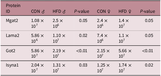
2,329 proteins were identified when comparing CON vs HFD males. Likewise, the same amount was identified when comparing CON vs HFD females. A smaller subset of proteins were identified as being differentially expressed. Specifically, 145 proteins exhibiting differential expression (DE) for CON vs HFD males were identified. Alternatively, 20 DE proteins were identified when comparing CON vs HFD females (Fig. 1a). Therefore, there were a total of 165 proteins identified by our analyses across both treatment comparisons and sexes. Out of the 165 total DE proteins, four were common to both male and female offspring (Mgat2, Lama2, Got2, Isyna1). Specifically, Mgat2, Got2, and Isyna1 were downregulated in HFD male offspring compared to CON male offspring (P ≤ 0.05; Table 1). This pattern of differential expression was not observed for these three proteins in HFD female offspring. Mgat2, Got2, and Isyna1 were all upregulated in HFD females when compared to CON females (P ≤ 0.05). The only protein that exhibited similar patterns of expression was Lama2. Lama2 was upregulated in HFD males when compared with CON males (P = 0.02; Table 1). Likewise, in HFD females, Lama2 expression was also upregulated when compared to CON females (P = 0.05; Table 1). This tendency for male HFD offspring to exhibit reduced protein expression was observed when evaluating the other DE proteins of this sex that were not outlined above. For example, of the 145 proteins identified in the male offspring of dams fed HFD, 131 were downregulated and the remaining 14 were upregulated (Fig. 1b). In contrast, the female offspring of dams fed HFD had 20 DE proteins, of those 13 were upregulated and the remaining seven were downregulated in the (Fig. 1c).
a–c: differentially expressed proteins in male and female offspring. Differentially expressed (DE) proteins in male (n = 4–6/treatment) and female (n = 4–6/treatment) offspring mice. (a) Number of DE proteins for CON vs. HFD females compared to the number of DE proteins for CON vs. HFD males and those found commonly between sexes. (b) Protein expression in CON vs HFD male offspring. (c) Protein expression in CON vs HFD female offspring. Proteins were considered DE when P ≤ 0.05.

Proteins common to ♂ and ♀ offspring

Table 1. Long description
A table comparing protein expression levels in male and female offspring under control (CON) and high-fat diet (HFD) conditions. The table has four rows and six columns. Column headers are Protein ID, CON δ, HFD δ, P-value, CON 9, HFD 9, and P-value. Row labels are Mgat2, Lama2, Got2, and Isyna1. Row 1: Mgat2, 1.08 x 10^7, 2.5 x 10^6, 0.05, 2.4 x 10^6, 1.4 x 10^7, 0.05. Row 2: Lama2, 5.86 x 10^6, 1.10 x 10^7, 0.02, 7.4 x 10^6, 1.1 x 10^7, 0.05. Row 3: Got2, 5.86 x 10^7, 2.19 x 10^7, <0.01, 2.15 x 10^7, 5.66 x 10^7, <0.01. Row 4: Isyna1, 2.04 x 10^7, 1.31 x 10^7, 0.03, 1.25 x 10^7, 1.74 x 10^7, 0.02. The table shows the expression levels of four proteins common to both male and female offspring under different dietary conditions, highlighting significant differences in expression levels indicated by P-values.
Average expression levels of proteins identified in both male (n = 4–6/treatment) and female offspring (n = 4–6/treatment); Mannosyl-glycoprotein 2-beta-N-acetylglucosaminyltransferase (Mgat2); Laminin subunit alpha 2 (Lama2); Glutamic oxaloacetic transaminase 2 (Got2); Inositol-3-phosphate synthase 1 (Isyna1). Proteins were considered DE when P ≤ 0.05.
Pathway analysis
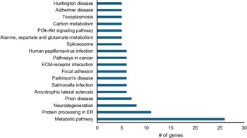
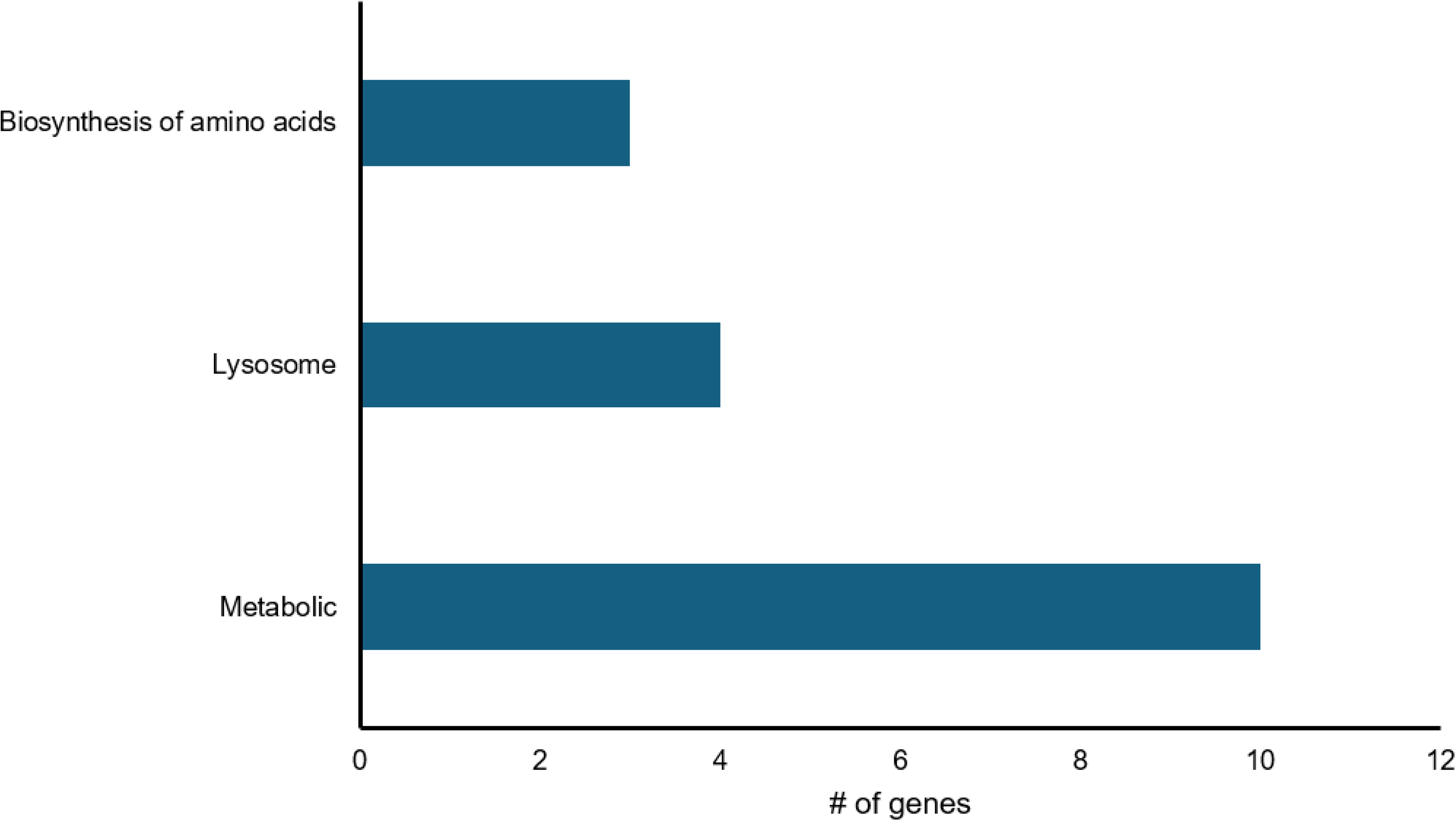
Evaluation of the pancreas tissue of male offspring from CON vs. HFD groups indicated 26 proteins within the metabolic pathway classified as differentially expressed. Notably, 22 of the 26 proteins exhibited decreased expression, while the four remaining proteins exhibited increased expression. For the protein processing pathway, 11 DE proteins were categorized as such and all of these proteins were downregulated. For the neurodegeneration pathway, eight proteins were differentially expressed, all of which had decreased expression Conversely, for pancreas tissue of female offspring from CON vs. HFD groups, 10 DE proteins were identified using pathway analyses. Among these, nine proteins exhibited increased expression for the HFD group), and the remaining protein was reduced.
Proteomic analysis
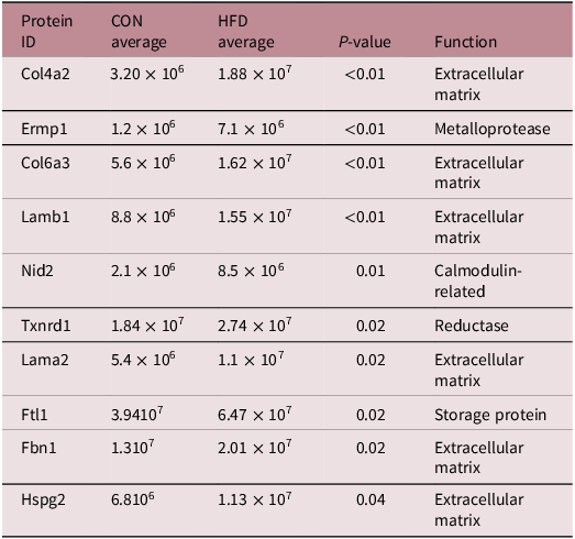
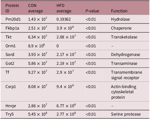
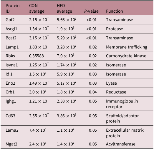
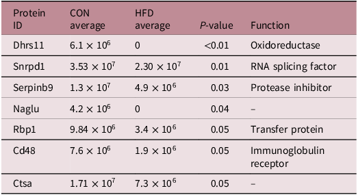
Proteins involved in key biological processes were upregulated for the HFD group. Notably, proteins such as Col4a2, Lamb1, and Col6a3, all integral to structural and signaling components of the extracellular matrix (ECM), exhibited increased expression (Fig. 2, Table 2, P < 0.01). Similarly, proteins involved in the stress response of the cell (Txnrd1 and Nid2) had increased expression (Fig. 2, Table 2, P < 0.02). In contrast, a number of metabolic proteins were downregulated in these male offspring. Proteins such as Pm20d1, Tkt, and Got2, exhibited decreased expression in HFD males when compared to CON males (Fig. 2, Table 3, P < 0.01). In the female offspring, a different pattern of protein expression was observed, with a subset of proteins involved in glycolysis and amino acid metabolism (Bcat2 and Isyna1) showing increased expression in response to the maternal HFD (Fig. 3, Table 4, P < 0.02). Conversely, proteins associated with immune regulation and RNA processing (Serpinb9 and Snrpd1) exhibited reduced expression (Fig. 3, Table 5, P < 0.03).
Male KEGG pathways. Male pathway analysis. Significant (P ≤ 0.05) were analyzed via Kegg pathway analysis.

Female KEGG pathways.Figure 3.Female pathway analysis. Significant (P ≤ 0.05) were analyzed via Kegg pathway.

Male upregulated protein expression

Table 2. Long description
A table with 10 rows and 5 columns comparing protein expression levels between two groups, CON and HFD. The columns are labeled Protein ID, CON average, HFD average, P-value, and Function. The table lists proteins such as Col4a2, Ermp1, Col6a3, Lamb1, Nid2, Txnrd1, Lama2, Ftl1, Fbn1, and Hspg2. Each row provides the average expression levels for the CON and HFD groups, the p-value indicating statistical significance, and the function of the protein. Notable trends include increased expression levels in the HFD group for proteins like Col4a2, Lamb1, and Col6a3, which are integral to the extracellular matrix, and increased expression of stress response proteins Txnrd1 and Nid2.
Average expression levels for upregulated proteins in CON vs HFD male offspring; 4–6 pups/treatment were used per treatment group; Collagen type 4 α chain 2 (Col4a2) Endoplasmic reticulum metallopeptidase 1 (Ermp1) Collagen type 6 α 3 chain (Col6a3) Laminin Subunit Beta 1 (Lamb1) Nidogen 2 (Nid2) Thioredoxin reductase 1 (Txnrd1) Laminin subunit alpha 2 (Lama2) Ferritin light polypeptide 1 (Ftl1) Fibrillin 1 (Fbn1) Heparan sulfate proteoglycan 2 (Hspg2). Proteins were considered differentially expressed when P ≤ 0.05.
Male down regulated protein expression

Table 3. Long description
A table comparing protein expression levels between two groups, CON and HFD. The table has 10 rows and 5 columns. The columns are labeled Protein ID, CON average, HFD average, P value, and Function. Each row lists a protein ID, its average expression levels in the CON and HFD groups, the P value indicating statistical significance, and the protein’s function. For example, the first row shows Protein ID Pm20d1 with CON average 1.43 x 10^7, HFD average 0.19362, P value <0.01, and Function Hydrolase. The table highlights proteins that are downregulated in the HFD group compared to the CON group.
Average expression levels for downregulated proteins in CON vs HFD male offspring; 4–6 pups/treatment were used per treatment group; Peptidase M20 domain containing 1 (Pm20d1) FK binding protein prolyl isomerase 1A (Fkbp1a) Transketolase (Tkt) Orosomucoid 1 (Orm1) Sorbitol dehydrogenase (Sord) Glutamic oxaloacetic transaminase 2 (Got2) Transcription factor (Tf) Cysteine and glycine rich protein 1 (Csrp1) Heterogeneous nuclear ribonucleoprotein (Hnrnpr) Trypsin 5 (Try5). Proteins were considered differentially expressed when P ≤ 0.05.
Female upregulated protein expression

Table 4. Long description
A table with 12 rows and 5 columns comparing protein expression levels between two groups, CON and HFD. The columns are labeled Protein ID, CON average, HFD average, P-value, and Function. Each row lists a specific protein ID, its average expression levels in the CON and HFD groups, the p-value indicating statistical significance, and the function of the protein. The table shows proteins involved in various biological processes, with some proteins upregulated and others downregulated in the HFD group compared to the CON group. Notable proteins include Got2, Asrgl1, Bcat2, Lamp1, Rbks, Isyna1, Idi1, Eno2, Crb1, Ighg1, Cd63, Lama2, and Mgat2, each with specific functions such as transaminase, protease, membrane trafficking, carbohydrate kinase, isomerase, lyase, reductase, immunoglobulin receptor, scaffold/adaptor protein, extracellular matrix protein, and acyltransferase.
Average expression levels for upregulated proteins in CON vs HFD female offspring; 4–6 pups/treatment were used per treatment group; Glutamic oxaloacetic transaminase 2 (Got2)Asparaginase and isoaspartyl peptidase 1 (Asrgl1) Branched chain amino acid transaminase 2 (Bcat2) Lysosome-associated membrane glycoprotein 1 (Lamp1) Ribokinase (Rbks) Inositol-3-phosphate synthase 1 (Isyna1) Isopentenyl-diphosphate delta isomerase 1 (Idi1) Enolase 2 (Eno2) Carbonyl reductase 1 (Cbr1) Immunoglobulin heavy constant gamma 1 (Ighg1) Cluster of differentiation 63 (Cd63) Laminin subunit alpha 2 (Lama2) Mannosyl-glycoprotein 2-beta-N-acetylglucosaminyltransferase (Mgat2). Proteins were considered differentially expressed when P ≤ 0.05.
Female downregulated protein expression

Table 5. Long description
A table titled ‘Female downregulated protein expression’ with eight rows and five columns. The columns are labeled ‘Protein ID’, ‘CON average’, ‘HFD average’, ‘P-value’, and ‘Function’. The table compares the average expression levels of various proteins between two groups, CON and HFD. Each row lists a protein ID, its average expression in the CON group, its average expression in the HFD group, the P-value indicating the significance of the difference, and the function of the protein. For example, the protein Dhrs11 has an average expression of 6.1 times 10 to the power of 6 in the CON group and 0 in the HFD group, with a P-value of less than 0.01 and a function as an oxidoreductase. The table highlights proteins that are downregulated in the female offspring.
Average expression levels for downregulated proteins in CON vs HFD female offspring;4–6 pups/treatment were used per treatment group; Dehydrogenase/reductase SDR family member 11 (Dhrs11) Small nuclear ribonucleoprotein D1 polypeptide (Snrpd1) Serpin family B member 9 (Serpinb9) N-Acetyl-Alpha-Glucosaminidase (Naglu) Retinol binding protein (Rbp1) Cluster of differentiation 48 (Cd48) Cathepsin A (Ctsa). Proteins were considered differentially expressed when P ≤ 0.05.
Discussion
Maternal nutrition contributes to proper fetal development, with the understanding that a poor maternal diet can have long-lasting adverse effects extending into adulthood. Reference Catalano and Shankar1 Specifically, excessive sugar and fat intake during pregnancy can impair the metabolism, pancreatic β-cell function, and adiposity in the offspring. Reference Moullé and Parnet14 Developmental programming research has focused on elucidating the mechanisms by which the maternal environment facilitates these permanent changes in the offspring. For example, epigenetic modifications, such as DNA methylation and histone modifications, result in an increased likelihood of children developing obesity, insulin resistance, and type 2 diabetes, due to epigenetic modifications of genes related to metabolism. Reference Eileen and Peterson12,Reference Lecoutre, Maqdasy and Breton15 Changes to DNA methylation patterns can lead to differences in the protein expression of key factors within the pancreas tissue of mouse pups born to HFD-fed dams. Reference Eileen and Peterson12 Herein we have focused exclusively on the effects of maternal HFD during pregnancy on the proteome of the pancreas of the resulting offspring. This approach enabled us to (1) identify specific targets of protein expression for future studies and (2). Gain insight about how developmental programming impairs pancreatic function of the offspring.
Proteomics via LC-MS allows for a broad analysis of DE proteins that are not as restrictive as performing western blots. Furthermore, LC-MS yields large subsets of proteomics data. As a result, pathway analyses are used as a way to categorize these large protein data sets based on their potential role in different processes. Reference Khatri, Sirota and Butte16 It also allows researchers to determine if there are differences in the predominance of certain pathways in response to different treatments. Reference Khatri, Sirota and Butte16 It is important to note that using LC-MS is not without its pitfalls. Challenges can arise with low abundance proteins, detection limits, and available sample size. Reference Knecht, Eberl and Kreisz17 Therefore, care must be taken when designing experiments to ensure adequate sample volume as well as the usage of proper bioinformatics methods. We have found that male and female offspring exhibit differential methylation profiles that correspond to differences in protein expression in response to alterations to the maternal diet during pregnancy. Reference Eileen and Peterson12 The authors would like to note that our work has been conducted using whole pancreas tissue. Therefore, our protein expression profile not only encompasses the endocrine cells of the pancreas but the acinar cells as well. This has allowed us to gain a broader picture of the changes in protein expression. While we do discuss the expression of our DE proteins in the context of specific cell types, our work highlights the critical need for additional research to be conducted in murine as well as human models looking specifically at the different cellular populations in the pancreas.
The findings that we have presented here are in agreement with this as well as the findings of others that have determined there are differences in the predominance of certain pathways when comparing between the sexes. Reference Curran, Fogarty Draper and Scott-Boyer18,Reference Cikic, Chandra and Harman19 Additionally it is well established that developmental programming can have different effects on the male and female offspring. Reference Hoffman20,Reference Peterson, Gauvin and Pillai21 These sex-specific differences in pancreatic protein expression may be influenced by the actions of sex hormones, including estrogen and testosterone, which regulate metabolic, inflammatory, and insulin signaling pathways. Reference Curran, Fogarty Draper and Scott-Boyer18,Reference Cikic, Chandra and Harman19 For example, estrogen has been shown to enhance insulin sensitivity, promote β-cell survival, and modulate immune responses, whereas testosterone can influence glucose metabolism, lipid homeostasis, and inflammatory signaling. Reference Curran, Fogarty Draper and Scott-Boyer18,Reference Cikic, Chandra and Harman19 Through their interactions with hormone receptors and downstream transcriptional networks, these hormones may contribute to differential protein expression patterns and distinct physiological responses observed between male and female offspring. Reference Curran, Fogarty Draper and Scott-Boyer18,Reference Cikic, Chandra and Harman19,Reference Yong, Toledo, Nowakowski and Wang22 Furthermore, a wide variety of studies performed in mice, rats, and cell culture models have demonstrated that these hormones can have impacts on male/female pancreas function and cellular composition vitro. Reference Wang, Gorelick and Bhargava23 For example, administration of estrodiol to male rats resulted in an increase in acinar cell enzyme content within the pancreas tissue. Reference Tangoku, Doi, Chowdhury, Blevins, Pasley and Rayford24 Data were also analyzed from the Human Islet Research Network’s Human Pancreas Analysis Program (HPAP), and were used to determine that the gene expression profiles in the endocrine cells of the pancreas were different between male and females. Reference Yong, Toledo, Nowakowski and Wang22 However, it is important to note that the underlying mechanisms by which these hormones facilitate the sexual dimorphisms observed in pancreas structure, function, and gene expression are poorly understood. Reference Wang, Gorelick and Bhargava23 Even within our present study, while we do observe key differences between the sexes, we do not know at what stage of the estrous cycle our females were sampled at. Therefore, additional work needs to be done with a more controlled study to better understand how these hormones and cyclicity in the female could impact the gene as well as protein expression in the pancreatic tissue.
Several of the DE proteins identified in this study have also been highlighted in other developmental programming research studies focused on pancreatic health and function. For example, Col4a2 which was upregulated in male offspring in the present study, has been previously identified as a biomarker for insulin resistance and islet dysfunction. Reference Woo, Sudhir and Zhang25 A study conducted by Pujar et al. (2019),using Gene omnibus biological repository, found that increased expression of Col4a2 results in structural alterations in the pancreatic ECM. Reference Pujar, Vastrad and Vastrad26 Similarly, the upregulation of Lama2 in male offspring is consistent with its established role in maintaining basement membrane integrity of pancreatic islets, which is integral for insulin secretion under metabolic stress conditions Reference Doumatey, Xu and Huang27 Lama2 is also a key structural protein involved in cell adhesion and ECM organization, suggesting that its increased. expression may reflect broader cell remodeling rather than direct functional adaptations to maternal nutrition. Reference Doumatey, Xu and Huang27 This aligns with its potential role in male-specific adaptive responses to metabolic stress. Proteins involved in ECM organization and repair, such as Col4a2, Lamb1, and Col6a3, were upregulated. These proteins play an important role in maintaining tissue structure and facilitating signaling pathways. Reference Pujar, Vastrad and Vastrad26 For instance, Col4a2 contributes to the integrity of the basement membrane, which is important for cell adhesion and signaling. Reference Pujar, Vastrad and Vastrad26 Similarly, Lamb1 and Col6a3 regulate ECM stability, supporting processes like cell migration and tissue repair. Reference Pujar, Vastrad and Vastrad26 Upregulation of ECM related proteins may act as a protective mechanism to counteract tissue damage or stress caused by excessive fat exposure, consistent with previous studies that maternal obesity disrupts tissue integrity and triggers inflammatory processes in the offspring. Reference Catalano and Shankar1 This potential adaptive response demonstrates that there may be interplay between structural integrity and metabolic disruption in offspring exposed to adverse uterine environments. Future work needs to be done to investigate the molecular mechanisms underlying ECM protein upregulation and exploring the dynamics during development.
Beyond ECM related adaptations, male offspring also exhibited upregulation of Txnrd1. The deletion of Txnrd1 in mice leads to metabolic disruptions, including increased β-cell apoptosis, reduced insulin secretion, and glucose intolerance. Reference Stancill, Hansen, Mathison, Schmidt and Corbett28 In pancreatic tissue, Txnrd1 regulates oxidative stress, supports cellular proliferation and differentiation, and mitigates inflammatory damage. Reference Stancill, Hansen, Mathison, Schmidt and Corbett28 The upregulation observed in our study may reflect a compensatory mechanism to counteract inflammatory stress, aiding in the preservation of β-cell health. Conversely, male offspring exhibited downregulation of proteins involved in energy metabolism, including mitochondrial function and carbohydrate metabolism. Observed reduction in proteins Tkt and Got2 suggest that these factors might mediate developmental programmed effects on cellular energy metabolism and metabolic efficiency. Furthermore, the downregulation of these proteins supports the hypothesis that developmental programming sets the stage for long-term metabolic disturbances in the offspring. Reference Barker11 The analysis also revealed an association between male HFD offspring with neurodegenerative pathways, including ALS, Parkinson’s, Alzheimer’s, and Huntington’s diseases. These findings suggest that maternal obesity may predispose offspring to neurodegenerative conditions through metabolic dysregulation. The brain and the pancreas share several common molecular and inflammatory pathways, such as insulin signaling, oxidative stress, and inflammatory cytokine responses, which are central to their function and linked to both metabolic and neurodegenerative diseases. Reference De Felice and Ferreira29 For example, disruptions in insulin signaling and chronic inflammation can simultaneously impair pancreatic β-cell function and promote neurodegeneration locally. Reference Freeman, Haley-Zitlin, Rosenberger and Granholm9,Reference Spinelli, Fusco and Mainardi30 The sex-specific differences observed in metabolic protein expression highlight how maternal diet impacts both metabolic and the neural health of the pancreas in male offspring. As a result, this may potentially predispose them to conditions related to neurodegeneration in the pancreas tissue.
In contrast to the metabolic dysregulation observed in the males, female offspring exhibited a distinct pattern of protein expression characterized by the upregulation of proteins involved in glycolysis and amino acid metabolism, such as Bcat2 and Isyna1. Bcat2 aids in the metabolism of branched chain amino acids, which are critical for maintaining insulin secretion by providing metabolic intermediates for energy production. Reference White, McGarrah, Herman, Bain, Shah and Newgard31 Similarly, Isyna1 is pivotal for insulin signaling pathways that regulate β-cell survival. Reference Watkins, Yong, Sharma and Chan32 This metabolic flexibility reflects resilience to environmental stressors, which has been observed in previous research. Reference Zhang, Xiao and Zheng33 Females often demonstrate enhanced adaptive capacity under adverse conditions, potentially due to sex-specific hormonal and molecular differences. Reference Baines and West34 Increased expression of these targets in females may reflect compensatory mechanisms to counteract the effects of maternal HFD induced stress, by enhancing energy production and maintaining metabolic homeostasis in response to nutrient overload. These findings support previous research that maternal HFD leads to upregulation of glycolysis and amino acid metabolism, potentially altering metabolic status in the offspring. Reference Zheng, Xiao, Zhang, Wang, Yu and Xu8,Reference Zhang, Xiao and Zheng33 This compensatory adaptation suggests a sex-specific metabolic plasticity, where females prioritize energy metabolism pathways to maintain systemic homeostasis under conditions of maternal dietary stress. Reference Zheng, Xiao, Zhang, Wang, Yu and Xu8,Reference Zhang, Xiao and Zheng33 More specifically, the increased expression of Eno2 suggests enhanced glycolic activity, consistent with the hypothesis that maternal obesity drives metabolic programming in the offspring. Reference Zheng, Xiao, Zhang, Wang, Yu and Xu8
Maternal HFD have been linked to alterations in mitochondrial gene expression and reduced activity of metabolic enzymes in male offspring, impairing oxidative phosphorylation and energy metabolism. Reference Latouche, Heywood and Henry35 These effects may stem from sex-specific epigenetic reprogramming, which has been shown to modulate the expression of genes associated with lipid metabolism, insulin signaling, and inflammation more so in males compared to females. Reference Nguyen, Chen, Pollock and Saad36 Additionally, male offspring exhibit greater sensitivity to intrauterine nutritional stress, possibly due to reduced compensatory mechanisms, such as mitochondrial adaptation, as seen in females. Reference Pileggi, Hedges and Segovia37 The downregulation of proteins in association with the metabolic pathways highlights the vulnerabilities of the male offspring to metabolic dysfunction, which could predispose them to long-term health conditions. These results highlight the importance of sex as a biological variable in understanding the impact of maternal obesity on offspring health. Evidence suggests that adaptive mechanisms differ between males and females, with females exhibiting enhanced resilience potentially due to differential gene expression. Reference Baines and West34 Further research should involve exploring the mechanisms underlying these sex-specific adaptations and examining whether the identified proteins contribute to long-term protection against metabolic dysfunction in female offspring
Conclusion
Our study provides evidence that maternal HFD during pregnancy induces sex-specific alterations in the pancreatic proteome of offspring, affecting pathways involved in metabolism, protein processing, and neurodegeneration. Male offspring exhibited changes in key metabolic proteins, suggesting impaired energy production and an increased risk of neurodegenerative conditions, while females displayed increased expression in compensatory glycolytic pathways and decreased expression of regulatory proteins, indicating adaptive responses to their environment. Future research should focus on identifying the long-term consequences of these proteomic changes and identifying potential therapeutic targets to mitigate the adverse effects of maternal obesity on offspring health.
Data availability statement
The raw data used in this study can be made available upon reasonable request via the corresponding author.
Acknowledgments
We would like to thank Jordan Lally for his assistance with this project as well as the URI vivarium staff, fiscal clerks (Katherine Faverau), and undergraduate researchers (Hannah Piracini).
Author contribution
M.P., conceptualization, methodology, validation, formal analysis, investigation, resources, data curation, writing – original draft preparation, writing – review and editing, visualization, supervision, project administration, funding acquisition. L.E., methodology, validation, formal analysis, investigation, data curation, writing – original draft preparation, writing – review and editing, visualization. D.T., writing – review and editing. All authors have read and agreed to the published version of the manuscript.
Financial support
Funding for this project was provided via the Rhode Island Foundation.
Competing interests
The authors declare no conflicts of interest. The funders had no role in the design of the study; in the collection, analyses, or interpretation of data; in the writing of the manuscript; or in the decision to publish the results.
Ethical standard
This study was performed in accordance with guidelines set forth by the University of Rhode Island Institutional Animal Care and Use Committee. Approval for this project was granted by the committee and the protocol number AN1819-018 provided.
Informed consent statement
Not applicable.


 Open access

