Cardiac valve formation is a complex process in cardiac morphogenesis that leaves the valves susceptible to abnormal development. Once malformation occurs, the pathophysiology results in the valve becoming either obstructive or incompetent. Acquired valvar cardiac disease, such as infective endocarditis or residual post-surgical lesions, can mimic congenital valve malformation and disruptive properties. Symptomatology is associated with the onset of valve disease and the degree of disruption to unidirectional flow by the valve. Management of valvar cardiac disease can be challenging in the paediatric critical care setting and is dependent on the ability to minimise the effects caused by obstruction or incompetence to the valve apparatus until intervention is necessary.
Atrioventricular valves maintain unidirectional blood flow from the atria to the ventricle, and valve dysfunction, by stenosis or regurgitation, will primarily disturb the morphology and function of the atria and venous capacitance circuits. Severe forms of atrioventricular valve dysfunction can lead to marked dilation of the atrial chambers, which can cause life-threatening atrial arrhythmias. In addition, venous capacitance circuits can become significantly volume loaded and lead to vascular congestion and associated oedema. Critical care management of these lesions can be challenging and typically focusses on the prevention of conditions that can worsen the effects of atrial dysfunction. Diuresis and closely monitoring the balance of fluids are the mainstays of therapy for both pathophysiological entities. Ultimately, once the atrioventricular valve dysfunction worsens to the point that symptoms can no longer be medically managed, patients are presented for procedural intervention, typically valve repair or replacement. A comprehensive description of valve-specific management of the two types of atrioventricular valve dysfunction, obstruction and incompetence, will be discussed.
Semilunar valves connect the ventricles to the great arteries and serve to maintain unidirectional blood flow to their respective circulatory systems. The disturbance of flow from the ventricle to the great arteries, by either stenosis or insufficiency, can lead to organ hypoperfusion. Over time, increased wall stress on the ventricular chamber can alter myocardial mechanics and lead to dilation or hypertrophy with associated systolic and/or diastolic dysfunction. Given the potential for obstruction to develop at several locations within the outflow tracts during morphogenesis, isolated semilunar valve obstruction is much more common than regurgitant lesions.Reference Spencer, Neill and Sank1 Outflow obstruction also occurs frequently in the presence of congenital heart anomalies with multiple other structural defects – for example, Shone’s complex and its associated multiple left-sided obstructive lesions. Early presentation of clinically significant semilunar valve obstruction often occurs in the neonatal period and may require maintaining the arterial duct with the use of prostaglandins to adequately perfuse the respective circulation. Regurgitant semilunar valve lesions are rare in the neonatal period; however, they can be clinically significant as is seen with truncal valve insufficiency in common arterial trunk malformation. This review discusses the preoperative critical care management of cardiac valve disease, with particular focus on mitral and tricuspid valve diseases. There will also be a brief discussion of pulmonary valve disease. Aortic valve disease will be reviewed elsewhere in this Supplement.
Mitral valve disease
Congenital mitral valve abnormalities are rare and occur in only four of 1000 children with CHD.Reference Hoffman and Kaplan2 Mitral valve disease becomes common when multiple left-sided structural heart abnormalities exist, such as in Shone’s complex and atrioventricular septal defects. It also occurs post-surgically and is common with atrioventricular septal defect repair and cleft mitral valve closure. The incidence of acquired mitral valve disease from rheumatic fever and rheumatic heart disease has significantly declined in developed countries because of the improvement in detection and treatment of group A streptococcal infection.Reference Massell, Chute and Walker3
The pathophysiology of mitral stenosis begins when there is obstruction to left atrial emptying and a pressure gradient develops between the left atrium and ventricle. With difficulty filling the left ventricle, the left atrial function is altered and the left atrial pressure increases with a subsequent increase in pulmonary venous pressure. The rise in venous pressure results in a two-fold problem. First, the left atrial dimensions can become markedly distended, which stretches the atrial tissue and increases the risk for atrial arrhythmias that further impede ventricular filling. Second, there is increased resistance of flow from the pulmonary capillary bed to the pulmonary venous channels, which results in perivascular oedema and subsequent luminal narrowing. This, in combination with pulmonary venous flow reversal, can lead to secondary pulmonary arterial hypertension and if not treated appropriately can result in right ventricular pressure overload and annular dilation with tricuspid valve regurgitation (volume overload). Right ventricular systolic dysfunction can then develop in the presence of a volume-loaded and pressure-loaded ventricle (Fig 1). The timing and manifestation of symptoms from mitral stenosis vary with the degree of obstruction to left ventricular inflow, the presence and type of associated lesions, and the growth rate of the infant. Echocardiography is the primary non-invasive diagnostic tool to determine the degree of stenosis. Mitral valve area is the best measure of obstruction; however, it is difficult to accurately measure when the valve is congenitally malformed.Reference Ruckman and Van Praagh4 Mean transmitral gradient is a relatively accurate substitution of mitral valve area and has been shown to be consistent with direct pullback measures obtained during simultaneous catheterisation.Reference David, Lang and Marcus5, Reference Nishimura, Rihal and Tajik6 A mean gradient >3 mmHg has been generally accepted to be significant mitral stenosis and there should be a low threshold to initiate medical management strategies in the presence of an increasing gradient and symptomatology.
Pathophysiology of mitral stenosis.

The critical care management of mitral stenosis focusses on minimising the effects of an increased left atrial pressure that leads to left atrial volume overload and decreased forward flow causing atrial arrhythmias and secondary pulmonary arterial hypertension. Preserving sinus rhythm to avoid atrioventricular dysynchrony and heart rate control to avoid tachycardia will enhance diastolic filling of the left ventricle and may prevent haemodynamic instability. Avoiding factors that may worsen pulmonary arterial hypertension, such as acidosis, hypoxia, and agitation, is important to prevent pulmonary hypertensive crises. If patients with mitral stenosis have continued symptoms despite these interventions, intravenous infusions such as calcium chloride or esmolol may be necessary. Caution must be exercised when using β blockade in the presence of acute congestive heart failure and pulmonary oedema. Mechanical ventilation may be required to stabilise the patient as a bridge to interventional therapy. Haemodynamics usually improve after mitral valve repair or replacement, because obstruction to left ventricular filling is resolved and left atrial pressure is normalised. If residual lesions are present, the patient should be managed similar to the preoperative setting with an emphasis on limiting left atrial hypertension. If the patient has significant preoperative pulmonary hypertension or right ventricular dysfunction, they may need vasoactive infusion support postoperatively. In addition, inhaled nitric oxide for pulmonary vasodilation (after obstruction is relieved) and adequate sedation with possible neuromuscular blockade may be necessary to aid right ventricular function and prevent pulmonary hypertensive crises.
Mitral regurgitation is more commonly found in paediatric patients and manifests through several mechanisms. Atrioventricular septal defects are the primary congenital cause of mitral regurgitation. Dilated cardiomyopathy can lead to annular dilatation and subsequent mitral regurgitation. Post-infectious causes of mitral regurgitation include endocarditis and rheumatic heart disease. The pathophysiology of left-sided regurgitant valve lesions leads to increased blood volume in the left atrium and subsequent atrial chamber dilation (Fig 2). The residual atrial blood volume leads to volume overload of the atrial and ventricular chambers. In an attempt to reduce wall stress caused by volume load, afterload reduction to maximise forward flow is the mainstay of therapy. In the case of acute left-sided atrioventricular valve regurgitation, rapid development of pulmonary venous congestion and left ventricular volume overload can be life threatening. Prompt initiation of diuretics, inotropic agents, and afterload reducing infusions can allow for quick stabilisation of the patient. The most commonly utilised inotrope to manage mitral regurgitation is milrinone because of its contractility, afterload reducing, and lusitropic effects on the myocardium. In addition, sodium nitroprusside is used as a primary or secondary agent to provide afterload reduction. Likewise, mechanical ventilation and sedation to reduce oxygen consumption and to aid in the management of pulmonary oedema/hypoxaemia may be necessary. Once the patient has stabilised, transition to oral afterload-reducing agents, such as angiotensin-converting enzyme inhibitors, will allow for medical therapy to be transitioned to the outpatient setting. If the degree of regurgitation is beyond medical management, surgical repair or replacement may be necessary.
Pathophysiology of mitral regurgitation.
Tricuspid valve disease
Tricuspid valve stenosis may be congenital but is more commonly acquired. The acquired stenosis is usually secondary to an inflammatory response from endocarditis or rheumatic valve disease and is rarely post-surgical after atrioventricular septal defect valve repair. The typical physiology in tricuspid stenosis occurs from the obstruction to antegrade flow across the valve. Over time, this leads to right atrial dilation and elevated central venous pressures with subsequent hepatic congestion (hepatomegaly), ascites, and possibly peripheral oedema. Atrial distention may also cause atrial arrhythmias. Critical care management of tricuspid valve disease focusses on unloading of the right atrium by providing diuresis and, if necessary, arrhythmia control. The ultimate treatment when clinically indicated is percutaneous balloon valvuloplasty,Reference Reddy, Ahmed and Alli7 or more recently tricuspid valve replacement using a percutaneous approach within the native tricuspid annular position.Reference Cullen, Cabalka and Alli8, Reference Tzifa, Momenah and Al Sahari9 The surgical options consist of surgical valvuloplasty or replacement if the valvular apparatus cannot be restored.Reference Songur, Simsek and Ozen10
Mild tricuspid regurgitation is expected in most healthy individuals, which can be secondary to change in loading conditions and anatomical characteristics inherent to the valve. There are situations, however, that could display higher degrees of regurgitation and are not directly related to the valve, such as pulmonary hypertension, where the tricuspid valve acts as a pop-off valve without disrupting the normal anatomy of the valve. Conversely, there are other conditions that are specific to the tricuspid valve, such as congenitally unguarded tricuspid valve, tricuspid valve dysplasia, and Ebstein’s anomaly. These conditions share a common physiological pathway characterised by overdistention of the right atrium secondary to the regurgitant jet and secondary volume loading. If the forward flow across the pulmonary valve is sufficient despite the degree of regurgitation, there will not be a low output state or cyanosis in the presence of an atrial level shunt. Often, these individuals are diagnosed at a later age because of right heart failure. On the other hand, if pulmonary blood flow is insufficient, the presentation will occur earlier with severe cyanosis after closure of the arterial duct. The critical care management for these patients consists of stabilisation and initiation of a prostaglandin E1 infusion to maintain ductal patency until repair or palliative procedure is completed. Providers should also be aware of the potential for development of circular shunts with congenitally unguarded tricuspid valve and Ebstein’s anomaly with a patent atrial level communication and pulmonary regurgitation after the arterial duct is re-opened.Reference Wald, Adatia and Van Arsdell11 This particular physiology may further overload the right ventricle with evidence of low output because of ductal runoff and inadequate systemic blood flow and may require urgent surgical intervention.Reference Rome12 Typically, however, tricuspid regurgitation is well tolerated in the absence of additional right-sided disease and does not require medical treatment beyond diuretics.
Pulmonary valve disease
Pulmonary stenosis has a prevalence of 25–30% of all patients with CHD.Reference Hayes, Gersony and Driscoll13 Valve stenosis is found in 80–90% of all patients with right ventricular outflow obstruction.Reference Driscoll, Michels and Gersony14 The main effect of valvar pulmonary stenosis is the increase in right ventricular pressure, which leads to increased muscle mass and, over time, right ventricular dilation and failure. Right ventricular failure may occur in infancy if severe neonatal obstruction is present. Any increase in oxygen demand, such as during feeding in infants, may result in frank peripheral cyanosis. In patients with an atrial level shunt, central cyanosis is observed as a result of right-to-left shunting when the right atrial pressure exceeds the left atrial pressure. When the degree of valvar pulmonary stenosis is severe enough to cause a decrease in foetal right ventricular output, a significant atrial right-to-left shunt is established in utero. After birth, if the atrial level shunt remains patent and shunts right-to-left, or if the arterial duct is required to maintain adequate pulmonary blood flow, the neonate is considered to have critical pulmonary stenosis.Reference Cho, Bah and Kim15 In the current era, many patients are identified prenatally; otherwise, the usual presentation can be up to 2 weeks of age when the arterial duct closes. Neonates are often cyanotic and have systemic or supra-systemic right ventricular pressure, which may be severe enough to be life threatening.Reference Nugent, Freedom and Nora16 Immediately after birth, these patients require initiation of a prostaglandin E1 infusion to maintain ductal patency. After the stenosis is relieved, right-to-left atrial shunting and cyanosis can persist for months. Noteworthy is the concept of a “suicidal RV”, in which patients with severe stenosis could develop a severe dynamic obstruction of the infundibulum immediately after balloon valvuloplasty, triggering a low output state, which requires the administration of fluid and esmolol to break the cycle.Reference Thapar and Rao17 Finally, it is important to highlight the frequent association of pulmonary stenosis with genetic syndromes that can have problems with other organ systems, which makes caring for these children more complex in the critical care environment. For instance, patients with RASopathies, such as Noonan and LEOPARD syndrome, can have feeding intolerance.Reference Pierpont, Magoulas and Adi18, Reference Rauen19 Patients with Williams–Beuren syndrome can have electrolyte abnormalities and higher incidence of acute kidney injury.Reference Weber, Souza and Ribeiro20, Reference Zarate, Lepard and Sellars21 Patients with Alagille syndrome can have both liver disease and airway anomalies due to facial dysmorphisms.Reference Kamath, Podkameni and Hutchinson22
Isolated pulmonary regurgitation is exceedingly rare in paediatric patients. It is most commonly associated with a pre-existent pulmonary valve lesion that requires a surgical valvotomy or interventional valvuloplasty. In conditions like tetralogy of Fallot, where a transannular patch is part of the repair strategy, most of these patients will have residual pulmonary regurgitation. This can be well tolerated for long periods of time until the compliant characteristics of the right ventricle are replaced by a remodelled, stiffer right ventricle, making the patient prone to sustained dysrhythmias or even sudden death.Reference Arya, Kovach and Singh23 There is minimal critical care management needed for these patients in the perioperative period beyond diuretic therapy.
Conclusions
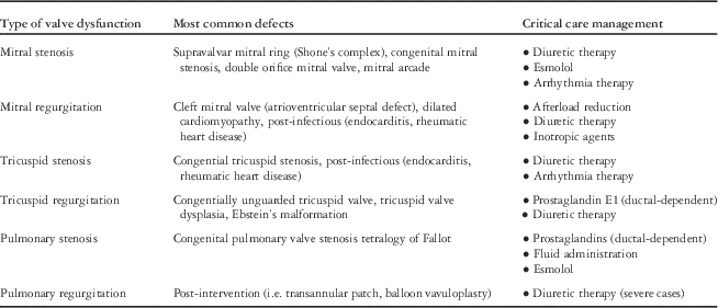
Although management of patients with valvar disease remains challenging, advances in diagnostic techniques and improvement in early recognition have significantly reduced morbidity and mortality in these populations. There continues to be room for improvement, with consideration towards more aggressive outpatient management to limit the need for pre-procedural intensive care admission. Future studies should focus on optimising and standardising in- and out-of-hospital care, with a goal of improving morbidity, mortality, and functional outcomes (Table 1).
Summary of types of valve dysfunction, common associated lesions, and critical care management
Acknowledgement
None.
Financial Support
This research received no specific grant from any funding agency, commercial or not-for-profit sectors.
Conflicts of Interest
None.
Ethical Standards
The authors assert that all procedures contributing to this work comply with the ethical standards of the relevant national guidelines.