Pulmonary hypertension is the most sinister cause of arterial desaturation in a patient with atrial septal defect. In some cases, however, arterial desaturation can occur despite normal pulmonary artery pressure. Therefore, for optimal management, it is important to identify the exact cause of arterial desaturation in every patient with atrial septal defect. Herein, we report yet another mechanism of arterial desaturation in a patient with atrial septal defect in absence of pulmonary hypertension.
Case summary
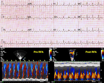
A 15-year-old schoolboy with a large atrial septal defect presented with exertional dyspnoea. Oxygen saturation on pulse oximetry was 92%, which dropped to 85% on exertion. A 12-lead electrocardiogram showed atrial flutter with variable atrioventricular conduction and an incomplete right bundle branch block (Fig 1a). Echocardiography confirmed the presence of a large ostium secundum atrial septal defect with a deficient inferior rim. The right atrium and right ventricle were dilated. Interestingly, the flow across the atrial septal defect was bidirectional (Online videos 1, 2). Colour motion mode across atrial septal defect revealed that the right-to-left shunt coincided with the flutter waves, which did not conduct to the ventricle (Fig 1b, yellow arrows). Cardiac catheterisation confirmed increased pulmonary blood flow with a ratio of pulmonary to systemic blood flow 2.5:1, and normal pulmonary artery pressure.
Twelve-lead ECG (a), colour M-mode echocardiogram pre- (b), and post-RFA (c) from a patient with atrial septal defect with arterial desaturation at presentation. The ECG (a) shows atrial flutter with variable atrioventricular conduction and incomplete right bundle branch block. Colour M-mode echocardiogram (b) demonstrates bidirectional flow across atrial septal defect with right-to-left shunt coinciding with non-conducted flutter waves (yellow arrow). The flow across atrial septal defect normalised following the resumption of sinus rhythm after radiofrequency ablation (c).

Electrophysiology study confirmed isthmus-dependent atrial flutter, which was successfully treated with radiofrequency ablation. Following the resumption of normal sinus rhythm, the echocardiography showed predominantly left-to-right shunt across atrial septal defect (Fig 1c, Online videos 3, 4). The effort tolerance of the patient also improved with the disappearance of arterial desaturation both at rest and on exertion. The patient underwent uneventful atrial septal defect surgical closure with no recurrence of atrial flutter.
Discussion
The flow across atrial septal defect has phasic variation. During the major part of the cardiac cycle, the blood flows from the left atrium to the right atrium, but the flow reverses briefly at the onset of ventricle systole and ventricular diastole. Reference Lin, Fu, Yeh and Wu1 This flow reversal coincides with a minor reversal of pressure gradient between the left atrium and right atrium, during z point, x descent, and y descent of left atrial pressure trace. Reference Lin, Fu, Yeh and Wu1 The flow across atrial septal defect is also affected by respiration. During inspiration, discrepant, out of phase venous return of systemic and pulmonary veins result in transient exaggeration of right-to-left shunt across atrial septal defect. Reference Rudolph and Rudolph2 A similar flow reversal across atrial septal defect is also observed during the release phase of the Valsalva maneuver, breath-holding episodes, and crying. Reference Rudolph and Rudolph2 Such transient exaggeration of a right-to-left shunt, however, does not result in arterial desaturation.
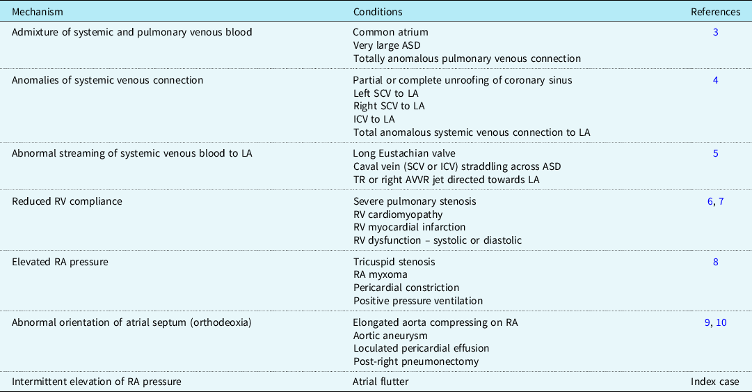
Arterial desaturation in atrial septal defect is commonly due to pulmonary hypertension, but can exist despite normal pulmonary artery pressure (Table 1). None of these mechanisms was present in our case.
Mechanism and causes of arterial desaturation in atrial septal defect with normal or mildly elevated pulmonary artery pressure

ASD = atrial septal defect; AVVR = atrioventricular valve regurgitation; ICV = inferior caval vein; LA = left atrium; RA = right atrium; RV = right ventricle; SCV = superior caval vein; TR = tricuspid regurgitation.
Atrial flutter is a common arrhythmia in the setting of atrial septal defect and is well known to cause clinical worsening. This case demonstrates atrial flutter induced right-to-left shunt across atrial septal defect despite normal pulmonary artery pressure. We postulate it to be the result of relatively higher pressure in the right atrium compared to the left atrium consequent to frequent contraction of the atriums against closed atrioventricular valves. The mechanism is also supported by normalisation of flow across atrial septal defect, the disappearance of arterial desaturation, and improved functional capacity following successful radiofrequency ablation. We believe that atrial flutter related arterial desaturation is yet another mechanism of clinical worsening in patients with atrial septal defect and is a potential target for treatment while the patient awaits closure of atrial septal defect.
Supplementary material
To view supplementary material for this article, please visit https://doi.org/10.1017/S104795112100010X.
Acknowledgements
The authors acknowledge the help received from Professor Shyam S. Kothari in the analysis and haemodynamic correlation of the case.
Summary
Atrial flutter exaggerates right-to-left shunt across atrial septal defect and is yet another mechanism of arterial desaturation despite normal pulmonary artery pressure and normal right ventricular compliance. The appreciation of this mechanism is important for optimal management.
Author contributions
Saurabh K. Gupta was involved in the concept and design of the manuscript. Sakshi Sachdeva drafted the manuscript. Saurabh K. Gupta critically reviewed and approved the final submitted manuscript. Late Dr Rajnish Juneja was significantly involved in patient management.
Financial support
This research received no specific grant from any funding agency, commercial, or not-for-profit sectors.
Conflicts of interest
None.
Ethical standards
The authors assert that all procedures contributing to this work comply with the ethical standards of the relevant national guidelines on human experimentation (please name) and with the Helsinki Declaration of 1975, as revised in 2008.