Introduction
The Amazon region harbours the largest freshwater drainage basin in the world, covering approximately 7,000,000 km2 and extending across all countries in northern South America, from the Andean foothills to the Atlantic Ocean (Val et al Reference Val, Almeida-Val, Fearnside, Santos, Piedade, Junk, Nozawa, Silva, Dantas and Tundisi2010). Corresponding to 6% of this area are the ‘floodplains’, which are regions located along white-water river margins that are subject to periodic flooding regulated by the flood pulse (Junk et al Reference Junk, Bayley, Sparks and Dodge1989). These are highly fertile and productive ecosystems, and the alternation between flood and drought periods influences a range of biological processes in the organisms inhabiting these environments, including fish and their parasites (Goulding et al Reference Goulding, Barthem and Ferreira2003; Moreira et al Reference Moreira, Oliveira, Morey and Malta2017).
Seasonality exerts a strong influence on the dynamics of aquatic communities, as it affects, among several factors, trophic interactions that regulate the life cycles of parasites and the availability of their intermediate and definitive hosts. Consequently, seasonal patterns may alter the transmission of these organisms, as well as the composition and structure of parasite infracommunities in aquatic hosts (Esch and Fernández Reference Esch and Fernández1993; Schade et al Reference Schade, Raupach and Wegner2016; Timi and Poulin Reference Timi and Poulin2020). In tropical regions, it has been reported that the organization of fish parasite populations may be differentially affected (Vasconcelos and Tavares-Dias Reference Vasconcelos and Tavares-Dias2014) as a result of water level fluctuations. In the case of endoparasites, the internal environment of their hosts may influence their distribution (Kennedy Reference Kennedy2006) and affect their preference for specific infection sites (Aguiar et al Reference Aguiar, Oliveira, Matos, Gomes, Costa and Silva2018; Matos et al Reference Matos, Oliveira, Farias, Silva and Malta2026).
Parasites are structural components of ecological communities, exhibiting non-random patterns of distribution and abundance influenced by environmental factors and host ecology. Thus, they reflect ecological interactions and can provide indirect information about ecosystem functioning (Poulin Reference Poulin1992). Neotropical freshwater fish may be infected by a wide diversity of endoparasites, comprising different taxonomic groups, such as the phylum Platyhelminthes (classes Trematoda and Cestoda), Nematoda, and Acanthocephala (Thatcher Reference Thatcher2006). Among these, acanthocephalan helminths are interesting biological models for investigating the effects of seasonality on host–parasite interactions, since their complex life cycles and population responses, which are sensitive to changes in the aquatic environment, make them excellent ecological indicators (Perrot-Minnot et al Reference Perrot-Minnot, Cozzarolo, Amin, Barčák, Bauer, Filipović Marijić, García-Varela, Hernández-Orts, Yen Le, Nachev, Orosová, Rigaud, Šariri, Wattier, Reyda and Sures2023).
The phylum Acanthocephala is a small monophyletic group composed of parasites known as ‘thorny-headed worms’. This group comprises almost 1,300 species, with estimates that may exceed 1,500 species, reflecting the continuous description of new taxa as well as taxonomic revisions based on recent molecular data (Bennet et al Reference Bennet, Poulin and Presswell2023). They are obligate endoparasites of the intestines of definitive hosts, with transmission occurring through trophic interactions involving intermediate hosts. Although the egg stage may persist in the environment, development to infective stages requires ingestion by an intermediate host. This is a highly successful and widely distributed group found in all classes of vertebrates, particularly in freshwater teleost fishes (Aguilar-Aguilar et al Reference Aguilar-Aguilar, González-García and García-Valera2026).
In temperate regions, seasonal changes in acanthocephalan dynamics are relatively well documented and influence the transmissibility of these parasites to their hosts, resulting in variation in infection levels, intestinal distribution patterns of these helminths, and population parameters such as sex ratio, size class distribution, and gonadal development (Aguiar et al Reference Aguiar, Oliveira, Matos, Gomes, Costa and Silva2018; Boping and Wenbin Reference Boping and Wenbin2007; Carpio-Hernández et al Reference Carpio-Hernández, Violante-González, Monks, Rojas-Herrera, García-Ibáñez, Toribio-Jiménez and Castro-Mondragón2020; Matos et al Reference Matos, Oliveira, Farias, Silva and Malta2026; Rauque et al Reference Rauque, Semenas and Viozzi2006). In contrast, studies on tropical and subtropical acanthocephalans remain comparatively scarce, despite growing evidence that seasonality also affects these populations in warmer regions (Aguiar et al Reference Aguiar, Oliveira, Matos, Gomes, Costa and Silva2018; Carpio-Hernández et al Reference Carpio-Hernández, Violante-González, Monks, Rojas-Herrera, García-Ibáñez, Toribio-Jiménez and Castro-Mondragón2020; Matos et al Reference Matos, Oliveira, Farias, Silva and Malta2026; Violante-González et al Reference Violante-González, Villalba-Vásquez, Monks, García-Ibáñez, Rojas-Herrera and Flores-Garza2017a, Reference Violante-González, Marquez-Silva, Monks, García-Ibañez, Pulido-Flores, Rojas-Herrera and Flores-Rodríguez2017b). For instance, Violante-González et al (Reference Violante-González, Villalba-Vásquez, Monks, García-Ibáñez, Rojas-Herrera and Flores-Garza2017a, Reference Violante-González, Marquez-Silva, Monks, García-Ibañez, Pulido-Flores, Rojas-Herrera and Flores-Rodríguez2017b) described the reproductive traits and population dynamics of Neoechinorhynchus brentnickoli in the Pacific fat sleeper (Dormitator latifrons) from a tropical lagoon in Mexico, demonstrating that seasonal fluctuations influence infection levels and reproductive timing even in tropical systems.
Octospiniferoides incognita Schmidt & Hugghins, Reference Schmidt and Hugghins1973 is an acanthocephalan belonging to the family Neoechinorhynchidae, with records associated with fish of the family Anostomidae, whose species are abundant in Amazonian floodplain lakes (Oliveira et al Reference Oliveira, Gonçalves, Ferreira, Pinheiro, Neves, Dias and Tavares-Dias2017). The frequent occurrence of these acanthocephalans in these hosts has been reported in studies that are predominantly taxonomic, including morphological descriptions of adult specimens (Schmidt and Huggins Reference Schmidt and Hugghins1973; Thatcher Reference Thatcher2006), records of their natural hosts, and infection levels expressed through parasitological indices (Matos et al Reference Matos, Oliveira, Farias, Silva and Malta2026; Silva de Souza et al Reference de Souza AK, Porto, Silva and Malta2017; Thatcher Reference Thatcher1998). Studies integrating the ecological dynamics of parasitism by O. incognita with the influence of seasonality imposed by the flood pulse in Amazonian floodplain ecosystems remain limited. Thus, in this study, we investigate the population structure and reproductive dynamics of O. incognita throughout seasonal water level fluctuations. We hypothesize that infection levels peak during high water, reproductive activity concentrates during rising and high water, and intestinal distribution varies seasonally between sexes. This approach is particularly relevant, considering that these environments are characterized by high variability in river discharge, a well-known structuring factor of ecological interactions in floodplain systems.
Materials and methods
The host
Rhytiodus microlepis is an herbivorous anostomid fish with a wide distribution in the Amazon Basin and frequent occurrence in floodplain environments associated with the flood pulse. The species has a predominantly herbivorous diet, feeding mainly on aquatic macrophytes, algae, seeds, and other plant material available in floodplain environments (Santos Reference Santos1981). Morphologically, the species has an elongated body and can reach up to 40 cm in total length. The mouth is slightly upturned, with four narrow-based incisiform teeth with broad edges on each jaw. Its lateral line has 85 to 95 scales, and its coloration is dark grey on the dorsal region and silvery on the ventral region (Queiroz et al Reference Queiroz, Torrente-Vilara, Ohara, Pires, Zuanon and Doria2013). Among anostomids, R. microlepis is the largest species commercially traded in fish markets in the Amazon region (Santos et al Reference Santos, Ferreira and Zuanon2006).
Sampling of hosts and acanthocephalans
183 specimens of R. microlepis were collected between July 2022 and November 2023 in the Catalão lake complex (Iranduba city, Amazonas, Brazil), located in a floodplain area near the confluence of the Negro and Solimões rivers (3°10′04″S, 59°54′45″W). Fish were sampled during the four seasonal phases of the water level fluctuations: rising water, which typically occurs between January and April; high water, between May and July; receding water, between August and September; and low water, between October and December (Bittencourt and Amadio Reference Bittencourt and Amadio2007; Espinoza-Villar et al Reference Espinoza-Villar, Guyot, Ronchail, Cochonneau, Filizola, Fraizy, Labat, Oliveira, Ordonez and Vauchel2009).
Fish were captured using gill nets with mesh sizes of 20, 25, and 30 mm between opposite knots, which were set in the lake for a standardized period of 10 hours during the daytime, with net checks performed every 4 hours. After capture, fish were anesthetized and euthanized by spinal cord sectioning, measured (cm), and weighed (g).
During necropsy, the host intestine was carefully removed and longitudinally opened for parasite recovery. Acanthocephalans were transferred to a Petri dish containing chilled distilled water and maintained at low temperature to allow full eversion of the proboscis and, in males, the copulatory bursa. After this process, a subsample of 693 parasites was fixed in 10% buffered formalin for a period of 48 hours.
Parasitic load, population structure, and intestinal distribution of acanthocephalans in hosts
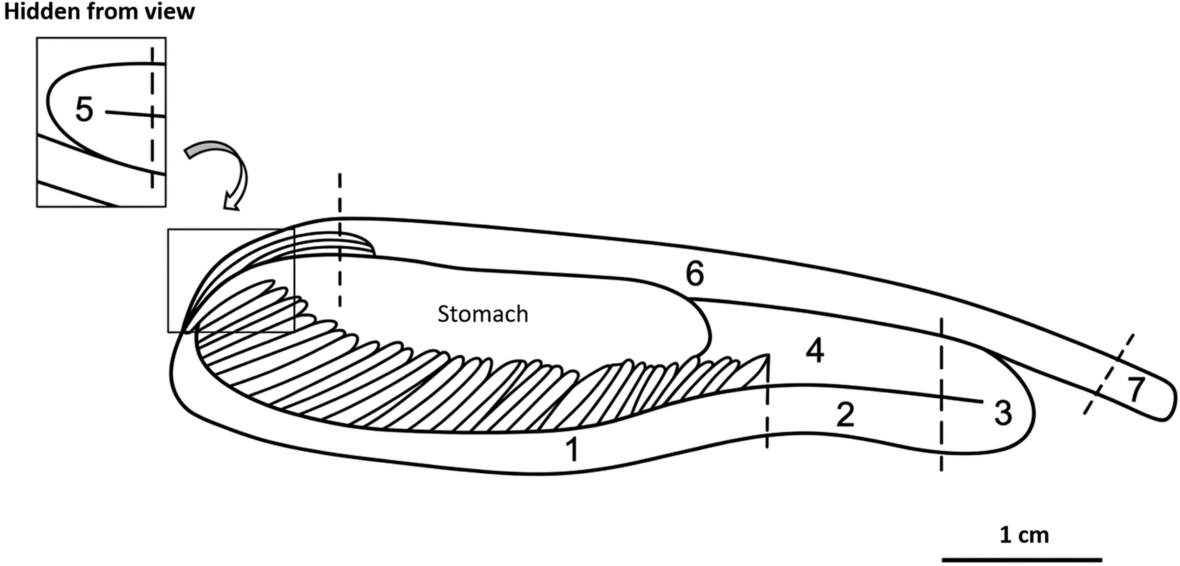
In each host, the number of acanthocephalans was recorded for the estimation of parasitological indices, including mean and total intensity, prevalence, and abundance, following the methods proposed by Bush et al (Reference Bush, Lafferty, Lotz and Shostak1997). Parasite density, size (mm), and sex were recorded for three intestinal regions: anterior intestine, comprising the first portions of the tube (P1 to P3); middle intestine, including the intermediate portions (P4 and P5); and posterior intestine, comprising the terminal portions of the intestinal tube (P6 to P7), as described by Matos et al (Reference Matos, Oliveira, Malta and Silva2021) (Figure 1).
Anatomical overview of the digestive tract of Rhytiodus microlepis, showing the delimitation of intestinal regions. P1–P3 (anterior intestine), P4–P5 (middle intestine), and P6–P7 (posterior intestine). Adapted from Matos et al (Reference Matos, Oliveira, Malta and Silva2021).

The stages of sexual maturation of acanthocephalans were determined under a stereomicroscope (LEICA EZ4) with specimens clarified using lactophenol to enhance visualization of internal features, and/or microscopic examination of the gonads and associated reproductive structures, such as the copulatory bursa in males and the copulatory cap in females (adapted from Boping & Wenbin, Reference Boping and Wenbin2007). Permanent slides of specimens at different developmental stages were prepared using carmine staining following Amato and Amato (Reference Amato, Amato, Von Matter, Queiroz Piacentini, Accordi and Cândido2010), and some samples were processed for histology using paraffin embedding. Sections of 4.5 μm were obtained using a semi-automatic microtome (Leica DM 2700) and stained with haematoxylin and eosin (H&E) for observation of reproductive structures.
Statistical analyses
Prevalence differences among seasonal phases of water level were tested using a binomial GLM with flood phases as the predictor. Abundance differences were analysed using a negative binomial GLM (accounting for overdispersion and zeros in the low-water phase). Sex ratio was determined as the proportion of females among all sexed individuals in each seasonal water-level phase, using only specimens for which sex could be identified. Individuals with undetermined sex were excluded from this calculation. The observed proportion of females and males in each phase was then compared against an expected 1:1 ratio using chi-square tests for proportions. Associations between reproductive stages and seasonal phases, and parasite distribution across intestinal sections (portions 1–7), were tested with chi-square tests (goodness-of-fit for homogeneity; independence for sex × section). All analyses were performed in R v4.3 (packages: MASS for GLM; stats for chi-square), α = 0.05.
Ethical statement
The collection of host and parasite specimens was previously authorized under a license issued by the Brazilian Biodiversity Authorization and Information System (SISBIO) (permit no. 81505-1). All procedures involving anaesthesia and euthanasia of hosts used in this study were approved by the Animal Use Ethics Committee (CEUA) of the National Institute for Amazonian Research (INPA) (protocol no. SEI 01280.000794/2022-99) and were conducted in accordance with the regulations of the Brazilian National Council for the Control of Animal Experimentation (CONCEA, 2018).
Results
Infection levels of Octospiniferoides incognita across seasonal water level fluctuations
A total of 3.983 acanthocephalans were recovered from the intestines of the examined hosts. The prevalence of O. incognita infection in R. microlepis was high during high-water periods, with 73% of fish infected during the rising water phase and 100% at the peak of high water. Statistically, seasonal water level fluctuations significantly affected prevalence (binomial GLM, LRT: χ2 = 114,83; df = 3; p < 0,001), with higher odds of infection during high water compared to rising water after model adjustment (OR = 39,33; 95% CI = 4,75–5128,37; p < 0,001), whereas receding water did not differ from rising water after adjustment (OR = 2,76; IC 95% = 0,86–10,22; p = 0,5406).
Abundance values indicate that throughout most of the seasonal water level fluctuations (rising, high, and receding water), host populations remained infected by O. incognita, with higher infection levels during high and receding water, when the highest intensity values were also recorded (Table 1). Mean abundance also differed among phases of these fluctuations (negative binomial GLM; LRT: χ2 = 290,48; df = 3; p < 0,001), being higher during high water compared to rising water (IRR = 16,33; 95% CI = 10,54–25,29; p < 0,001).
Parasitic indices of Octospiniferoides incognita in Rhytiodus microlepis collected across the seasonal water level fluctuations of Catalão Lake, Iranduba, Amazonas, Brazil

Table 1. Long description
The table consists of five columns and seven rows. The columns represent the Parasitic index followed by four seasonal water phases: Rising water (23.7 plus or minus 2.5 m), High water (27.9 plus or minus 0.9 m), Receding water (23.4 plus or minus 3.1 m), and Low water (16.4 plus or minus 2.5 m).
* Prevalence (percent): Rising 73; High 100; Receding 89; Low 0.
* Mean Intensity plus or minus D P: Rising 4 plus or minus 3.6; High 47.7 plus or minus 48.4; Receding 45.1 plus or minus 47.7; Low 0.
* Intensity range: Rising 1 to 16; High 2 to 246; Receding 1 to 166; Low 0.
* Abundance: Rising 2.9; High 47.7; Receding 40.1; Low 0.
* Total parasites: Rising 108; High 2,431; Receding 1,444; Low 0.
* Total hosts (mean plus or minus S D): Rising 37 (26 plus or minus 2.67); High 51 (25.8 plus or minus 2.31); Receding 36 (25.8 plus or minus 2.31); Low 33 (24.6 plus or minus 2.67).
Mean values ± standard deviation of water level for the sampling period of this study are shown in parentheses.
Size structure and spatial distribution of acanthocephalans in the intestine of R. microlepis
The relative frequency distribution across body length classes revealed marked sexual size dimorphism throughout the seasonal water level fluctuations (Figure 2). During the rising water phase, males were more frequent in the smallest size classes, with peaks between 0,4 and 0,6 cm, whereas females exhibited a broader size distribution, with higher occurrence in intermediate and larger classes, especially between 0,8 and 1,6 cm. In the high water phase, greater overlap between sexes was observed, although males still predominated in the smallest classes. Females maintained higher relative frequencies in intermediate classes, resulting in a more homogeneous distribution compared to the rising water phase. During the receding water phase, sexual segregation remained evident. Males continued to be concentrated in the smallest length classes, whereas females showed higher frequencies in the larger classes, reaching the maximum body sizes recorded in the study during this phase.
Relative frequency distribution of total length (cm) of male and female Octospiniferoides incognita during the rising, high, and receding water phases of Catalão Lake, Iranduba, Amazonas, Brazil.

Figure 2. Long description
A multi-panel bar chart with three vertical sections. A legend at the top identifies blue bars as Male and purple bars as Female.
* Axes: The x-axis represents Total length in centimeters ranging from 0 to 2.6. The y-axis represents Relative frequency percentage ranging from 0% to 50%.
* Top Panel (Rising water): Male frequency peaks sharply at 0.4 cm (nearly 50%) and 0.6 cm (over 40%), with no males recorded beyond 0.8 cm. Female frequency is more widely distributed, peaking at 1.0 cm (20%) and extending up to 2.4 cm.
* Middle Panel (High water): Male frequency peaks at 0.4 cm (over 40%) and decreases steadily toward 1.0 cm. Female frequency is spread across the range, with a primary peak at 0.4 cm (nearly 20%) and smaller peaks at 1.0 cm and 1.4 cm, extending to 2.2 cm.
* Bottom Panel (Receding water): Male frequency shows three distinct peaks at 0.4 cm (35%), 0.6 cm (28%), and 0.8 cm (28%). Female frequency peaks at 1.0 cm (nearly 20%) and 1.4 cm (over 10%), with the distribution ending at 2.0 cm.
Across all phases, males are consistently smaller and more concentrated in lower length classes, while females exhibit a broader range and larger maximum lengths.
The intermediate region of the intestine was the main site of occupation by O. incognita in both sexes (Figure 3). The distribution of females along the intestine varied significantly among water level fluctuations (χ2 = 56.53, p < 0.001). During rising water, females predominated in the anterior intestinal region (60%), whereas during high and receding water they were concentrated in the intermediate and posterior regions, with no significant difference in frequency between these phases (p = 1.000). Among males, variation was less pronounced. A significant difference was observed only between rising and high water phases (p = 0,029). During rising water, males were mainly concentrated in the anterior region (33%) and absent from the posterior region; during high water, some individuals shifted to the posterior region (24%). All other comparisons were not significant (p > 0,30). Males and females differed only during rising water (p = 0,047), when the proportion of females in the anterior region (60%) exceeded that of males (33%). During high and receding water, spatial distribution was similar between sexes, with predominance in the intermediate region (62%–69%).
Relative frequency of male and female Octospiniferoides incognita in the anterior (P1–P4) and posterior (P5–P7) intestinal regions of Rhytiodus microlepis across the seasonal water level fluctuations in an Amazonian floodplain lake.

Figure 3. Long description
A multi-panel heat map grid with three main columns representing seasonal water levels: Rising water, High water, and Receding water.
Axes and Legend:
* Vertical Y-axis: Intestinal region divided into Anterior (P 1 to P 2), Intermediate (P 3 to P 5), and Posterior (P 6 to P 7).
* Horizontal X-axis: Sex, divided into Males and Females for each seasonal panel.
* Legend: A vertical color scale for Relative frequency ranging from light tan (0 percent) to dark brown (60 percent).
Data Breakdown:
1. Rising water panel:
* Anterior: Males 33 percent, Females 60 percent.
* Intermediate: Males 67 percent, Females 35 percent.
* Posterior: Males show a grey cell indicating absence of records, Females 5 percent.
2. High water panel:
* Anterior: Males 15 percent, Females 13 percent.
* Intermediate: Males 62 percent, Females 69 percent.
* Posterior: Males 24 percent, Females 19 percent.
3. Receding water panel:
* Anterior: Males 20 percent, Females 15 percent.
* Intermediate: Males 64 percent, Females 64 percent.
* Posterior: Males 16 percent, Females 22 percent.
Statistical notes at the bottom indicate Chi-square tests for region by period by sex, with significant p-values for both males and females across seasonal transitions.
Maturation stages, L50 and reproductive dynamics of O. incognita
Three reproductive morphotypes were identified in males of O. incognita (Figures 4 and 5): (1) initial maturation stage: smaller specimens in which testes, sperm ducts, cement gland, and cement reservoir were not clearly distinguishable macroscopically. In this morphotype, microscopic examination revealed these structures, but with absence of sperm in the seminiferous tubules; (2) advanced maturation stage: males without an everted copulatory bursa, with testes, sperm ducts, cement gland, and cement reservoir clearly distinguishable macroscopically. Histological analysis revealed testes containing sperm in the seminiferous tubules, indicating spermatogenic activity; and (3) active mature stage: males with an everted copulatory bursa, with testes, sperm ducts, cement gland, and cement reservoir clearly visible macroscopically. As in morphotype 2 males, seminiferous tubules contained sperm.
Macroscopic overview of reproductive morphotypes of male and female Octospiniferoides incognita (Acanthocephala: Neoechinorhynchidae) obtained from the host Rhytiodus microlepis in Catalão Lake, Iranduba, Amazonas, Brazil. Panels A–C represent males, and D–F represent females. T, testes; CG, cement gland; CR, cement reservoir; OB, ovarian balls; CC, copulatory cap; E, eggs.

Figure 4. Long description
A collection of six micrographs labeled A through F.
* Panel A shows a male parasite in a curled, C-shaped posture with a scale bar of 0.3 cm.
* Panel B shows a straightened male. Internal labels from top to bottom identify two T, a C G, and a C R. Scale bar is 0.4 cm.
* Panel C shows a male with a posterior inset box magnifying the tail region. The inset has an 800 micro-m scale bar. Main scale bar is 0.4 cm.
* Panel D shows a female parasite. A square in the mid-body corresponds to a bottom-left inset labeled O B, showing granular ovarian balls with a 400 micro-m scale bar. Main scale bar is 0.4 cm.
* Panel E shows a female with a posterior inset box magnifying the tail tip, labeled C C. The inset has an 800 micro-m scale bar. Main scale bar is 0.4 cm.
* Panel F shows a female with a posterior inset box magnifying the lower body, labeled E, showing elongated eggs. The inset has no separate scale bar, but the main scale bar is 0.5 cm.
Histology of reproductive morphotypes in male Octospiniferoides incognita (Acanthocephala: Neoechinorhynchidae) obtained from the host Rhytiodus microlepis in Catalão Lake, Iranduba, Amazonas, Brazil. Panel A shows a specimen in initial maturation (morphotype 1). Panels B and C show morphotypes 2 and 3, respectively, highlighting testes with seminiferous tubules containing sperm. Panel D shows a detail of the everted copulatory bursa in males of morphotype 3. T, testes; ST, seminiferous tubules; S, sperm; CB, copulatory bursa.

Figure 5. Long description
A four-panel histological series stained in pink and purple hues.
Panel A, top-left, shows a cross-section of a male in initial maturation. A 200 micro-meter scale bar is in the bottom right. Two large, rounded lobes labeled lowercase t for testes occupy the upper central cavity, surrounded by a thick outer body wall. A structure labeled p is visible near the bottom right of the internal cavity.
Panel B, top-right, is a higher magnification view of morphotype 2. A 50 micro-meter scale bar is in the bottom right. The field is filled with numerous dark purple, granular clusters labeled lowercase s t for seminiferous tubules.
Panel C, bottom-left, provides a high-detail view of morphotype 3. A 20 micro-meter scale bar is in the bottom right. It shows seminiferous tubules labeled lowercase s t containing fibrous, lighter-stained material labeled lowercase s for sperm.
Panel D, bottom-right, shows the posterior end of a morphotype 3 male. A 200 micro-meter scale bar is in the bottom right. The central feature is an everted, folded muscular structure labeled lowercase c b for the copulatory bursa, extending from the body opening.
For females of O. incognita, three morphotypes were also identified based on the organization and composition of the content within the pseudocoelomic cavity (Figures 4 and 6): (1) initial maturation stage: characterized by the presence of ovarian balls arranged within the pseudocoelom, macroscopically visible, with or without a copulatory cap. Microscopic analysis of this morphotype confirmed only the occurrence of aggregated ovarian balls, with no evidence of oocytes or eggs, suggesting a very early reproductive condition with no active production of offspring; (2) females in intermediate maturation stage: characterized by more dispersed ovarian balls, associated with the presence of oocytes and/or free eggs occupying the pseudocoelomic cavity, with or without a copulatory cap; and (3) females in advanced maturation stage: characterized by the absence of ovarian balls, with a predominance of eggs, frequently embryonated, occupying the entire pseudocoelomic cavity, with or without the presence of a copulatory cap. Across the analysed morphotypes, the copulatory cap was observed in some females regardless of their reproductive condition. This structure was considered evidence of mating occurrence, although its presence does not necessarily indicate successful fertilization.
Histology of reproductive morphotypes in female Octospiniferoides incognita (Acanthocephala: Neoechinorhynchidae) obtained from the host Rhytiodus microlepis in Catalão Lake, Iranduba, Amazonas, Brazil. The reproductive structures shown in Panels A–C were observed in the middle region of the body (metassoma). Panel A shows a specimen in initial maturation (morphotype 1), characterized by the presence of ovarian balls only. Panel B shows an intermediate-maturation female (morphotype 2), with ovarian balls and oocytes visible in the pseudocoelom. Panel C shows a morphotype 3 female. Panel D shows a detail of the copulatory cap. OB, ovarian balls; O, oocytes; CC, copulatory cap.

Figure 6. Long description
A four-panel histological series labeled A through D.
Panel A, top-left, shows morphotype 1 at a 50 micro-meter scale. The field is densely packed with O B, ovarian balls, which appear as dark purple, granular clusters of cells held within a light-colored matrix.
Panel B, top-right, shows morphotype 2 at a 50 micro-meter scale. The O B are larger and more distinct, surrounded by numerous elongated, spindle-shaped structures labeled e, representing oocytes, scattered throughout the pseudocoelom.
Panel C, bottom-left, shows morphotype 3 at a 50 micro-meter scale. This panel is entirely filled with a high density of mature oocytes, with the O B no longer visible.
Panel D, bottom-right, shows a lower magnification view at a 200 micro-meter scale. It depicts the posterior end of the female with a prominent C C, copulatory cap. The C C is a dark purple, bulbous mass attached to the folded, lighter purple tegument of the worm’s body.
The morphotype structure of O. incognita varied across seasonal water level fluctuations (Figure 7), with a consistent predominance of females in intermediate maturation (F2) across all periods, indicating that the population is largely composed of individuals in a functionally reproductive stage. During the rising water phase, a higher proportion of individuals in advanced maturation (morphotype 3 males and females) was observed, with post-copulatory characteristics, suggesting that this period concentrates the peak reproductive activity of acanthocephalans. At high water, there was a strong presence of females in intermediate maturation; however, a lower proportion of females bearing a copulatory cap was recorded. During the receding water phase, there was a marked reduction in the proportion of males with an everted copulatory bursa, suggesting, for both sexes, a post-reproductive transitional phase between high and receding water periods. Overall, morphotype assessment indicates that the population structure of O. incognita exhibits variation across seasonal water level fluctuations, with higher reproductive activity concentrated during rising water, followed by a maintenance phase at high water and a reduction in sexual activity indicators during receding water. Nevertheless, the continuous predominance of mature females across all periods suggests the maintenance of a stable reproductive contingent within the population, independent of these phases.
Proportion of reproductive morphotypes of Octospiniferoides incognita across seasonal water level fluctuations. Composition differed significantly among phases (χ2(6) = 18,46; p = 0,005), with the only significant pairwise difference observed between rising and receding water phases (Bonferroni; p = 0,002).

Figure 7. Long description
The Y-axis represents Proportion in percent from 0 to 60. The X-axis categorizes data into four morphotypes: M 2 (light blue), M 3 (dark blue), F 2 (tan), and F 3 (brown).
* In the Rising water panel, M 2 is 18 percent, M 3 is 16 percent, F 2 is 49 percent, and F 3 is 16 percent.
* In the High water panel, M 2 increases to 28 percent, M 3 decreases to 10 percent, F 2 increases to 54 percent, and F 3 decreases to 9 percent.
* In the Receding water panel, M 2 increases to 39 percent, M 3 decreases to 5 percent, F 2 is 52 percent, and F 3 decreases to 4 percent.
A legend on the right defines the morphotypes: M 2 is Advanced maturation male (sperm, no copulatory bursa); M 3 is Active mature male (sperm plus everted copulatory bursa); F 2 is Intermediate maturation female (ovarian balls plus oocytes or eggs, plus or minus cap); F 3 is Advanced maturation female (oocytes or eggs only, plus or minus cap).
The size at first sexual maturity (L50) of O. incognita was estimated at 0,12 cm, indicating the onset of the adult stage at a reduced body size, with a significant effect of total length on the probability of maturation (p < 0,001) (Figure 8). The logistic curve showed a sharp transition: individuals smaller than 0,1 cm had a probability of maturation below 25%, whereas from approximately 0,3 cm onward the probability exceeded 90%, with nearly all individuals above 0,4 cm classified as mature or post-copulatory. Immature individuals were recorded up to 0,7 cm in length, suggesting that maturation occurs early and that the persistence of immature stages at larger body sizes is uncommon.
Probability of sexual maturation of Octospiniferoides incognita as a function of total length (cm), estimated by logistic regression. The L50 (dashed vertical line) was 0.12 cm (p < 0.001). Points represent individuals in initial maturation (pink) and advanced/mature stages (blue); the shaded area indicates the 95% confidence interval.

Figure 8. Long description
The X axis represents Total length in centimeters from 0.0 to 0.6. The Y axis represents P open parenthesis adult close parenthesis as a percentage from 0 percent to 100 percent.
Data points:
- Pink circles representing Juveniles are clustered along the 0 percent line, mostly between 0.0 and 0.4 centimeters.
- Blue circles representing Adults are clustered along the 100 percent line, starting from approximately 0.1 centimeters and extending to 0.7 centimeters.
Trends and Markers:
- A solid dark blue logistic curve rises from left to right, starting near 25 percent at 0.0 centimeters and reaching an asymptote near 100 percent at 0.6 centimeters.
- A light blue shaded area around the curve represents the 95 percent C I.
- A horizontal dotted line marks the 50 percent probability level.
- A vertical dashed line intersects the curve at the 50 percent mark, labeled as L 50 equals 0.116 centimeters.
- Two vertical dotted lines flank the L 50 line, representing the 95 percent C I range from 0.054 to 0.178 centimeters.
- A text box in the center-left provides the specific values: L 50 equals 0.116 centimeters and 95 percent C I [0.054, 0.178].
Discussion
In the present study, variation in the parasitic load of the acanthocephalan Octospiniferoides incognita in the intestine of its natural host, Rhytiodus microlepis, corroborates the findings of Matos et al (Reference Matos, Oliveira, Farias, Silva and Malta2026), who evaluated the influence of seasonal water level fluctuations on infection dynamics in the same host–parasite system. The high prevalence of infected hosts during inundation periods and the marked increase in mean abundance during high water suggest that this phase provides more favourable environmental conditions for the establishment and proliferation of acanthocephalans. In Amazonian flood pulse environments, the high-water period is associated with a marked expansion of flooded areas, which may influence host–parasite interactions by modifying habitat availability and trophic relationships. Although the intermediate host of O. incognita remains unknown, previous studies have shown that aquatic invertebrates, including ostracods and other crustaceans, can act as intermediate hosts for acanthocephalans in freshwater ecosystems (Fanton et al Reference Fanton, Franquet, Logez, Cavalli and Kaldonski2022; Gallhammer et al Reference Gallhammer, Grimm, Reier and Sefc2025). For example, Cypridopsis vidua (Ostracoda) has been reported as the intermediate host of Neoechinorhynchus buttnerae, a species also belonging to the family Neoechinorhynchidae (Lourenço et al Reference Lourenço, Morey and Malta2018). Therefore, the increase in infection levels observed during high water may be associated with changes in environmental conditions that favour trophic transmission, but further studies investigating potential intermediate hosts are necessary to confirm this hypothesis. On the other hand, the absence of hosts infected by O. incognita during the low water period may be associated with a disruption of the transmission chain, since the decrease in water level reduces habitat connectivity and negatively affects the abundance and diversity of aquatic invertebrates, which are essential for heteroxenous life cycles (Poulin Reference Poulin2011) typical of acanthocephalan biology.
By expanding the investigation of the host–parasite relationship through the assessment of reproductive morphotype composition across the seasonal water level fluctuations, the temporal dynamics of reproduction in the acanthocephalan population were revealed, representing an aspect still unexplored for these parasites in Amazonian floodplain environments. During rising water, when O. incognita infrapopulations in hosts are small, as evidenced by the low parasite abundance in this period, the proportion of reproductively active males and females is high, indicating that copulation begins at this stage. The presence of the copulatory cap in adult females provides evidence of male insemination, as this structure is formed by secretions from the male cement glands that seal the female gonopore after copulation (Crompton Reference Crompton, Crompton and Nickol1985; Dezfuli and Biaggi Reference Dezfuli and Biaggi2000). Although direct observations of copulation are rarely reported in acanthocephalans, the copulatory cap is a well-documented reproductive structure among several species, functioning as a post-copulatory plug that may prevent subsequent inseminations and reduce sperm competition (Dezfuli et al Reference Dezfuli, Pironi, Rossetti and Herlyn2024; Leidenberg et al Reference Leindenberg, Boström and Wayland2019). However, the rising water phase marks the onset of copulatory activity rather than its reproductive peak. During high and receding water, although the proportion of post-copulatory morphotypes declines, the absolute number of mature females is higher: 54% of 508 individuals during high water compared to 49% of only 61 individuals during rising water. Thus, effective egg maturation is concentrated during high water and the early receding water phase, when the high abundance of mature females may coincide with the environmental conditions that favour the availability of potential intermediate hosts. In practical terms, copulation in acanthocephalans begins during rising water, but it is during high water that most eggs are effectively produced and potentially fertilized. We acknowledge that egg production was not quantified in this study; future fecundity estimates would provide a more direct measure of peak reproductive output. This temporal separation between copulation and peak egg production has also been reported for Acanthocephalus tumescens in Patagonian lakes, where low temperatures delay reproductive processes following recruitment (Rauque et al Reference Rauque, Semenas and Viozzi2006). The sex ratio biased toward females during rising water, with a tendency toward equilibrium during receding water, is consistent with the greater longevity of females within the host, a pattern commonly reported in acanthocephalans (Crompton and Whitfield Reference Crompton and Whitfield1968; Poulin and Morand Reference Poulin and Morand2004). The rebalancing observed during receding water, accompanied by an increase in mature males, confirms the influx of recruits that characterizes this phase. Therefore, in Catalão Lake, a typical Amazonian floodplain lake, the occurrence of mature acanthocephalans throughout the entire period, together with a higher proportion of sexually active individuals during rising water, suggests temporal variation in the reproductive activity of the species. This pattern is consistent with the acanthocephalan ecology literature, which shows that environmental factors and host availability influence population dynamics and reproductive processes, often resulting in seasonal variation in parasite activity (Carpio-Hernández et al Reference Carpio-Hernández, Violante-González, Monks, Rojas-Herrera, García-Ibáñez, Toribio-Jiménez and Castro-Mondragón2020; Poulin Reference Poulin2011).
The distribution of O. incognita within the intestine is not random. Concentration in the intermediate portions of the organ evidences microhabitat selection, which may be driven by gradients of pH, nutrients, and mucosal characteristics (Dezfuli et al Reference Dezfuli, Pironi, Campisi, Shinn and Giari2010; Richardson and Nickol Reference Richardson and Nickol2000). Our results suggest that females respond more directly to intestinal microenvironmental conditions throughout the cycle, possibly seeking optimal sites for maturation and egg release, which is consistent with observations in Paulisentis missouriensis, where gravid females aggregate in the anterior intestinal region, and males follow this distribution (Lawlor et al Reference Lawlor and Nickol1990; McCormick and Nickol Reference McCormick and Nickol2004). The expansion of females into more posterior intestinal regions during the transition from rising to high water may be related to density-dependent displacement, as previously reported by Kennedy et al (Reference Kennedy, Broughton and Hine1976), who observed that with increasing parasite load during high water, larger females of Pomphorhynchus laevis are pushed into adjacent regions away from the optimal site. In contrast, male stability is interpreted as a dispersal strategy aimed at maximizing encounters with mates (Nickol Reference Nickol and Woo2006). The sexual size dimorphism (males 0,2–0,8 cm; females up to 2,6 cm), expected for acanthocephalans (Poulin and Morand Reference Poulin and Morand2004), supports this interpretation, suggesting that females, being larger and metabolically more demanding, may be more susceptible to spatial constraints within the intestine. In dioecious helminths such as Acanthocephala and Nematoda, female size is primarily related to egg production, which should be considered when interpreting size-related patterns in parasite populations.
O. incognita reaches sexual maturity at a small body size. The logistic curve shows a rapid transition, with less than 25% probability of maturation below 0,1 cm and more than 90% above 0,3 cm. There are no published estimates of L50 for O. incognita. Violante-González et al. (Reference Violante-González, Villalba-Vásquez, Monks, García-Ibáñez, Rojas-Herrera and Flores-Garza2017a) estimated L50 values of 2.9 mm for females and 2.3 mm for males of Neoechinorhynchus brentnickoli from a tropical coastal lagoon in Mexico. The L50 of 0.12 cm (1.2 mm) estimated for O. incognita in the present study is lower, indicating an exceptionally early maturation relative to a congeneric species from a comparable tropical system. This difference may reflect an adaptive response to the seasonality of the Amazonian flood pulse, where the compressed window of favourable conditions for transmission may select for rapid reproductive activation at minimal body sizes. In contrast, the comparatively stable environmental conditions of Tres Palos Lagoon allow N. brentnickoli to maintain continuous reproduction year-round (Violante-González et al Reference Violante-González, Villalba-Vásquez, Monks, García-Ibáñez, Rojas-Herrera and Flores-Garza2017a, Reference Violante-González, Marquez-Silva, Monks, García-Ibañez, Pulido-Flores, Rojas-Herrera and Flores-Rodríguez2017b), potentially relaxing the selective pressure for extremely early maturation. The rapid maturation of O. incognita is also consistent with the accelerated developmental pattern reported for Neoechinorhynchus buttnerae, whose complete larval development in the intermediate host is achieved in only 29 days (Lourenço et al Reference Lourenço, Morey and Malta2018), suggesting that early reproductive activation may be a shared trait among Neoechinorhynchidae.
The significant predominance of females during the rising water (sex ratio: 0,54) and high-water (sex ratio: 0,60) phases is common in acanthocephalans and may be interpreted as an adaptive strategy to maximize reproductive success under favourable environmental conditions, since females are metabolically more costly and produce thousands of eggs (Kennedy Reference Kennedy2006). Males, being generally smaller, may be produced in lower numbers, as a single male is capable of fertilizing multiple females (Nickol Reference Nickol and Woo2006).
The copulatory cap in mature females bearing fertilized eggs indicates successful insemination, occurring when males produce secretions from the cement glands that seal the female genital pore immediately after copulation (Crompton Reference Crompton, Crompton and Nickol1985; Dezfuli and Biaggi Reference Dezfuli and Biaggi2000; Boping and Wenbin Reference Boping and Wenbin2007). It is important to note that the copulatory cap is a post-copulatory structure exclusive to females; adult males do not possess this structure, but instead display an everted copulatory bursa and cement glands that produce the secretions forming the cap on the female (Crompton Reference Crompton, Crompton and Nickol1985; Nickol Reference Nickol and Woo2006). The higher proportion of reproductively active adult males and females indicates that the receding water phase is the period of active reproduction of O. incognita in the host intestine. The maintenance of mature individuals in subsequent phases of the seasonal water level may be associated with increased resource availability during high water, when river margins are inundated and a large number of intermediate hosts are released into the environment, facilitating fish infection and the recruitment of new parasites (Casali and Takemoto Reference Casali and Takemoto2016; Negreiros et al Reference Negreiros, Pereira, Tavares-Dias and Tavares2018).
Conclusions
These results advance the understanding of host–parasite dynamics in Amazonian floodplain environments by integrating population structure, reproductive morphology, and spatial distribution of acanthocephalans within the host intestine across a complete fluvial cycle. This study is the first to characterize the reproductive morphotypes, estimate the size at first sexual maturity, and relate these parameters to seasonal water level fluctuations for Octospiniferoides incognita in a floodplain lake. The synchronized patterns of infection and reproductive activity with the flood pulse suggest that fluvial seasonality is a key structuring force for this host-parasite system. While the incorporation of spatial replicates and experimental identification of the intermediate host will be important to broaden these findings, the present data demonstrate that the integrated approaches combining multiple population parameters provide critical insights into how parasites with complex life cycles respond to natural environmental fluctuations in Neotropical floodplain ecosystems.
Acknowledgements
The authors would like to thank the Conselho Nacional de Desenvolvimento Científico e Tecnológico (CNPq) for providing the scholarship that enabled the execution of this study, the Coordenação de Aperfeiçoamento de Pessoal de Nível Superior (CAPES/Brazil) – Finance Code 001, and the Fundação de Amparo a Pesquisa do Estado do Amazonas (FAPEAM), within the scope of the POSGRAD program.
Competing interests
The authors declare that they have no conflict of interest.